. 2015 Aug 27;34(35):4570-80.
doi: 10.1038/onc.2014.385. Epub 2014 Dec 15.

Claudin-7 expression induces mesenchymal to epithelial transformation (MET) to inhibit colon tumorigenesis

Affiliations

Claudin-7 expression induces mesenchymal to epithelial transformation (MET) to inhibit colon tumorigenesis

A A Bhat et al. Oncogene. .

Abstract

In normal colon, claudin-7 is one of the highly expressed claudin proteins and its knockdown in mice results in altered epithelial cell homeostasis and neonatal death. Notably, dysregulation of the epithelial homeostasis potentiates oncogenic transformation and growth. However, the role of claudin-7 in the regulation of colon tumorigenesis remains poorly understood. Using a large colorectal cancer (CRC) patient database and mouse models of colon cancer, we found claudin-7 expression to be significantly downregulated in cancer samples. Most notably, forced claudin-7 expression in poorly differentiated and highly metastatic SW620 colon cancer cells induced epithelial characteristics and inhibited their growth in soft agar and tumor growth in vivo. By contrast, knockdown of claudin-7 in HT-29 or DLD-1 cells induced epithelial-to-mesenchymal transition (EMT), colony formation, xenograft-tumor growth in athymic mice and invasion. Importantly, a claudin-7 signature gene profile generated by overlapping the DEGs (differentially expressed genes in a high-throughput transcriptome analysis using claudin-7-manipulated cells) with human claudin-7 signature genes identified high-risk CRC patients. Furthermore, Rab25, a colon cancer suppressor and regulator of the polarized cell trafficking constituted one of the highly upregulated DEGs in claudin-7 overexpressing cells. Notably, silencing of Rab25 expression counteracted the effects of claudin-7 expression and not only increased proliferation and cell invasion but also increased the expression of p-Src and mitogen-activated protein kinase-extracellular signal-regulated kinase 1/2 that were suppressed upon claudin-7 overexpression. Of interest, CRC cell lines, which exhibited decreased claudin-7 expression, also exhibited promoter DNA hypermethylation, a modification associated with transcriptional silencing. Taken together, our data demonstrate a previously undescribed role of claudin-7 as a colon cancer suppressor and suggest that loss of claudin-7 potentiates EMT to promote colon cancer, in a manner dependent on Rab25.


Conflict of interest statement

CONFLICT OF INTEREST

The authors declare no conflict of interest.

Figures
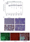
Figure 1
Figure 1
Claudin-7 expression is reduced in human colon cancer. (A) Normalized expression for claudin-7 in 10 normal adjacent specimens versus six adenoma specimens and 250 colon adenocarcinoma samples by microarray analysis. Stage I =33; Stage II =76; Stage III = 82; Stage IV =59; All stages: 250. Claudin-7 expression was significantly downregulated in the adenoma and adenocarcinoma groups compared with the adjacent normals (P<0.001). Median and s.d. are shown. (B) The expression pattern of claudin-7 was examined by immunohistochemical staining using 11 human colon cancer tissue samples. Decreased claudin-7 expression was observed in paraffin-embedded sections of normal and colonic tumor samples from the same patients. (C) Representative immunofluorescence images of tumors from APCmin mice immunostained for β-catenin and claudin-7.
Figure 2
Figure 2
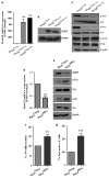
Manipulation of claudin-7 expression alters EMT and functional characteristics. SW620, SW620control, SW620claudin-7(1) and SW620claudin-7(2) cells (overexpressing claudin-7) were examined for ((a) (i and ii)) expression of EMT markers, including E-cadherin, Vimentin, N-cadherin, ZO-1, β-catenin, Zeb-1 and Snail-1. Actin was used as a loading control. (b) Localization of claudin-7, E-cadherin, β-catenin and ZO-1 was examined using immunofluorescence. (c and d) Anchorage-independent growth assay. Colonies were counted from three individual plates for each sample and were photographed 2 weeks after plating of the cells. The number of soft agar colonies presented is the mean of colony counts from three different experiments. ***P<0.001 compared with SW620control. (e and f) Cell invasion assay. Vector and claudin-7-expressing cells were grown on 24-well transwells coated with collagen type I (100 μg/ml). After 72 h of plating, cells from the top of the filter were removed, and the cells that had invaded the coated membrane were fixed and counted. Data are presented as mean colony counts in six × 20 microscopic fields from triplicate wells. *P<0.01, ***P<0.001, as compared with respective controls.
Figure 3
Figure 3
Loss of claudin-7 induces mesenchymal features in epithelial cells. HT29control, HT29shRNA, DLD-1control and DLD-1shRNA cells (inhibition of claudin-7) were utilized to determine ((a) (i and ii)) expression of EMT markers, including E-cadherin, Vimentin, N-cadherin, ZO-1, β-catenin, ZEB-1 and Snail-1, by immunoblotting. Actin was used as a loading control. (b) Localization of claudin-7, E-cadherin, β-catenin and ZO-1 was examined using immunofluorescence. (c and d) Anchorage-independent growth assay. Colonies were counted from three individual plates for each sample and were photographed 2 weeks after plating of the cells. The number of soft agar colonies presented is the mean of colony counts from three different experiments. ***P<0.001 compared with control cells. (e and f) Cell invasion assay. Vector and claudin-7 knockdown cells were grown on 24-well transwells coated with collagen type I (100 μg/ml). After 72 h of plating, cells from the top of the filter were removed, and the cells that had invaded the coated membrane were fixed and counted. Data are presented as mean colony counts in six × 20 microscopic fields from triplicate wells. **P<0.01. ***P<0.001, as compared with respective controls.
Figure 4
Figure 4
Effect of modulation of claudin-7 expression on tumor xenograft in vivo. SW620control or SW620Claudin-7 cells were subcutaneously injected into the left and right sides of the nude mice (n =7 mice per group). Circles indicate the tumors generated subcutaneously in nude mice. The nude mice were killed 4 weeks after the injection, and the tumors were removed and weighed. Claudin-7 expressing cell-induced tumors in nude mice were smaller in size compared with those of control cells (a and b). Conversely, HT29shRNA expressing cell-induced tumors in nude mice were bigger in size cells (c and d). Tumors were evaluated for markers of proliferation (Ki67), apoptosis (TUNEL) as well as claudin-7 and E-cadherin expression by immunostaining (e (i) and f (i)). Tumors were also immunoblotted for cleaved caspase-3, claudin-7 and E-cadherin (e (ii) and f (ii)). **P<0.01, ***P<0.001.
Figure 5
Figure 5
Claudin-7 expression in human CRC and association with clinical outcomes. (a) Unsupervised hierarchical clustering analysis for claudin-7. The claudin-7 gene expression profile (113 probes) separates 250 CRC patients into three clusters. (b and c) Analysis of clinical outcomes demonstrates that the claudin-7 gene signature is significantly associated with overall and disease-free survival in 250 CRC patients. Cluster 2 (blue) patients demonstrate better overall survival and less recurrent disease than clusters 3 and 1 patients (P =0.005 and <0.001, respectively). No association was noted with grade or adjuvant treatment; however, a significant association was noted between the clusters and the stage of the patients (P =0.02).
Figure 6
Figure 6
Claudin-7 effects are mediated through Rab25 signaling. (a and b) Expression of Rab25 was confirmed at the mRNA and protein levels in claudin-7-manipulated cells using quantitative real-time PCR (qRT–PCR) analysis and immunoblot analysis. Results are plotted as mean ±s.d. from three independent experiments and are presented as fold change. (c) Signaling proteins upregulated or downregulated in claudin-7-manipulated cells were determined by immunoblot analysis. (d) Silencing of Rab25 expression in SW620claudin-7 cells was confirmed by qRT–pCR. (e) Effect of the inhibition of Rab25 expression in SW620claudin-7 cells on ERK/Src signaling, cell proliferation (f) and cell invasion (g). Cellular proliferation was measured using the MTT assay after 48 h of transfection. Cell invasion was performed as described in Materials and methods. Results were plotted as mean ±s.d. from three independent experiments and presented as fold change. **P<0.01, ***P<0.001 when compared with control.
Figure 7
Figure 7
Suppression of claudin-7 expression is associated with the claudin-7 promoter hypermethylation in SW620 CRC cells. (a) Expression of claudin-7 in CRC cell lines determined by western blotting analysis. Actin was used as a loading control. ((b) (i and ii)) Methylation status of claudin-7 in colon cancer cell lines. Methylated (M) and unmethylated (U) gene sequences were amplified individually by methylation-specific PCR (MSP) using sodium-bisulfite-treated DNA from the indicated colon cancer cell lines. MSP products were resolved by electrophoresis on a 1.5% agarose gel. ((b) (ii)) Re-expression of claudin-7 in SW620 cells following treatment with the demethylating agent 5-aza-dC. Real-time PCR was performed for claudin-7, and PCR products were resolved by electrophoresis on a 1.5% agarose gel. (c) Immunoblotting of claudin-7 in SW620 cells following treatment with the demethylating agent 5-aza-dC. (d) SW480 and HCT15 cells were transfected with the pcDNA-3 expression plasmid containing the full-length human DNMT3A or vector alone and immuonoblotted for DNMT3A and claudin-7 after 48 h of transfection. Actin was used as a loading control.

References

    1. Tsukita S, Furuse M, Itoh M. Multifunctional strands in tight junctions. Nat Rev Mol Cell Biol. 2001;2:285–293. - PubMed
    1. Schneeberger EE, Lynch RD. The tight junction: a multifunctional complex. Am J Physiol Cell Physiol. 2004;286:C1213–C1228. - PubMed
    1. Soler AP, Miller RD, Laughlin KV, Carp NZ, Klurfeld DM, Mullin JM. Increased tight junctional permeability is associated with the development of colon cancer. Carcinogenesis. 1999;20:1425–1431. - PubMed
    1. Turksen K, Troy TC. Barriers built on claudins. J Cell Sci. 2004;117:2435–2447. - PubMed
    1. Sheehan GM, Kallakury BV, Sheehan CE, Fisher HA, Kaufman RP, Jr, Ross JS. Loss of claudins-1 and -7 and expression of claudins-3 and -4 correlate with prognostic variables in prostatic adenocarcinomas. Hum Pathol. 2007;38:564–5692007. - PubMed

Publication types

MeSH terms