Review
. 2015 Jan;36(1):88-101.
doi: 10.1038/aps.2014.133. Epub 2014 Dec 15.

A structural perspective on nuclear receptors as targets of environmental compounds

Affiliations
Review

A structural perspective on nuclear receptors as targets of environmental compounds

Vanessa Delfosse et al. Acta Pharmacol Sin. 2015 Jan.

Abstract

Nuclear receptors (NRs) are members of a large superfamily of evolutionarily related transcription factors that control a plethora of biological processes. NRs orchestrate complex events such as development, organ homeostasis, metabolism, immune function, and reproduction. Approximately one-half of the 48 human NRs have been shown to act as ligand-regulated transcription factors and respond directly to a large variety of endogenous hormones and metabolites that are generally hydrophobic and small in size (eg, retinoic acid or estradiol). The second half of the NR family comprises the so-called orphan receptors, for which regulatory ligands are still unknown or may not exist despite the presence of a C-terminal ligand-binding domain, which is the hallmark of all NRs. Several chemicals released into the environment (eg, bisphenols, phthalates, parabens, etc) share some physicochemical properties with natural ligands, allowing them to bind to NRs and activate or inhibit their action. Collectively referred to as endocrine disruptors or endocrine-disrupting chemicals (EDCs), these environmental pollutants are highly suspected to cause a wide range of developmental, reproductive, neurological, or metabolic defects in humans and wildlife. Crystallographic studies are revealing unanticipated mechanisms by which chemically diverse EDCs interact with the ligand-binding domain of NRs. These studies thereby provide a rational basis for designing novel chemicals with lower impacts on human and animal health. In this review, we provide a structural and mechanistic view of endocrine disrupting action using estrogen receptors α and β, (ERα/β), peroxisome proliferator activated receptor γ (PPARγ), and their respective environmental ligands as representative examples.


Figures

Figure 1
Figure 1
The position of helix H12 determines receptor activity. Upon binding of an agonist molecule, H12 adopts a stable active position, which allows the receptor to interact with transcriptional coactivators (CoA) (active form, bottom left). Binding of an antagonist ligand redirects H12 to the coactivator binding site, precluding coactivator interaction (inactive form, bottom middle). Inverse agonist compounds stabilize the interaction with corepressors (CoR) (repressive form, bottom right). Molecular representations were generated using the PyMOL software (http://www.pymol.org/).
Figure 2
Figure 2
Xenoestrogens use diverse binding modes. Close-up views of the ligand-binding pocket of ERα in complex with estradiol (A), benzophenone-2 (B), resveratrol (C), propylparaben (D), HPTE (E), and benzyl-butyl-phthalate (F). Ligands and side chains of residues interacting with them are displayed in stick representation. The three key interacting residues Glu353, Arg394, and His524 are in blue, hydrophobic residues in light orange, and Thr347 in magenta. Oxygen, nitrogen, and chlorine atoms are displayed in red, blue, and green, respectively, and hydrogen bonds are indicated with dashed lines.
Figure 3
Figure 3
Structural determinants of xenoestrogens activity. Interaction network of bisphenol-A (A, in green) and bisphenol-C (B, in pink) with residues of the ERα ligand-binding pocket compared to that of estradiol (in teal), showing the agonist and antagonist position of bisphenols. Structure superposition of E2-bound ERβ LBD (in teal) with ferutinine-bound ERα LBD (C, in orange) or chlordecone-bound ERα LBD (D, in yellow). The presence of I373 in ERβ instead of M421 in ERα will induce a shift of bulky ligands towards helix H12, thus lowering the stability of AF-2. Oxygen, nitrogen, sulfur, and chlorine atoms are shown in red, blue, yellow, and green, respectively, and the interactions are indicated with dashed lines.
Figure 4
Figure 4
PPARγ bound to pharmaceutical and natural ligands. Close-up views of the ligand-binding pocket of PPARγ in complex with rosiglitazone (A) and the fatty acid 9-HODE (B), showing the two sub-pockets that can be differentially occupied. Oxygen, carbon and nitrogen atoms are shown in red, yellow, and blue, respectively. The dashed lines depict hydrogen bonds.
Figure 5
Figure 5
Binding modes of organotins and halogenated bisphenol A. Close-up views of the ligand-binding pocket of RXRα (A) and PPARγ (B, C) in complex with the tributyltin (TBT), tripropyltin (TPT) and tetrabromobisphenol A (TBBPA), respectively. Oxygen, carbon, sulfur, and tin atoms are shown in red, magenta, yellow, and gray, respectively. The dashed lines depict hydrogen bonds. Water molecules are displayed as red spheres.
Figure 6
Figure 6
Differential binding modes of natural and pharmaceutical compounds. Close-up views of the ligand-binding pocket of PPARγ in complex with magnolol (A), luteolin (B), and RU-486 (C). Oxygen, carbon, and nitrogen atoms are displayed in red, magenta, and blue, respectively. The dashed lines depict hydrogen bonds. Water molecules are displayed as red spheres.
Table 1
Table 1
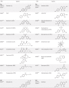
Structures of ERα and ERβ natural and environmental ligands.
Table 2
Table 2
Structures of PPARγ natural and environmental ligands.

References

    1. Germain P, Staels B, Dacquet C, Spedding M, Laudet V. Overview of nomenclature of nuclear receptors. Pharmacol Rev 2006; 58: 685–704. - PubMed
    1. Gronemeyer H, Gustafsson JA, Laudet V. Principles for modulation of the nuclear receptor superfamily. Nat Rev Drug Discov 2004; 3: 950–64. - PubMed
    1. Rastinejad F, Huang P, Chandra V, Khorasanizadeh S. Understanding nuclear receptor form and function using structural biology. J Mol Endocrinol 2013; 51: T1–T21. - PMC - PubMed
    1. Li Y, Lambert MH, Xu HE. Activation of nuclear receptors: a perspective from structural genomics. Structure 2003; 11: 741–6. - PubMed
    1. Bourguet W, Germain P, Gronemeyer H. Nuclear receptor ligand-binding domains: three-dimensional structures, molecular interactions and pharmacological implications. Trends Pharmacol Sci 2000; 21: 381–8. - PubMed