. 2015 Jan;125(1):386-402.
doi: 10.1172/JCI76738. Epub 2014 Dec 15.

Intestinal farnesoid X receptor signaling promotes nonalcoholic fatty liver disease

Intestinal farnesoid X receptor signaling promotes nonalcoholic fatty liver disease

Changtao Jiang et al. J Clin Invest. 2015 Jan.

Abstract

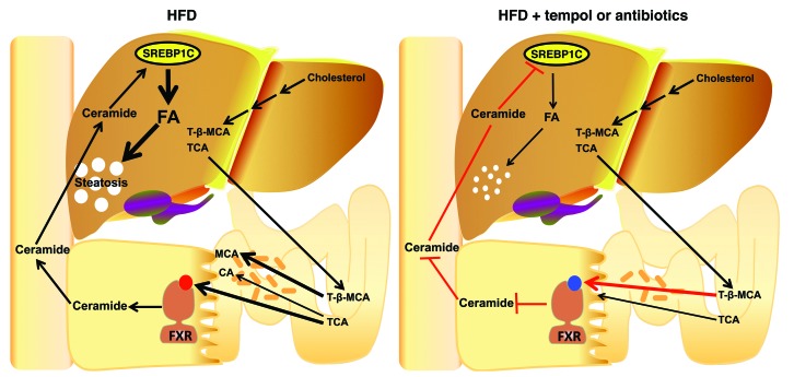
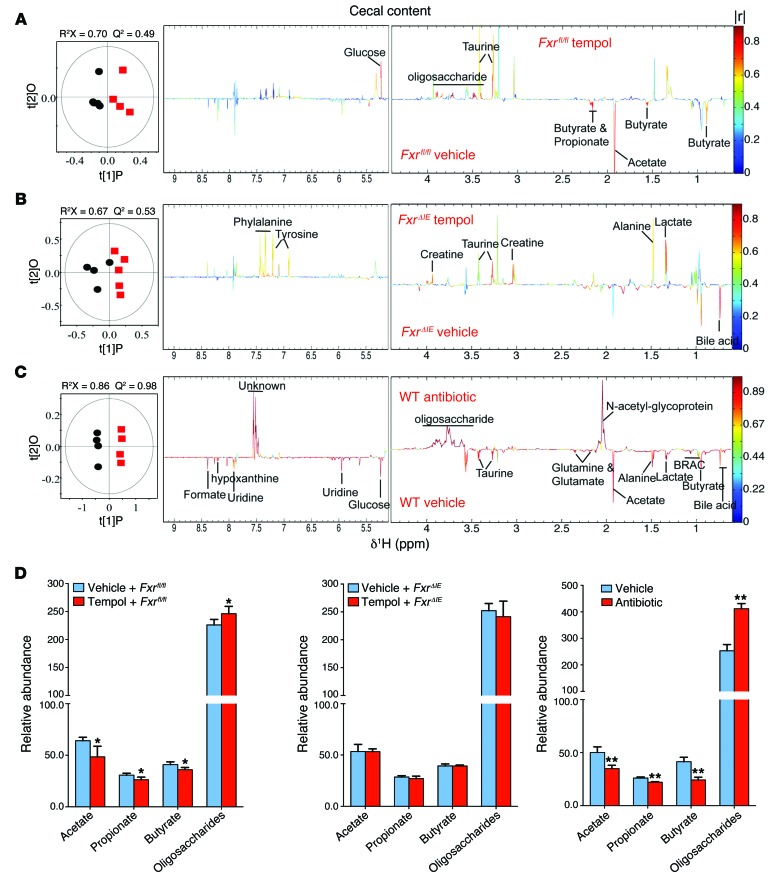
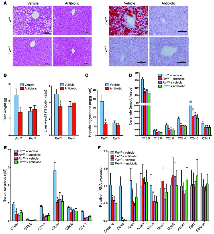
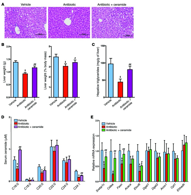
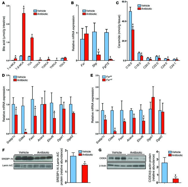
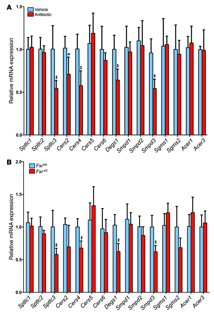
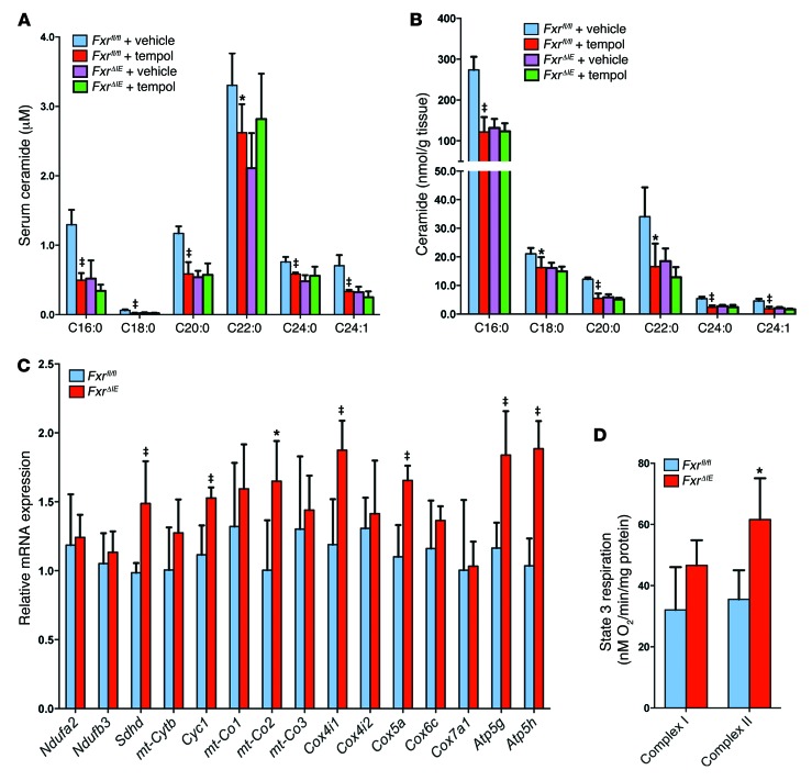
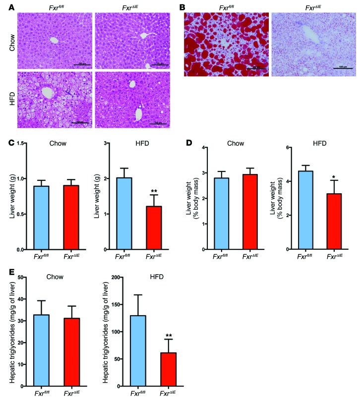
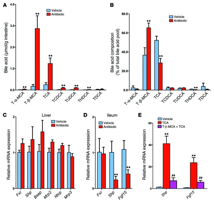
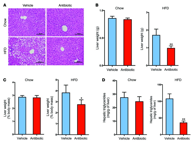
Nonalcoholic fatty liver disease (NAFLD) is a major worldwide health problem. Recent studies suggest that the gut microbiota influences NAFLD pathogenesis. Here, a murine model of high-fat diet-induced (HFD-induced) NAFLD was used, and the effects of alterations in the gut microbiota on NAFLD were determined. Mice treated with antibiotics or tempol exhibited altered bile acid composition, with a notable increase in conjugated bile acid metabolites that inhibited intestinal farnesoid X receptor (FXR) signaling. Compared with control mice, animals with intestine-specific Fxr disruption had reduced hepatic triglyceride accumulation in response to a HFD. The decrease in hepatic triglyceride accumulation was mainly due to fewer circulating ceramides, which was in part the result of lower expression of ceramide synthesis genes. The reduction of ceramide levels in the ileum and serum in tempol- or antibiotic-treated mice fed a HFD resulted in downregulation of hepatic SREBP1C and decreased de novo lipogenesis. Administration of C16:0 ceramide to antibiotic-treated mice fed a HFD reversed hepatic steatosis. These studies demonstrate that inhibition of an intestinal FXR/ceramide axis mediates gut microbiota-associated NAFLD development, linking the microbiome, nuclear receptor signaling, and NAFLD. This work suggests that inhibition of intestinal FXR is a potential therapeutic target for NAFLD treatment.


Figures

Figure 10
Figure 10. Summary of the gut microbiota–mediated development of NAFLD through a bile acid/intestinal FXR/ceramide axis.
T-β-MCA produced in the liver from the oxidation of cholesterol was secreted into the intestine. BSH, a bacterial enzyme that hydrolyzes T-β-MCA to MCA, is substantially reduced after tempol or antibiotic treatment. T-β-MCA thereby accumulates in the ileum, which inhibits FXR signaling. The inhibition of intestinal FXR signaling elicits an improvement in mitochondrial function and repression of ceramide synthesis, resulting in decreased serum ceramide levels. Decreased circulation ceramides downregulate hepatic SREBP1C and CIDEA expression, resulting in decreased hepatic steatosis.
Figure 9
Figure 9. NMR metabolomic analysis of mouse cecal content extracts.
(A) OPLS-DA scores (left) and correlation coefficient–coded loadings plots for the models (right) from NMR spectra of cecal content aqueous extracts from vehicle-treated Fxrfl/fl mice and tempol-treated Fxrfl/fl mice fed a HFD for 16 weeks. n = 5 mice per group. (B) OPLS-DA scores (left) and correlation coefficient–coded loadings plots for the models (right) from NMR spectra of cecal content aqueous extracts from vehicle-treated FxrΔIE mice and tempol-treated FxrΔIE mice fed a HFD for 16 weeks. n = 4–5 mice per group. (C) OPLS-DA scores (left) and correlation coefficient–coded loadings plots for the models (right) from NMR spectra of cecal content aqueous extracts from vehicle-treated and antibiotic-treated mice fed a HFD for 7 weeks. The correlation coefficient values indicating significantly changed metabolites are shown in Supplemental Table 3. |r| cutoff value is 0.755, n = 5, P < 0.05; CV-ANOVA: P = 0.04, 0.02 and 1.18 × 10⁻⁴, respectively. n = 4–5 mice per group. (D) Relative abundance of SCFAs (acetate, propionate, and butyrate) and oligosaccharides in cecal content extracts from vehicle-treated Fxrfl/fl mice and tempol-treated Fxrfl/fl mice fed a HFD for 16 weeks, vehicle-treated FxrΔIE mice and tempol-treated FxrΔIE mice fed a HFD for 16 weeks, and vehicle-treated and antibiotic-treated mice fed a HFD for 7 weeks. n = 4–5 mice per group. Data are presented as the mean ± SD. *P < 0.05, **P < 0.01 (2-tailed Student’s t test) compared with vehicle-treated mice.
Figure 8
Figure 8. Antibiotic treatment improved NAFLD via the inhibition of intestinal FXR.
(A) Representative H&E staining (left panel) and Oil red O staining (right panel) of liver sections from vehicle- and antibiotic-treated Fxrfl/fl and FxrΔIE mice fed a HFD for 14 weeks. n = 5 mice per group. Lipids stained positive (red color) with Oil red O. Scale bars: 100 μm. (B) Liver weights and liver weight/body weight ratios of vehicle- and antibiotic-treated Fxrfl/fl and FxrΔIE mice fed a HFD for 14 weeks. n = 5 mice per group. (C) Liver triglyceride content in vehicle- and antibiotic-treated Fxrfl/fl and FxrΔIE mice fed a HFD for 14 weeks. n = 5 mice per group. (D) Ileal ceramide profiles after 14 weeks of antibiotic treatment in HFD-fed mice. n = 5 mice per group. (E) Serum ceramide profiles after 14 weeks of antibiotic treatment in HFD-fed mice. n = 5 mice per group. (F) mRNA levels of fatty acid synthesis–, triglyceride synthesis–, and fatty acid catabolism–related genes in the livers of vehicle and antibiotic-treated Fxrfl/fl mice and FxrΔIE mice on a HFD for 14 weeks. n = 5 mice per group. (BF) Data are presented as the mean ± SD. One-way ANOVA with Tukey’s correction. *P < 0.05 and P < 0.01 compared with vehicle-treated mice of the same genotype.
Figure 7
Figure 7. Administration of ceramide attenuates the effects of antibiotic treatment on the development of HFD-induced steatohepatitis.
All mice were fed a HFD for 7 weeks and treated with no antibiotic (Vehicle) or a 0.1% antibiotic cocktail in their drinking water (Antibiotic). A group of mice on the antibiotic cocktail were i.p. injected with C16:0 ceramide as described in the Methods. (A) Representative H&E staining of liver sections. n = 5 mice per group. Scale bars: 100 μm. (B) Liver weights and liver weight/body weight ratios. n = 5 mice per group. (C) Liver triglyceride content. n = 5 mice per group. (D) Serum ceramide profiles. n = 5 mice per group. (E) Hepatic mRNA levels of fatty acid synthesis–, triglyceride synthesis–, and fatty acid catabolism–related genes. n = 5 mice per group. Expression was normalized to 18S RNA. All data are presented as the mean ± SD. One-way ANOVA with Tukey’s correction. P < 0.01 compared with vehicle-treated mice; #P < 0.05 and ##P < 0.01 compared with antibiotic-treated mice.
Figure 6
Figure 6. Intestinal FXR signaling influences the ceramide/SREBP1C/CIDEA pathway.
(A) Ileal bile acid profiles after 3 days of antibiotic treatment in HFD-fed mice. n = 5 mice per group. (B) Fxr, Shp, and Fgf15 mRNA levels in the ileum after 3 days of antibiotic treatment in HFD-fed mice. n = 5 mice per group. (C) Ileal ceramide profiles after 3 days of antibiotic treatment in HFD-fed mice. n = 5 mice per group. (D) mRNA levels of fatty acid synthesis– and triglyceride synthesis–related enzymes in the liver of HFD-fed mice after 7 weeks of antibiotic treatment. n = 5 mice per group. (BD) Expression was normalized to 18S RNA. (AD) Data are presented as the mean ± SD. *P < 0.05 and P < 0.01 (2-tailed Student’s t test) compared with vehicle-treated mice. (E) mRNA levels for encoding enzymes involved in fatty acid and triglyceride synthesis in the livers of Fxrfl/fl and FxrΔIE mice fed a HFD for 14 weeks. n = 5 mice per group. Expression was normalized to 18S RNA. Data are presented as the mean ± SD. *P < 0.05 and P < 0.01 (2-tailed Student’s t test) compared with Fxrfl/fl mice. (F) Western blot analysis of liver nuclear SREBP1-N expression after 7 weeks of antibiotic treatment in HFD-fed mice and quantitation of SREBP1-N expression. n = 3 mice per group. (G) Western blot analysis of liver CIDEA expression after 7 weeks of antibiotic treatment in HFD-fed mice and quantitation of SREBP1-N expression. n = 3 mice per group. (F and G) Data are presented as the mean ± SD. *P < 0.05 and P < 0.01 (2-tailed Student’s t test) compared with vehicle-treated mice.
Figure 5
Figure 5. Inhibition of intestinal FXR alters expression of mRNAs encoding ceramide synthesis– and catabolism–related enzymes.
(A) mRNA levels for ceramide synthesis– and catabolism–related enzymes in the ileum after 7 weeks of antibiotic treatment in HFD-fed mice. n = 5 mice per group. Data are presented as the mean ± SD. *P < 0.05 and P < 0.01 (2-tailed Student’s t test) compared with vehicle-treated mice. (B) mRNA levels for ceramide synthesis– and catabolism–related enzymes in the ileum of Fxrfl/fl and FxrΔIE mice fed a HFD for 14 weeks. n = 5 mice per group. Expression was normalized to 18S RNA. Data are presented as the mean ± SD. *P < 0.05 and P < 0.01 (2-tailed Student’s t test) compared with Fxrfl/fl mice.
Figure 4
Figure 4. Intestine-specific Fxr disruption promotes mitochondrial biogenesis in the intestine.
(A) Serum ceramide levels in vehicle- and tempol-treated Fxrfl/fl and FxrΔIE mice after 16 weeks of HFD feeding. n = 5 mice per group. (B) Ileal ceramide levels in vehicle- and tempol-treated Fxrfl/fl and FxrΔIE mice after 16 weeks of HFD feeding. n = 5 mice per group. (A and B) Data are presented as the mean ± SD. One-way ANOVA with Tukey’s correction. *P < 0.05 and P < 0.01 compared with vehicle-treated mice of the same genotype. C, side chain carbon number. (C) mRNA levels of mitochondrial oxidative phosphorylation–related enzymes in ileal mucosa from Fxrfl/fl and FxrΔIE mice after 14 weeks of HFD feeding. Expression was normalized to 18S RNA. n = 5 mice per group. mt, mitochondrial. (D) Measurement of state 3 respiration for complex I– and complex II–dependent respiration from the ileal mucosa of Fxrfl/fl and FxrΔIE mice after 14 weeks of HFD feeding. n = 4 mice per group. (C and D) Data are presented as the mean ± SD. *P < 0.05 and P < 0.01 (2-tailed Student’s t test) compared with Fxrfl/fl mice.
Figure 3
Figure 3. Intestine-specific Fxr disruption protects against HFD-induced NAFLD.
(A) Representative H&E staining of liver sections from Fxrfl/fl and FxrΔIE mice after 14 weeks of chow or HFD feeding. Scale bars: 100 μm. n = 4–5 mice per group. (B) Representative Oil red O staining of liver sections from Fxrfl/fl and FxrΔIE mice after 14 weeks of HFD feeding. Lipids stained positive (red color) with Oil Red O. Scale bars: 100 μm. n = 5 mice per group. (C) Liver weights of Fxrfl/fl and FxrΔIE mice after 14 weeks of chow or HFD feeding. n = 4–5 mice per group. (D) Liver weight/body weight ratios of Fxrfl/fl and FxrΔIE mice after 14 weeks of chow or HFD feeding. n = 4–5 mice per group. (E) Liver triglyceride contents in Fxrfl/fl and FxrΔIE mice after 14 weeks of chow or HFD feeding. n = 4–5 mice per group. All data are presented as the mean ± SD. *P < 0.05 and **P < 0.01 (2-tailed Student’s t test) compared with Fxrfl/fl mice.
Figure 2
Figure 2. Gut microbiota regulate bile acid composition.
(A) Individual taurine-conjugated bile acid levels in the ileum of HFD-fed mice after 7 weeks of antibiotic treatment. n = 4–5 mice per group. (B) Proportion of individual taurine-conjugated bile acids, expressed as a percentage of the total bile acid pool, in ileum from the vehicle- and antibiotic-treated groups fed a HFD for 7 weeks. n = 4–5 mice per group. (C) mRNA levels for FXR signaling–related genes in the liver after 7 weeks of antibiotic treatment in mice fed a HFD. n = 4–5 mice per group. (D) mRNA levels of Shp and Fgf15 in ileum after 7 weeks of antibiotic treatment in HFD-fed mice. n = 5 mice per group. (C and D) Expression was normalized to 18S RNA. (AD) Data are presented as the mean ± SD. **P < 0.01 (2-tailed Student’s t test) compared with vehicle-treated mice. (E) mRNA levels of Shp and Fgf15 in ileum after 24 hours of T-β-MCA with TCA treatment in HFD-fed mice. mRNA expression was normalized to 18S RNA. n = 6 mice per group. Data are presented as the mean ± SD. One-way ANOVA with Tukey’s correction. **P < 0.01 compared with vehicle; ##P < 0.01 compared with TCA treatment.
Figure 1
Figure 1. Antibiotic treatment ameliorates the development of NAFLD.
(A) Representative H&E staining of liver sections from vehicle- and antibiotic-treated mice on a chow diet or a HFD for 7 weeks. Scale bars: 100 μm. n = 5 mice per group. (B) Liver weights of vehicle- and antibiotic-treated mice on a chow diet or a HFD for 7 weeks. n = 5 mice per group. (C) Ratios of liver weight/body weight of vehicle- and antibiotic-treated mice on a chow diet or a HFD for 7 weeks. n = 5 mice per group. (D) Liver triglyceride content of vehicle- and antibiotic-treated mice on a chow diet or a HFD for 7 weeks. n = 5 mice per group. All data are presented as the mean ± SD. *P < 0.05 and **P < 0.01 (2-tailed Student’s t test) compared with vehicle-treated mice.

Comment in

References

    1. Sattar N, Forrest E, Preiss D. Non-alcoholic fatty liver disease. BMJ. 2014;349:g4596. doi: 10.1136/bmj.g4596. - DOI - PMC - PubMed
    1. Dyson J, Day C. Treatment of non-alcoholic fatty liver disease. Dig Dis. 2014;32(5):597–604. doi: 10.1159/000360511. - DOI - PubMed
    1. Bhatia LS, Curzen NP, Calder PC, Byrne CD. Non-alcoholic fatty liver disease: a new and important cardiovascular risk factor? Eur Heart J. 2012;33(10):1190–1200. doi: 10.1093/eurheartj/ehr453. - DOI - PubMed
    1. Baffy G, Brunt EM, Caldwell SH. Hepatocellular carcinoma in non-alcoholic fatty liver disease: an emerging menace. J Hepatol. 2012;56(6):1384–1391. doi: 10.1016/j.jhep.2011.10.027. - DOI - PubMed
    1. Park JS, Seo JH, Youn HS. Gut microbiota and clinical disease: obesity and nonalcoholic Fatty liver disease. Pediatr Gastroenterol Hepatol Nutr. 2013;16(1):22–27. doi: 10.5223/pghn.2013.16.1.22. - DOI - PMC - PubMed

Publication types

MeSH terms

Substances