Review
. 2014 Dec 9;19(12):20580-93.
doi: 10.3390/molecules191220580.

Au-nanomaterials as a superior choice for near-infrared photothermal therapy

Affiliations
Review

Au-nanomaterials as a superior choice for near-infrared photothermal therapy

Fahmida Jabeen et al. Molecules. .

Abstract

Photothermal therapy (PPT) is a platform to fight cancer by using multiplexed interactive plasmonic nanomaterials as probes in combination with the excellent therapeutic performance of near-infrared (NIR) light. With recent rapid developments in optics and nanotechnology, plasmonic materials have potential in cancer diagnosis and treatment, but there are some concerns regarding their clinical use. The primary concerns include the design of plasmonic nanomaterials which are taken up by the tissues, perform their function and then clear out from the body. Gold nanoparticles (Au NPs) can be developed in different morphologies and functionalized to assist the photothermal therapy in a way that they have clinical value. This review outlines the diverse Au morphologies, their distinctive characteristics, concerns and limitations to provide an idea of the requirements in the field of NIR-based therapeutics.


Conflict of interest statement

The authors declare no conflict of interest.

Figures

Figure 1
Figure 1
Morphology of Au-nanoparticles: (a) TEM image of Au-nanorods (GNRs) (b) High-resolution TEM image of Au/Fe2O3 nanoparticles coated with amorphous SiO2 shells, (c) TEM of GSS nanoparticles showing the speckled silica surface; inset z-contrast digital TEM, (d) TEM images of nanocomposite and Au@SiO2 (inset) (e) SEM image of nanomatryoshkas and (f) TEM image of self-assembly structured Au-nanoparticles. (reproduced from [25] (a), [26] (b), [27] (c), [22] (d), [28] (e), [29] (f) with permission).
Figure 2
Figure 2
(a) Representative confocal microscope picture of lung A549 cells labeled with the FITC doped GSS nanoparticles showing the presence of nanoparticles (green) near the nucleus (blue—stained with Hoechst). (b) A z-position cross section showing the localization of GSS nanoparticles adjacent to the nuclear boundary. (c) Trypan blue stained dead cells as ablated selectively along the path of the NIR laser and unharmed surrounding cells. (d) Higher magnification of trypan blue stained dead cells (reproduced from [38] with permission from The Royal Society of Chemistry).
Figure 3
Figure 3
(A) The temperature rise curves of aqueous solutions of GAs. (B) The UV/Vis/NIR spectra of GA7 before (left) and after (right) NIR illumination. (C) SEM images of GA7 before and after NIR illumination. (D) TEM images of GA7 before and after incubation with esterase solution at 12 h, 24 h and 36 h respectively (reproduced from [41], an open-access article from Ivyspring International Publisher).
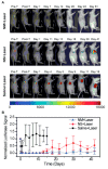
Figure 4
Figure 4
Evaluation of tumor response to photothermal therapy by bioluminescence imaging. The bioluminescence signal is generated only in living cancer cells as a result of luciferase activity. (A) Representative mice of each experimental group showing the luciferase activity in the tumor. The mice injected with nanomatryoshkas or nanoshells and treated with laser experienced loss of bioluminescence in the area illuminated by the laser as seen after therapy. Mice were euthanized when tumor volume reached 1,500 mm3 or if the tumor persisted at 60 days after treatment. (B) Mean luciferase activity in the tumor with standard deviations. The luciferase signal was normalized to the signal before treatment (reprinted with permission from [49]; copyright (2014) American Chemical Society).

References

    1. Jaque D., Martínez Maestro L., del Rosal B., Haro-Gonzalez P., Benayas A., Plaza J.L., Martín Rodrígueza E., García Soléa J. Nanoparticles for photothermal therapies. Nanoscale. 2014;6:9494–9530. doi: 10.1039/C4NR00708E. - DOI - PubMed
    1. Chen S., Deng J., Yuan Y., Flachenecker C., Mak R., Hornberger B., Jin Q., Shu D., Lai B., Maser J., et al. The Bionanoprobe: Hard X-ray fluorescence nanoprobe with cryogenic capabilities. J. Synchrotron Radiat. 2014;21:66–75. doi: 10.1107/S1600577513029676. - DOI - PMC - PubMed
    1. Howes P.D., Rana S., Stevens M.M. Plasmonic nanomaterials for biodiagnostics. Chem. Soc. Rev. 2014;43:3835–3853. doi: 10.1039/c3cs60346f. - DOI - PMC - PubMed
    1. Khlebtsov B.N., Panfilova E.V., Terentyuk G.S., Maksimova I.L., Ivanov A.V., Khlebtsov N.G. Plasmonic nanopowders for photothermal therapy of tumors. Langmuir. 2012;28:8994–9002. doi: 10.1021/la300022k. - DOI - PubMed
    1. Huang N., Wang H., Zhao J., Lui H., Korbelik M., Zeng H. Single-wall carbon nanotubes assisted photothermal cancer therapy: Animal study with a murine model of squamous cell carcinoma. Lasers Surg. Med. 2010;42:638–648. doi: 10.1002/lsm.20968. - DOI - PubMed