. 2014 Dec 11;10(12):e1004559.
doi: 10.1371/journal.pgen.1004559. eCollection 2014 Dec.

Early mesozoic coexistence of amniotes and hepadnaviridae

Affiliations

Early mesozoic coexistence of amniotes and hepadnaviridae

Alexander Suh et al. PLoS Genet. .

Abstract

Hepadnaviridae are double-stranded DNA viruses that infect some species of birds and mammals. This includes humans, where hepatitis B viruses (HBVs) are prevalent pathogens in considerable parts of the global population. Recently, endogenized sequences of HBVs (eHBVs) have been discovered in bird genomes where they constitute direct evidence for the coexistence of these viruses and their hosts from the late Mesozoic until present. Nevertheless, virtually nothing is known about the ancient host range of this virus family in other animals. Here we report the first eHBVs from crocodilian, snake, and turtle genomes, including a turtle eHBV that endogenized >207 million years ago. This genomic "fossil" is >125 million years older than the oldest avian eHBV and provides the first direct evidence that Hepadnaviridae already existed during the Early Mesozoic. This implies that the Mesozoic fossil record of HBV infection spans three of the five major groups of land vertebrates, namely birds, crocodilians, and turtles. We show that the deep phylogenetic relationships of HBVs are largely congruent with the deep phylogeny of their amniote hosts, which suggests an ancient amniote-HBV coexistence and codivergence, at least since the Early Mesozoic. Notably, the organization of overlapping genes as well as the structure of elements involved in viral replication has remained highly conserved among HBVs along that time span, except for the presence of the X gene. We provide multiple lines of evidence that the tumor-promoting X protein of mammalian HBVs lacks a homolog in all other hepadnaviruses and propose a novel scenario for the emergence of X via segmental duplication and overprinting of pre-existing reading frames in the ancestor of mammalian HBVs. Our study reveals an unforeseen host range of prehistoric HBVs and provides novel insights into the genome evolution of hepadnaviruses throughout their long-lasting association with amniote hosts.


Conflict of interest statement

The authors have declared that no competing interests exist.

Figures
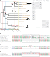
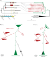
Figure 1
Figure 1. Non-avian hepatitis B paleovirus endogenization events.
(A) Simplified chronogram of non-mammalian amniotes based on molecular dates of phylogenetic relationships among amniotes , squamate lepidosaurs , , turtles , birds , and crocodilians , . Icosahedrons denote endogenization events, the asterisk indicates previously studied avian endogenizations , and the colored time axis corresponds to the International Stratigraphic Chart (http://www.stratigraphy.org/ICSchart/StratChart2010.pdf). All HBV EVE endogenization events were reconstructed based on their respective presence/absence patterns (“+”: presence, “−” absence; “?”: missing data or sequence could not be aligned). This is with the exception of cobra eSNHBVs where we could not ascertain presence/absence states in other squamates. The early Mesozoic eTHBV paleovirus (B) is present in orthologous locations in both pleurodiran and cryptodiran turtles, but absent in crocodilians. All crocodilians to the exclusion of the alligator (i.e., Longirostres [41]) share the Cretaceous eCRHBV1 insertion (C), while the Paleogene eCRHBV2 insertion (D) is present in saltwater and dwarf crocodile (i.e., Crocodylidae), but absent in orthologous positions in gharial and alligator. HBV-derived sequence residues are boxed.
Figure 2
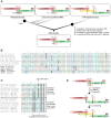
Figure 2. Non-avian hepatitis B paleovirus genome organization and features of viral replication.
(A) The fragments of the crocodilian and turtle HBV EVEs described herein extend over ∼81% (eCRHBV1), ∼52% (eCRHBV2), ∼21% (eTHBV), ∼18% (eSNHBV2), and ∼14% (eSNHBV1) of the circular 3,024-bp extant DHBV genome from ducks (AY494851). Genomic regions missing from these fragments are indicated by dashed grey lines. The reconstructed start (arrow) and stop (asterisk) codon positions for the ORFs of the polymerase protein (pol; red), the presurface/surface protein (preS/S; blue), and the precore/core protein (preC/C; green) indicate a highly similar genome organization among Hepadnaviridae. Genomic features related to viral replication are direct repeats (DR; purple vertical lines) and the RNA encapsidation signal (ε; orange box); these are contained in the crocodilian eCRHBV1 fragment. Comparison of DR sequence and ε RNA secondary structure of this crocodilian HBV EVE (B) with homologous structures in the Mesozoic avian HBV EVE (C) and human HBV (D) shows conservation of the priming bulge (bold), whereas the rest of the stably base-pared ε hairpin structure exhibits little sequence similarity between avian and mammalian HBVs, and also the crocodilian HBV (see also S1 Figure for an alignment of ε sequences). In extant HBVs , the conserved tyrosine (Y) residue of the terminal protein domain (numbers indicate the tyrosine amino acid site) of the pol ORF is attached to a DNA primer (grey letters) that binds to the ε priming bulge and the 5′ end of the DR. Arrows depict the direction of minus DNA synthesis.
Figure 3
Figure 3. Phylogeny of Hepadnaviridae and their amniote hosts.
(A) Plotting the HBV endogenization events (red boxes) reconstructed in this study and ref. on a dated consensus phylogeny of amniotes , suggests temporary Mesozoic coexistence of Hepadnaviridae with birds, crocodilians, and turtles, respectively. Extant coexistence with birds and mammals is denoted by green boxes and the undated evidence for snake HBV endogenization events is indicated by a dashed box. The rooted (A) and unrooted (B) phylograms (see S4 Figure for phylograms including the short fragments of eSNHBV1 and eSNHBV2) of a maximum likelihood (ML) analysis of the polymerase protein from hepadnaviruses and reverse-transcribing outgroups (caulimoviruses, retroviruses and retrotransposons) exhibit a phylogenetic placement of crocodilian eHBVs that recapitulates the deep phylogeny of their amniote hosts. The precore/core protein ML phylogram (C) on the same ingroup sampling (plus eTHBV) topologically indicates a bird+crocodilian grouping, too, as well as an affinity of these HBVs to the turtle eHBV. Nevertheless, the resolution of the PreC protein on deep HBV relationships remains limited as suggested by low bootstrap support of some internodes and different topologies with regards to the outgroup (i.e., grouping them within Orthohepadnaviruses) as well as the branching order of avian eHBVs. ML bootstrap values are shown in % on respective nodes. Note the long internodes leading to non-avian eHBV branches; these further support the distinctiveness of the protein sequences of these ancient hepadnaviral lineages. All ML trees were generated via RAxML 7.4.7 using the JTT+G model and 1000 bootstrap replicates.
Figure 4
Figure 4. An evolutionary scenario for the emergence of the oncogenic X gene.
(A) The genome of the HBV ancestor contained neither an X nor X-like ORF, given that avian and crocodilian eHBVs lack an X-like ORF, despite the fact that it is thought to be expressed in some closely related avihepadnaviruses . X (+2 frame) and X-like (+3 frame) are encoded in different reading frames relative to the part of pol they overlap with (+1 frame), which strongly suggests that these ORFs are non-homologous and the X-like protein emerged in the ancestor of avihepadnaviruses. Independently, the X protein arose in orthohepadnaviruses via overprinting (4.) after a segmental duplication (1.), a partial deletion (2.), and a frameshifting mutation (3.) in one region of the HBV genome. Only the part of the HBV genome between the ribonuclease H (RNH) and the terminal protein (TP) domains of the pol ORF is shown, including structural elements such as direct repeats (DR; purple vertical lines) and the RNA encapsidation signal (ε; orange box). (B) Translated sequence alignment of the X-like ORF sensu Chang et al. indicates presence of multiple internal stop codons in avian and crocodilian eHBVs, resulting in potential translation products <30 aa. Stop codon positions (asterisks) are highlighted with grey boxes if they are conserved between eHBVs, start codon positions for the longest possible ORF are highlighted by circles. Even when assuming that nonconventional start codons are used as suggested for DHBV , potential eHBV X-like proteins would comprise just a portion of the DHBV X-like protein. (C) Sequence similarity between translated preC/C 5′ end region (incl. in-frame aa sites upstream of the start codon) and translated central region of the preC/C ORF might be a potential remnant of an ancient segmental duplication of the first two thirds of the preC/C ORF. Amino acid residues with dark grey background are conserved between the start and the middle part of the preC/C ORF and thus constitute a potentially duplicated amino acid motif. (D) Schematic illustration of the proposed evolutionary steps of X ORF emergence [(1.) to (4.)] described in (A) that potentially led to the extant genome organization of orthohepadnaviruses. Black rectangles illustrate the location of the duplicated amino acid motif shown in (C).

References

    1. Weiss RA, Stoye JP (2013) Our viral inheritance. Science 340: 820–821. - PubMed
    1. Katzourakis A, Gifford RJ (2010) Endogenous viral elements in animal genomes. PLoS Genetics 6: e1001191. - PMC - PubMed
    1. Johnson WE (2010) Endless forms most viral. PLoS Genetics 6: e1001210. - PMC - PubMed
    1. Holmes EC (2011) The evolution of endogenous viral elements. Cell Host & Microbe 10: 368–377. - PMC - PubMed
    1. Feschotte C, Gilbert C (2012) Endogenous viruses: insights into viral evolution and impact on host biology. Nature Reviews Genetics 13: 283–296. - PubMed

Publication types

MeSH terms