

Gene expression profile analysis identifies metastasis and chemoresistance-associated genes in epithelial ovarian carcinoma cells
- PMID: 25502083
- PMCID: PMC4262766
- DOI: 10.1007/s12032-014-0426-5
Gene expression profile analysis identifies metastasis and chemoresistance-associated genes in epithelial ovarian carcinoma cells
Abstract
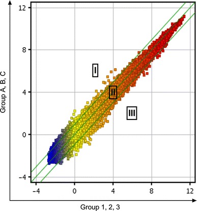
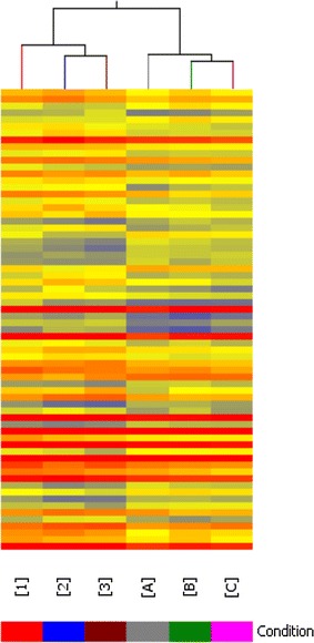
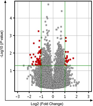
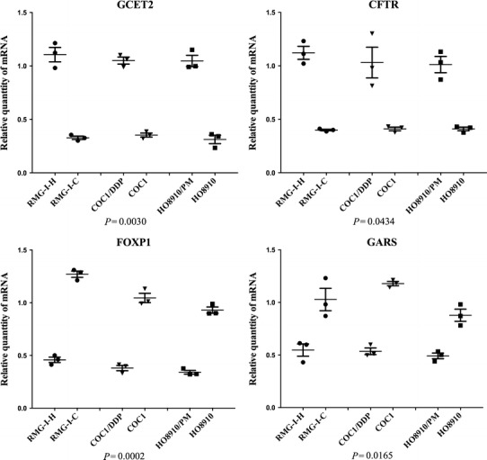
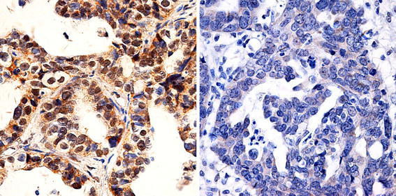
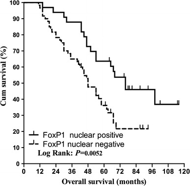
The purpose of this study was to identify genes that associated with higher ability of metastasis and chemotherapic resistance in epithelial ovarian carcinoma (EOC) cells. An oligonucleotide microarray with probe sets complementary to 41,000(+) unique human genes and transcripts was used to determine whether gene expression profile may differentiate three epithelial ovarian cell lines (RMG-I-C, COC1 and HO8910) from their sub-lines (RMG-I-H, COCI/DDP and HO8910/PM) with higher ability of metastasis and chemotherapic resistance. Quantitative real-time PCR and immunohistochemical staining validated the microarray results. Hierarchic cluster analysis of gene expression identified 49 genes that exhibited ≥2.0-fold change and P value ≤0.05. Highly differential expression of GCET2, NLRP4, FOXP1 and SNX29 genes was validated by quantitative PCR in all cell line samples. Finally, FOXP1 was validated at the protein level by immunohistochemistry in paraffin embedded ovarian tissues (i.e., for metastasis, 15 primary EOC and 10 omental metastasis [OM]; for chemoresistance, 13 sensitive and 13 resistant EOC). The identification of higher ability of metastasis and chemotherapic resistance-associated genes may provide a foundation for the development of new type-specific diagnostic strategies and treatment for metastasis and chemotherapic resistance in epithelial ovarian cancer.
Figures
Similar articles
-
[Whole Genome Expression Profiling Analysis of Metastasis and Drug-resistance-related Genes in Epithelial Ovarian Cancer Cells].Zhongguo Yi Xue Ke Xue Yuan Xue Bao. 2015 Dec;37(6):662-73. doi: 10.3881/j.issn.1000-503X.2015.06.006. Zhongguo Yi Xue Ke Xue Yuan Xue Bao. 2015. PMID: 26725389 Chinese.
-
FOXM1 expression is significantly associated with chemotherapy resistance and adverse prognosis in non-serous epithelial ovarian cancer patients.J Exp Clin Cancer Res. 2017 May 8;36(1):63. doi: 10.1186/s13046-017-0536-y. J Exp Clin Cancer Res. 2017. PMID: 28482906 Free PMC article.
-
Gene expression profile analysis in response to α1,2-fucosyl transferase (FUT1) gene transfection in epithelial ovarian carcinoma cells.Tumour Biol. 2016 Sep;37(9):12251-12262. doi: 10.1007/s13277-016-5080-4. Epub 2016 May 30. Tumour Biol. 2016. PMID: 27240592
-
miRNA-200a/c as potential biomarker in epithelial ovarian cancer (EOC): evidence based on miRNA meta-signature and clinical investigations.Oncotarget. 2016 Dec 6;7(49):81621-81633. doi: 10.18632/oncotarget.13154. Oncotarget. 2016. PMID: 27835595 Free PMC article. Review.
-
Mechanisms and Therapeutic Targets of microRNA-associated Chemoresistance in Epithelial Ovarian Cancer.Curr Cancer Drug Targets. 2016;16(5):429-41. doi: 10.2174/1568009616666160404121105. Curr Cancer Drug Targets. 2016. PMID: 27040353 Review.
Cited by
-
Functional Transcription Factor Target Networks Illuminate Control of Epithelial Remodelling.Cancers (Basel). 2020 Sep 30;12(10):2823. doi: 10.3390/cancers12102823. Cancers (Basel). 2020. PMID: 33007944 Free PMC article.
-
The NLR family pyrin domain-containing 11 protein contributes to the regulation of inflammatory signaling.J Biol Chem. 2018 Feb 23;293(8):2701-2710. doi: 10.1074/jbc.RA117.000152. Epub 2018 Jan 4. J Biol Chem. 2018. PMID: 29301940 Free PMC article.
-
Inflammatory pathway genes associated with inter-individual variability in the trajectories of morning and evening fatigue in patients receiving chemotherapy.Cytokine. 2017 Mar;91:187-210. doi: 10.1016/j.cyto.2016.12.023. Epub 2017 Jan 19. Cytokine. 2017. PMID: 28110208 Free PMC article.
-
CYLD downregulates Livin and synergistically improves gemcitabine chemosensitivity and decreases migratory/invasive potential in bladder cancer: the effect is autophagy-associated.Tumour Biol. 2016 Sep;37(9):12731-12742. doi: 10.1007/s13277-016-5157-0. Epub 2016 Jul 22. Tumour Biol. 2016. PMID: 27448305
-
Overexpression of HE4 (human epididymis protein 4) enhances proliferation, invasion and metastasis of ovarian cancer.Oncotarget. 2016 Jan 5;7(1):729-44. doi: 10.18632/oncotarget.6327. Oncotarget. 2016. PMID: 26575020 Free PMC article.
References
-
- Liu CM. Cancer of the ovary. N Engl J Med. 2005;352:1268–9; author reply-9. - PubMed
-
- Iwamori M, Tanaka K, Kubushiro K, Lin B, Kiguchi K, Ishiwata I, et al. Alterations in the glycolipid composition and cellular properties of ovarian carcinoma-derived RMG-1 cells on transfection of the alpha1,2-fucosyltransferase gene. Cancer Sci. 2005;96:26–30. doi: 10.1111/j.1349-7006.2005.00005.x. - DOI - PMC - PubMed
-
- Lin B, Hao YY, Wang DD, Zhu LC, Zhang SL, Saito M, et al. Transfection of alpha1, 2-fucosyltransferase gene increases the antigenic expression of Lewis y in ovarian cancer cell line RMG-I. Zhongguo yi xue ke xue yuan xue bao Acta Academiae Medicinae Sinicae. 2008;30:284–289. - PubMed
-
- Yan LM, Lin B, Zhu LC, Hao YY, Qi Y, Wang CZ, et al. Enhancement of the adhesive and spreading potentials of ovarian carcinoma RMG-1 cells due to increased expression of integrin alpha5beta1 with the Lewis Y-structure on transfection of the alpha1,2-fucosyltransferase gene. Biochimie. 2010;92:852–857. doi: 10.1016/j.biochi.2010.02.012. - DOI - PubMed
Publication types
MeSH terms
LinkOut - more resources
Full Text Sources
Other Literature Sources
Medical
Research Materials
