. 2014 Dec 15:5:5758.
doi: 10.1038/ncomms6758.

Arteries are formed by vein-derived endothelial tip cells

Affiliations

Arteries are formed by vein-derived endothelial tip cells

Cong Xu et al. Nat Commun. .

Abstract

Tissue vascularization entails the formation of a blood vessel plexus, which remodels into arteries and veins. Here we show, by using time-lapse imaging of zebrafish fin regeneration and genetic lineage tracing of endothelial cells in the mouse retina, that vein-derived endothelial tip cells contribute to emerging arteries. Our movies uncover that arterial-fated tip cells change migration direction and migrate backwards within the expanding vascular plexus. This behaviour critically depends on chemokine receptor cxcr4a function. We show that the relevant Cxcr4a ligand Cxcl12a selectively accumulates in newly forming bone tissue even when ubiquitously overexpressed, pointing towards a tissue-intrinsic mode of chemokine gradient formation. Furthermore, we find that cxcr4a mutant cells can contribute to developing arteries when in association with wild-type cells, suggesting collective migration of endothelial cells. Together, our findings reveal specific cell migratory behaviours in the developing blood vessel plexus and uncover a conserved mode of artery formation.


Figures
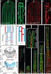
Figure 1
Figure 1. Visualizing blood vessels in the regenerating zebrafish fin.
(a,a′) Tg(fli1a:EGFP)y1 labels all fin blood vessels. (b,b′) Tg(−0.8flt1:RFP)hu5333 preferentially labels arterial endothelial cells. (c,d) Schematic drawings of the zebrafish fin vasculature, indicating the ray bifurcation, arteries (A) and veins (V). (e) Schematic of zebrafish fin, indicating plane of amputation and imaged area. (f) Overlay of Tg(fli1a:EGFP)y1 and Tg(−0.8flt1:RFP)hu5333 at different stages of fin regeneration highlighting the medially located arterial cells in yellow. Inset (f′) shows remodelling vascular plexus, which is located proximal to the sprouting front and distally to the established arteries and veins. Scale bar in a is 80 μm, in a′ is 20 μm and 200 μm in f. Representative images from a total of three zebrafish imaged per stage.
Figure 2
Figure 2. Time-lapse imaging of regenerating blood vessels reveals contribution of vein-derived tip cells to the forming artery.
(a) Still images taken every 6 h from a 24-h time-lapse movie on 9 d.p.a. fin regenerate of wild-type fish. (b) Tracking of individual cells deriving form the lateral vein (white dots), the artery (pink dots) or the medial vein (blue dots). While arterial cells hardly contribute to the advancing front, vein-derived cells contribute to the newly forming artery (inset at 23:45 h time point). (c) Tracks of labelled cells in (b). White arrows indicate migration paths of lateral vein-derived endothelial cells, while blue arrows indicate migration paths of medial vein-derived endothelial cells. Note change in migration direction. (d) Time stamp of tracked endothelial cells. (e) Schematic drawing of vessel front in the regenerated fin of wild-type fish at 10 d.p.a. Arteries and veins are indicated. (f) Schematic drawing of wild-type fin vasculature regenerated within 24 h from 9 d.p.a. Scale bar, 100 μm; d.p.a.=days post amputation. Representative movie of a total of three movies is shown.
Figure 3
Figure 3. Genetic lineage tracing of tip cells in the mouse retinal vascular plexus.
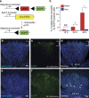
(a) Schematic of genetic lineage tracing and transgenic lines used. (b) Isolectin B4 staining of the retinal vasculature at P6. Bracket indicates vascular front. (c) Location of EGFP expressing cells at the vascular front 12 h after tamoxifen administration. (d) Overlay of isolectin B4 and EGFP channels. Remodelling arteries (A) and veins (V) are marked. (e) Isolectin B4 staining of the retinal vasculature at P6. (f) Location of EGFP expressing cells at the vascular front 48 h after tamoxifen administration. (g) Overlay of isolectin B4 and EGFP channels. Remodelling arteries (A) and veins (V) are marked. Arrows indicate EGFP-positive endothelial cells in arteries, while arrowheads indicate EGFP-negative veins. (h) Percentage of EGFP-expressing endothelial cells in arteries and veins at different time points after tamoxifen administration. Scale bar, 100 μm. For each time point five mice were analysed. *P<0.05; t-test, error bars indicate s.e.m.
Figure 4
Figure 4. The chemokine receptor cxcr4a and its ligand cxcl12a are required for proper arterial patterning during fin regeneration.
Dashed lines indicate amputation planes in ac′′. (a) Tg(cxcr4a:YFP)mu104 fish reveal YFP expression in individual cells in the centre of the regenerating fin ray. (a′) Tg(−0.8flt1:RFP)hu5333 labelled blood vessels. (a′′) Overlay of red and yellow channels reveals expression of YFP in individual endothelial cells (arrows), while neighbouring endothelial cells do not express YFP (arrowheads). (b) CFP expression in the centre of the regenerating fin in Tg(cxcl12a:CFP)mu146 transgenic zebrafish. (b′) Tg(−0.8flt1:RFP)hu5333 labelled blood vessels. (b′′) Overlay of red and blue channels. (c) YFP expression in filamentous structures (arrowheads) extending into the regenerating fin in Tg(cxcl12b:YFP)mu105 transgenic zebrafish. (c′) Tg(−0.8flt1:RFP)hu5333 labelled blood vessels. (c′′) Overlay of red and yellow channels. (d) Fin vasculature in wild-type sibling 14 d.p.a. Arrows indicate Tg(−0.8flt1:RFP)hu5333-positive endothelial cells in the centre of the fin ray. (e) cxcr4aum20 mutant; arrowheads indicate ectopic Tg(−0.8flt1:RFP)hu5333-positive endothelial cells. (f) Quantification of artery formation defects in cxcr4aum20 mutants. Endothelial cell numbers, vessel coverage and length are reduced in the centre. (g) Fin vasculature in wild-type sibling 14 d.p.a. Arrows indicate Tg(−0.8flt1:RFP)hu5333-positive endothelial cells in the centre of the fin ray. (h) cxcl12at30516 mutant; arrowheads indicate ectopic Tg(−0.8flt1:RFP)hu5333-positive endothelial cells. Scale bar (ac,e,h), 100μm. (i) Quantification of artery formation defects in cxcl12at30516 mutants. Endothelial cell numbers, vessel coverage and length are reduced in the centre. NS, not significant, **P<0.01, ****P<0.0001; Mann–Whitney U-test. Twenty individual fin rays from five fish were analysed.
Figure 5
Figure 5. Time-lapse imaging of regenerating blood vessels in cxcr4aum20 mutant fish.
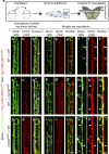
(a) Still images taken every 6 h from a 24 h time-lapse movie on 9 d.p.a. fin regenerate of cxcr4aum20 fish. (b) Tracking of individual cells deriving form the lateral vein (white dots), the artery (pink dots) or the medial vein (blue dots). (c) Tracks of labelled cells in (b). White arrows indicate migration paths of lateral vein-derived endothelial cells, while blue arrows indicate migration paths of medial vein-derived endothelial cells. Note persistent migration of tracked endothelial cells in the direction of the growing regenerate. (d) Time stamp of tracked endothelial cells. (e) Schematic drawing of vessel front in the regenerated fin of cxcr4aum20 fish at 10 d.p.a. Arteries and veins are indicated. (f) Schematic drawing of cxcr4aum20 fin vasculature regenerated within 24 h from 9 d.p.a. Scale bar, 100 μm; d.p.a., days post amputation. Representative movie of a total of three movies.
Figure 6
Figure 6. Analysis of cxcr4a function in chimeric fin vasculature.
(a) Schematic drawing of transplantation procedure. Cells from double transgenic Tg(fli1a:EGFP)y1; Tg(−0.8flt1:RFP)hu5333 donors were transplanted into single Tg(−0.8flt1:RFP)hu5333 hosts. Two different scenarios were observed: Either the vasculature of the whole ray was donor-derived (bd′′), or a mosaic ray vasculature, consisting of donor and host-derived endothelial cells formed (ej′′). (bb′′) In wild type to wild-type transplants, normal fin blood vessels formed. Arrow in b′′ marks artery (n=8 adult zebrafish). (cc′′) When wild-type cells were transplanted into cxcr4aum20 mutant hosts, normal arteries could form (arrow in c″, n=2 adult zebrafish). (dd′′) When cxcr4aum20 mutant cells were transplanted into wild-type hosts and formed the vasculature of an entire fin ray, arteries showed similar patterning defects as in cxcr4aum20 mutants (arrow in d′′, n=3 adult zebrafish). (ef′′) In a control mosaic situation, both donor (white arrowhead, ff′′) and host-derived (blue arrowhead, ff′′) wild-type cells contributed to forming arteries. Dashed box in (e) indicates magnified area in ff′′ (n=8 adult zebrafish). (gh′′) In a mosaic situation, both donor-derived wild-type cells (white arrowheads in hh′′) and host-derived cxcr4aum20 mutant cells (hh′′, blue arrowheads) contributed to forming arteries. Dashed box in (g) indicates magnified area in hh′′ (n=2 adult zebrafish). (ij′′) In a mosaic situation, both donor-derived cxcr4aum20 mutant cells (white arrowheads in j–j’’) and host-derived wild-type cells (jj′′, blue arrowheads) contributed to forming arteries. Dashed box in i indicates magnified area in jj′′ (n=3 adult zebrafish). Scale bars are 50 μm in i′′ and 20 μm in j′′.
Figure 7
Figure 7. Global overexpression of Cxcl12a-mCherry rescues the vascular phenotype of cxcl12t30516 mutant fish.
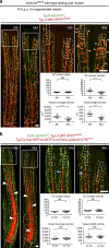
White dashed boxes indicate central areas quantified. Fin vasculature was analysed at 14 d.p.a. Dashed lines indicate amputation planes. (a) Heat-shocked control wild type and cxcl12at30516 mutant fish not carrying the Tg(Cry.kop.HSP:mutSDF1a.mCherry.globin3UTR)mu4 transgene. White arrowheads indicate ectopic Tg(−0.8flt1:RFP)hu5333-positive endothelial cells in cxcl12at30516 mutants. Endothelial cell numbers, vessel coverage and vessel length are reduced in cxcl12at30516 mutant fish. (b) Heat-shocked wild type and cxcl12at30516 mutants carrying the Tg(Cry.kop.HSP:mutSDF1a.mCherry.globin3UTR)mu4 transgene. None of the assayed parameters differs between heat-shocked wild type and cxcl12at30516 mutants. Note accumulation of ubiquitously overexpressed Cxcl12a-mCherry protein in bony rays (white arrowheads) but not in the joints (blue arrowheads) between bone segments. ****P<0.0001; Mann–Whitney U-test, NS, not significant; n=8 adult zebrafish per stage. Scale bar, 100 um.

References

    1. Eilken H. M. & Adams R. H. Dynamics of endothelial cell behavior in sprouting angiogenesis. Curr. Opin. Cell. Biol. 22, 617–625 (2010). - PubMed
    1. Herbert S. P. & Stainier D. Y. Molecular control of endothelial cell behaviour during blood vessel morphogenesis. Nat. Rev. Mol. Cell. Biol. 12, 551–564 (2011). - PMC - PubMed
    1. Potente M., Gerhardt H. & Carmeliet P. Basic and therapeutic aspects of angiogenesis. Cell 146, 873–887 (2011). - PubMed
    1. Wacker A. & Gerhardt H. Endothelial development taking shape. Curr. Opin. Cell. Biol. 23, 676–685 (2011). - PubMed
    1. DiPietro L. A. Angiogenesis and scar formation in healing wounds. Curr. Opin. Rheumatol. 25, 87–91 (2013). - PubMed

Publication types

MeSH terms

LinkOut - more resources