. 2014 Dec 11;10(12):e1004819.
doi: 10.1371/journal.pgen.1004819. eCollection 2014 Dec.

A massively parallel pipeline to clone DNA variants and examine molecular phenotypes of human disease mutations

Affiliations

A massively parallel pipeline to clone DNA variants and examine molecular phenotypes of human disease mutations

Xiaomu Wei et al. PLoS Genet. .

Abstract

Understanding the functional relevance of DNA variants is essential for all exome and genome sequencing projects. However, current mutagenesis cloning protocols require Sanger sequencing, and thus are prohibitively costly and labor-intensive. We describe a massively-parallel site-directed mutagenesis approach, "Clone-seq", leveraging next-generation sequencing to rapidly and cost-effectively generate a large number of mutant alleles. Using Clone-seq, we further develop a comparative interactome-scanning pipeline integrating high-throughput GFP, yeast two-hybrid (Y2H), and mass spectrometry assays to systematically evaluate the functional impact of mutations on protein stability and interactions. We use this pipeline to show that disease mutations on protein-protein interaction interfaces are significantly more likely than those away from interfaces to disrupt corresponding interactions. We also find that mutation pairs with similar molecular phenotypes in terms of both protein stability and interactions are significantly more likely to cause the same disease than those with different molecular phenotypes, validating the in vivo biological relevance of our high-throughput GFP and Y2H assays, and indicating that both assays can be used to determine candidate disease mutations in the future. The general scheme of our experimental pipeline can be readily expanded to other types of interactome-mapping methods to comprehensively evaluate the functional relevance of all DNA variants, including those in non-coding regions.


Conflict of interest statement

The authors have declared that no competing interests exist.

Figures

Figure 1
Figure 1. Schematic of our comparative interactome-scanning pipeline.
Our pipeline begins with Clone-seq (a), a massively-parallel low-cost site-directed mutagenesis pipeline leveraging next-generation sequencing. This is followed by a high-throughput GFP assay (b) to determine protein stability, and a high-throughput Y2H assay (c), along with SILAC-based mass spectrometry (d) to determine the impact of DNA coding variants on protein interactions.
Figure 2
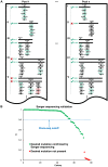
Figure 2. Identifying usable clones from Clone-seq.
(a) Schematic illustrating criteria used to determine which of the clones generated by our Clone-seq pipeline are usable for further assays – green ticks indicate usable clones, while red crosses indicate clones that cannot be used. (b) Variation of S across different mutagenesis attempts that either contain or do not contain the desired mutation as confirmed by Sanger sequencing.
Figure 3
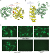
Figure 3. Examples of disease mutations in different structural loci of protein-protein interactions and examples of our GFP assay results.
(a) Crystal structure (PDB id: 3W4U) depicting a D100Y mutation (on Hbb) at an interface residue and a F104L mutation in the interface domain for the Hbb-Hbz interaction. (b) Crystal structure (PDB id: 1G3N) depicting a V31L mutation (on Cdkn2c) away from the Cdkn2c-Cdk6 interaction interface. (c) GFP assays that determine the stability of wild-type Rrm2b and the R41P and L317V mutations on Rrm2b that are at an interface residue and away from the interface for the Rrm2b-Rrm2b interaction; GFP assays that determine the stability of wild-type Hprt1 and the C206Y mutation on Hprt1 that is away from the interaction interface of Hprt-Hprt1. Empty vector was used as a negative control.
Figure 4
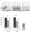
Figure 4. Effect of disease mutations on protein stability and protein-protein interactions.
(a) Western blotting with anti-GFP antibody confirming the protein expression levels of wild-type Rrm2b, Actn2, Hprt1, Pnp, Tpk1, Gnmt, Gale, Fbp1, Klhl3, Tp53, Pnp, Smad4, and corresponding mutant alleles. β-tubulin and γ-tubulin were used as loading controls. Red denotes “interface residue” mutations, orange denotes “interface domain” mutations and blue denotes “away from the interface” mutations. (b) Likelihood of disruption of interactions by “interface residue”, “interface domain” and “away from the interface” mutations – overall and for stable mutants only; likelihood of a disease mutation disrupting a given interaction in the absence of structural information. Error bars indicate +SE. (N = 204 mutations).
Figure 5
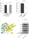
Figure 5. Relationships between molecular phenotypes and disease phenotypes.
(a) Fraction of mutation pairs on the same gene that cause the same disease: for the same and different effects on protein stability. (b) Fraction of mutation pairs on the same gene that cause the same disease: for the same and different interaction disruption profiles. Error bars indicate +SE. (c) Crystal structure (PDB id: 1U7F) depicting the Y353S and R361C mutations (on Smad4) at interface residues for the Smad4-Smad3 interaction. (d) Y2H analysis of the effects of Smad Y353S, R361, and N13S mutations on its interactions with Smad3, Lmo4, Rassf5, and Smad9. Western blotting with anti-GFP antibody confirming the protein expression levels of wild-type Smad4 and its 3 mutant alleles – Y353S, R361C and N13S. γ-tubulin was used as a loading control.
Figure 6
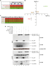
Figure 6. Identifying interactions of Mlh1 that are affected by the I107R mutation using SILAC-based mass spectrometry.
(a) Schematic illustrating criteria used to identify interactions that are lost/weakened, unchanged, and gained/enhanced due to the I107R mutation on Mlh1. Blue denotes samples cultured in light media and black denotes samples cultured in heavy media. (b) Scatter plot illustrating fold change (FC; log scale) in the amount of protein pulled down by wild-type Mlh1 and mutant Mlh1 (I107R). Values are computed based on the wild-type (heavy) vs. mutant (light) (X-axis) and mutant (heavy) vs. wild-type (light) (Y-axis) experiments. Green denotes enhancement of interaction, red denotes weakening of interaction, and gold denotes no change. Mlh1 is shown in grey. (c) Fold changes and read counts (r) for interactors of Mlh1 that can be reliably identified as weakened, unchanged, and enhanced due to the I107R mutation. (d) Anti-HA immunoprecipitation followed by Western blotting with anti-V5 antibody confirming that the Mlh1-Brip1 interaction remains unchanged and that the Mlh1-Hspa8 interaction is dramatically enhanced due to the I107R mutation.

References

    1. Stenson PD, Mort M, Ball EV, Howells K, Phillips AD, et al. (2009) The Human Gene Mutation Database: 2008 update. Genome Med 1: 13. - PMC - PubMed
    1. Consortium TGP (2012) An integrated map of genetic variation from 1,092 human genomes. Nature 491: 56–65. - PMC - PubMed
    1. Fu W, O'Connor TD, Jun G, Kang HM, Abecasis G, et al. (2013) Analysis of 6,515 exomes reveals the recent origin of most human protein-coding variants. Nature 493: 216–220. - PMC - PubMed
    1. Hindorff LA, Sethupathy P, Junkins HA, Ramos EM, Mehta JP, et al. (2009) Potential etiologic and functional implications of genome-wide association loci for human diseases and traits. Proc Natl Acad Sci U S A 106: 9362–9367. - PMC - PubMed
    1. Vidal M, Cusick ME, Barabasi AL (2011) Interactome networks and human disease. Cell 144: 986–998. - PMC - PubMed

Publication types

MeSH terms

LinkOut - more resources