

CX3CL1/CX3CR1 and CCL2/CCR2 chemokine/chemokine receptor complex in patients with AMD
- PMID: 25503251
- PMCID: PMC4266494
- DOI: 10.1371/journal.pone.0112473
CX3CL1/CX3CR1 and CCL2/CCR2 chemokine/chemokine receptor complex in patients with AMD
Abstract
Purpose: The chemokine receptors CX3CR1 and CCR2 have been implicated in the development of age-related macular degeneration (AMD). The evidence is mainly derived from experimental cell studies and murine models of AMD. The purpose of this study was to investigate the association between expression of CX3CR1 and CCR2 on different leukocyte subsets and AMD. Furthermore we measured the plasma levels of ligands CX3CL1 and CCL2.
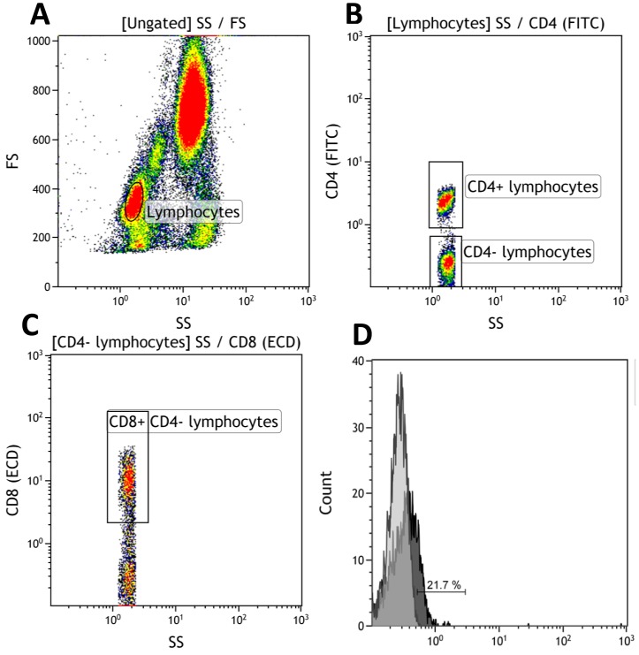
Methods: Patients attending our department were asked to participate in the study. The diagnosis of AMD was based on clinical examination and multimodal imaging techniques. Chemokine plasma level and chemokine receptor expression were measured by flow-cytometry.
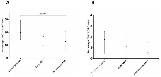
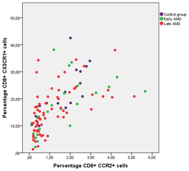
Results: A total of 150 participants were included. We found a significantly lower expression of CX3CR1 on CD8+ T cells in the neovascular AMD group compared to the control group (p = 0.04). We found a significant positive correlation between CCR2 and CX3CR1 expression on CD8+ cells (r = 0.727, p = 0.0001). We found no difference in plasma levels of CX3CL1 and CCL2 among the groups.
Conclusions: Our results show a down regulation of CX3CR1 on CD8+ cells; this correlated to a low expression of CCR2 on CD8+ cells. Further studies are needed to elucidate the possible role of this cell type in AMD development.
Conflict of interest statement
Figures
Similar articles
-
Chemokine-chemokine receptor CCL2-CCR2 and CX3CL1-CX3CR1 axis may play a role in the aggravated inflammation in primary biliary cirrhosis.Dig Dis Sci. 2014 Feb;59(2):358-64. doi: 10.1007/s10620-013-2920-6. Epub 2013 Nov 2. Dig Dis Sci. 2014. PMID: 24185682
-
Roles for the CX3CL1/CX3CR1 and CCL2/CCR2 Chemokine Systems in Hypoxic Pulmonary Hypertension.Am J Respir Cell Mol Biol. 2017 May;56(5):597-608. doi: 10.1165/rcmb.2016-0201OC. Am J Respir Cell Mol Biol. 2017. PMID: 28125278
-
CCL2/CCR2 and CX3CL1/CX3CR1 chemokine axes and their possible involvement in age-related macular degeneration.J Neuroinflammation. 2010 Dec 2;7:87. doi: 10.1186/1742-2094-7-87. J Neuroinflammation. 2010. PMID: 21126357 Free PMC article. Review.
-
Local vs. systemic mononuclear phagocytes in age-related macular degeneration and their regulation by CCL2-CCR2 and CX3CL1-CX3CR1 chemokine signalling.Adv Exp Med Biol. 2012;723:17-22. doi: 10.1007/978-1-4614-0631-0_3. Adv Exp Med Biol. 2012. PMID: 22183310 Review. No abstract available.
-
Wnt signaling in age-related macular degeneration: human macular tissue and mouse model.J Transl Med. 2015 Oct 17;13:330. doi: 10.1186/s12967-015-0683-x. J Transl Med. 2015. PMID: 26476672 Free PMC article.
Cited by
-
Chemokine Receptor Profiles of T Cells in Patients with Age-Related Macular Degeneration.Yonsei Med J. 2022 Apr;63(4):357-364. doi: 10.3349/ymj.2022.63.4.357. Yonsei Med J. 2022. PMID: 35352887 Free PMC article.
-
Expression of CCL2 and its receptor in activation and migration of microglia and monocytes induced by photoreceptor apoptosis.Mol Vis. 2017 Nov 1;23:765-777. eCollection 2017. Mol Vis. 2017. PMID: 29142497 Free PMC article.
-
Circulating monocytes and B-lymphocytes in neovascular age-related macular degeneration.Clin Ophthalmol. 2017 Jan 17;11:179-184. doi: 10.2147/OPTH.S121332. eCollection 2017. Clin Ophthalmol. 2017. PMID: 28176950 Free PMC article.
-
Associations of the Adaptive Immune System and Age-Related Macular Degeneration.Adv Exp Med Biol. 2025;1468:3-7. doi: 10.1007/978-3-031-76550-6_1. Adv Exp Med Biol. 2025. PMID: 39930164 Review.
-
Radiation Retinopathy: Microangiopathy-Inflammation-Neurodegeneration.Cells. 2025 Feb 18;14(4):298. doi: 10.3390/cells14040298. Cells. 2025. PMID: 39996770 Free PMC article.
References
-
- Xu H, Chen M, Forrester JV (2009) Para-inflammation in the aging retina. Prog Retin Eye Res 28:348–368. - PubMed
-
- Buschini E, Piras A, Nuzzi R, Vercelli A (2011) Age related macular degeneration and drusen: neuroinflammation in the retina. Prog Neurobiol 95:14–25. - PubMed
-
- Rollins BJ (1997) Chemokines. Blood 90:909–928. - PubMed
-
- Rossi D, Zlotnik A (2000) The biology of chemokines and their receptors. Annu Rev Immunol 18:217–242. - PubMed
Publication types
MeSH terms
Substances
LinkOut - more resources
Full Text Sources
Other Literature Sources
Medical
Research Materials
Miscellaneous
