. 2014 Dec 11;9(12):e114466.
doi: 10.1371/journal.pone.0114466. eCollection 2014.

Poly(A) binding protein 1 enhances cap-independent translation initiation of neurovirulence factor from avian herpesvirus

Affiliations

Poly(A) binding protein 1 enhances cap-independent translation initiation of neurovirulence factor from avian herpesvirus

Abdessamad Tahiri-Alaoui et al. PLoS One. .

Abstract

Poly(A) binding protein 1 (PABP1) plays a central role in mRNA translation and stability and is a target by many viruses in diverse manners. We report a novel viral translational control strategy involving the recruitment of PABP1 to the 5' leader internal ribosome entry site (5L IRES) of an immediate-early (IE) bicistronic mRNA that encodes the neurovirulence protein (pp14) from the avian herpesvirus Marek's disease virus serotype 1 (MDV1). We provide evidence for the interaction between an internal poly(A) sequence within the 5L IRES and PABP1 which may occur concomitantly with the recruitment of PABP1 to the poly(A) tail. RNA interference and reverse genetic mutagenesis results show that a subset of virally encoded-microRNAs (miRNAs) targets the inhibitor of PABP1, known as paip2, and therefore plays an indirect role in PABP1 recruitment strategy by increasing the available pool of active PABP1. We propose a model that may offer a mechanistic explanation for the cap-independent enhancement of the activity of the 5L IRES by recruitment of a bona fide initiation protein to the 5' end of the message and that is, from the affinity binding data, still compatible with the formation of 'closed loop' structure of mRNA.


Conflict of interest statement

Competing Interests: The authors have declared that no competing interests exist.

Figures

Figure 1
Figure 1. Effect of deletions of the internal homo-polymeric sequences on the activity of the 5L IRES and mutation analysis of affinity interaction between the 5L IRES and PABP1.
(A) Partial sequence from the full length 5L IRES from the immediate-early 1.8-kb mRNA that encodes pp14b isoform from Marek’s disease virus serotype 1. The 5L IRES spans nucleotides 129339-129798 (acc: AF243348). The internal poly-pyrimidine sequences C13 and U11 are boxed as well as the internal poly(A) sequences A11 and A9. (B) Schematic of the DNA constructs used for the luciferase reporter assay. In this vector the translation of R-Luc is controlled by the 5L IRES whereas the F-Luc is under the control of the intercistronic IRES (ICR). This configuration mimics the dual IRES bicistronic 1.8-kb mRNA from MDV1. DF-1 cells were transiently transfected with the indicated DNA vectors and after 24 h the cells were lysed and the luciferase activities were measured. The results are expressed as per cent change in luciferase activity relative to the control wild type sequence (5Lwt). (C) Northern blotting was performed on total RNA extracted from cells transfected with DNA constructs depicted in B. Hybridization was done with a random-primed 32P-labelled DNA fragment corresponding to the 5' end of the F-Luc open reading frame. Ethidium bromide-staining of the gel used for Northern blot is shown below the blot with 18S/28S rRNAs as size markers and loading control. (D) The mutated nucleotides within the internal poly(A) from the 5L IRES are underlined. The corresponding DNA vectors were used to transfect DF-1 cells as described in B. For simplicity, only the R-Luc values are shown as the F-Luc follows the same trend due to the coevolved synergistic functional relationship between the 5L IRES and the ICR IRES. The results are expressed as per cent change relative to the control wild type sequence (5Lwt). The experiment was repeated three times and the SEM is shown. (E) Purified recombinant human PABP1 (0.5 µM) was incubated with 32P-end labelled 5L IRES RNAs from wild type or from the indicated mutants and separated on a native 6% polyacrylamide gel by Electrophoretic Mobility Shift Assay (EMSA). It should be noted that the RNA was obtained by in vitro transcription and that it has no 3' poly (A) tail. The complex between PABP1 and the 5L IRES RNA was visualized by autoradiography using phosphor screen. The complex 5L IRES/PABP1 is observed in all combinations except with the mutants 5Lmt2 and 5Lmt2&3.
Figure 2
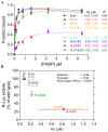
Figure 2. Effect of mutations within the internal poly(A) sequences on the binding affinities of the 5L IRES to the PABP1.
(A) Binding affinities between recombinant PABP1 and 32P-end labelled wild type and mutant 5L IRES sequences showing the fraction bound for each concentration of PABP1. The apparent dissociation constants are shown to the right with SEM from three repeats. (B) Correlations between binding affinities in panel A and the R-Luc activity as determined by transfection and reporter assay from panel D of Fig. 1.
Figure 3
Figure 3. PABP1 knockdown and functional analysis of the interplay between 5L IRES internal poly(A) and poly(A) tail.
(A) DF-1 cells were co-transfected with the depicted DNA constructs and with the PABP1 siRNA or nonsilencing siRNA control. The cells were lysed after 48 h incubation and used for luciferase assays. The results are presented as per cent change relative to nonsilencing siRNA control. The experiment was performed six times and the error bars indicate the SEM. Northern blotting was performed on total RNA extracted from transfected cells. Hybridization was done with a random-primed 32P-labelled DNA fragment corresponding to the 5' end of the F-Luc open reading frame. Ethidium bromide-staining of the gel used for Northern blot is shown below the blot with 18S/28S rRNAs as size markers and loading control. (B) Total proteins were harvested 48 h posttransfection and analysed by immunoblotting with the indicated antibodies. Quantification of the immunoblots from panel B using ImageQuant software is shown to the right. (C) DF-1 cells were transfected for 1 h with the indicated bicistronic dual IRES mRNA reporters and subsequently washed (0 hour); then 6 hours posttransfection the luciferase activity was measured and expressed as per cent change relative to capped and polyadenylated 5Lwt-R/ICR-F mRNA. The experiment was performed four times and the error bars indicate the SEM. (D) Total RNA was extracted from the transfected cells and the integrity of the bicistronic dual IRES mRNA reporters was analysed by Northern blotting and 32P-lablled probe against R-Luc, followed by phosphor screen autoradiography. As in panel A, Ethidium bromide-staining of the gel used for Northern blot is shown below the blot with 18S/28S rRNAs as size markers and loading control.
Figure 4
Figure 4. Effect of MDV1 infection on paip2 expression, PAPB1 level and localization.
(A) Chicken embryo fibroblasts (CEF) were transfected with oncogenic BAC clone pRB1B5 of MDV1 or mock-transfected for 72 h. Total proteins were harvested and analysed by immunoblotting with the indicated antibodies. Quantification of the immunoblots from panel A using ImageQuant software is shown to the right. The results are from two independent experiments each in duplicate. (B) Total proteins were extracted from control samples or from samples taken from chicken infected with the oncogenic BAC clone pRB1B5 derived from archive samples. Proteins were analysed by immunoblotting as in panel A. Quantification of the immunoblots from panel B using ImageQuant software is shown to the right. The results are repeats from two different archive samples derived from the same chicken challenge experiment. (C) Indirect immunofluorescence of pRB1B5-infected CEF 72 h posttransfection. A series of optical sections were taken sequentially for each channel along the z-axis using a step size of 0.290 µm. The resulting 3D confocal image was reconstructed using IMARIS software. DAPI-staining shows the nucleus in blue, PABP1 in red and pp14 in green, the scale bar: 10 µm.
Figure 5
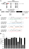
Figure 5. MDV1-encoded miRNAs target PABP1-interacting protein 2, paip2.
(A) The paip2 transcript showing paip2 open reading frame (ORF), the 3' untranslated region (UTR) and the miRNA response elements (MRE). (B) Schematic of the reporter construct containing individual or combined MREs sequences downstream of the simian virus 40 promoter-driven Renilla luciferase cassette from psiCHECK-2 vector. (C) The predicted duplexes between paip2 mRNA and MDV1 miRNAs. The mutated nucleotides within the seed regions of paip2 mRNA are underlined. (D) Luciferase-based miRNA reporter assay. The full length region from the paip2 mRNA that contains all the MREs or the individual MREs and their mutated versions were made as synthetic oligonucleotides and sub-cloned into the sensor plasmid downstream of the Renilla luciferase in psiCHECK-2 vector. The resulting constructs were used to transfect MSB1; an MDV1-transformed CD4+ T-cell line derived from a spleen lymphoma induced by BC-1 strain of MDV1 constitutively expressing viral miRNAs. As positive control for assay validation we have used MRE-M4 that was previously shown to be targeted by MDV1 miRNA-M4. The normalized Renilla luciferase activities from five experiments are shown with the error bars (SEM) relative to that seen for the empty vector psiCHECK-2 which value is set to 1.
Figure 6
Figure 6. Reverse genetic mutation analysis shows that MDV1 miRNAs from Lat-cluster are responsible for paip2 repression.
(A) Schematic representation of the bicistronic transcripts that we and others have cloned as cDNA and that encode for pp14a and pp14b isoforms, modified from Tahiri-Alaoui et al, J. Virol. Dec. 2009, Vol.83, No. 24, p12769-12778. (B) & (C) Chicken embryo fibroblasts (CEF) were transfected with BAC clone pRB1B5 Lat-miR-Revertant or pRB1B5 Lat-miR- deletion, respectively. RNA and proteins were simultaneously extracted using Trizol at the indicated time points. Viral and host proteins were detected by immunoblotting with the indicated antibodies. (D) Quantitative RT-PCR of host (paip2) and of viral transcripts (pp14a and pp14b isoforms) at the indicated time points. GAPDH is used as the endogenous control and time zero is used as the calibrator. All experiments were repeated three times and the error bars indicate the SEM.
Figure 7
Figure 7. Depletion of paip2 by RNA interference in cells infected with Lat-miR BAC mutants.
Chicken Embryo Fibroblasts were co-transfected with BAC clone of pRB1B5 Lat-miR-Revertant, pRB1B5 Lat-miR- deletion, si-paip2 RNA or siRNA control as indicated. The cells were lysed 72 hours post-transfection and the extracts were analysed by immunoblotting with the indicated antibodies against viral as well as host proteins.
Figure 8
Figure 8. Model depicting the closed loop topology for the bicistronic immediate-early transcript (IE) that encodes RLORF9 and the pp14 from MDV1.
In this model, only the pp14b isoform is shown, which is under the translation control of the 5L IRES. The internal poly(A) of the 5L IRES recruits PABP1 to the 5' end of the mRNA, which may be concomitant with the recruitment of PABP1 to the poly(A) tail of the message, leading to circularization of the bicistronic dual IRES IE-mRNA. A subset of viral miRNAs down-regulate the expression level of paip2 which is a well-known inhibitor of PABP1. This down regulation of paip2 indirectly contributes to an increased level of the available pool of active PABP1 which interacts with the internal poly(A) sequence of the 5L IRES hence leading to increased IRES activity.

Similar articles

Cited by

References

    1. Buchkovich NJ, Yu Y, Zampieri CA, Alwine JC (2008) The TORrid affairs of viruses: effects of mammalian DNA viruses on the PI3K-Akt-mTOR signalling pathway. Nat Rev Microbiol 6:266–275. - PMC - PubMed
    1. Walsh D, Mohr I (2011) Viral subversion of the host protein synthesis machinery. Nat Rev Microbiol 9:860–875. - PMC - PubMed
    1. Burgess HM, Gray NK (2010) mRNA-specific regulation of translation by poly(A)-binding proteins. Biochem Soc Trans 38:1517–1522. - PubMed
    1. Wells SE, Hillner PE, Vale RD, Sachs AB (1998) Circularization of mRNA by eukaryotic translation initiation factors. Mol Cell 2:135–140. - PubMed
    1. Jackson RJ, Hellen CU, Pestova TV (2010) The mechanism of eukaryotic translation initiation and principles of its regulation. Nat Rev Mol Cell Biol 11:113–127. - PMC - PubMed

Publication types

MeSH terms