. 2015 Jan 15;142(2):352-62.
doi: 10.1242/dev.115642. Epub 2014 Dec 11.

UPF2, a nonsense-mediated mRNA decay factor, is required for prepubertal Sertoli cell development and male fertility by ensuring fidelity of the transcriptome

Affiliations

UPF2, a nonsense-mediated mRNA decay factor, is required for prepubertal Sertoli cell development and male fertility by ensuring fidelity of the transcriptome

Jianqiang Bao et al. Development. .

Abstract

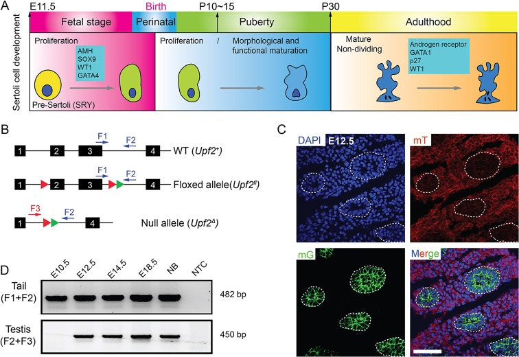
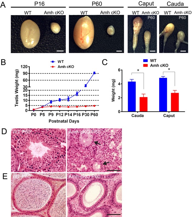
Nonsense-mediated mRNA decay (NMD) represents a highly conserved RNA surveillance mechanism through which mRNA transcripts bearing premature termination codons (PTCs) are selectively degraded to maintain transcriptomic fidelity in the cell. Numerous in vitro studies have demonstrated the importance of the NMD pathway; however, evidence supporting its physiological necessity has only just started to emerge. Here, we report that ablation of Upf2, which encodes a core NMD factor, in murine embryonic Sertoli cells (SCs) leads to severe testicular atrophy and male sterility owing to rapid depletion of both SCs and germ cells during prepubertal testicular development. RNA-Seq and bioinformatic analyses revealed impaired transcriptomic homeostasis in SC-specific Upf2 knockout testes, characterized by an accumulation of PTC-containing transcripts and the transcriptome-wide dysregulation of genes encoding splicing factors and key proteins essential for SC fate control. Our data demonstrate an essential role of UPF2-mediated NMD in prepubertal SC development and male fertility.

Keywords: 3′UTR shortening; Alternative splicing; Gonocyte; Mouse; Nonsense-mediated mRNA decay; Premature termination codon; RNA-Seq; Sertoli cell; Spermatogenesis; Sterility; Testis.


Figures

Fig. 1
Fig. 1
. Generation of Sertoli cell-specific Upf2 cKO mice. (A) Timeline of key developmental stages of murine Sertoli cells (SCs). (B) Structures of the floxed (Upf2fl) and null (Upf2Δ) Upf2 alleles after Cre-mediated recombination. Arrows indicate the positions of primer pairs used for genotyping. Red triangles represent loxP cassettes and green triangles depict FRT sites. Exons are numbered. (C) Cre activities represented by specific membrane-tagged eGFP (mG) signals were detected exclusively in SCs in Amh-Cre;Rosa26mTmG+/tg males at E12.5, whereas membrane-tagged Tomato red (mT) fluorescence was constitutively expressed in all cell types. Three individual mice were analyzed and representative confocal microscopy images are shown. Boundaries of the testis cords are outlined. Scale bar: 60 µm. (D) Genomic DNA PCR analyses showing Cre-mediated excision of exons 2-3 from E12.5 onwards. Sizes of PCR products (see B for primers) are as follows: F1/F2 primer pair: WT, 391 bp and floxed, 482 bp; F2/F3 primer pair: null/delete, 450 bp. NB, newborn; NTC, non-template control.
Fig. 2.
Fig. 2.
Inactivation of Upf2 in embryonic SCs leads to postnatal testicular atrophy and male sterility. (A) Gross morphology of the testis and the epididymis in WT and SC-specific Upf2 cKO (Amh-cKO) mice at P16 and P60. (B) Testis weights in WT and Amh-cKO mice during postnatal development. Data are presented as mean±s.d. (n=6). (C) Weights of both caput and cauda epididymides were significantly lower in Amh-cKO mice than in WT controls at P60. Data are shown as mean±s.d. (n=6 per genotype). *P<0.05. (D) Histology of WT and Amh-cKO testes at P60. Full spermatogenesis was observed in the seminiferous tubules in WT testes, whereas seminiferous tubules were rare and germ cells were almost completely depleted in Amh-cKO testes. Arrows indicate germ cell-depleted tubules. (E) Histology of the cauda epididymis in WT and Amh-cKO mice. The WT cauda epididymis contained abundant mature spermatozoa, whereas no spermatozoa were present in the Amh-cKO cauda epididymis. Scale bars: 1 cm in A; 40 µm in D,E.
Fig. 3.
Fig. 3.
Loss of SCs and male germ cells during postnatal testicular development in Amh-cKO mice. (A) Average number of seminiferous tubules per transverse section in WT and Amh-cKO testes at birth (P0), P5, P12, P16 and P60. At least 30 sections were scored for each of three mice per genotype at each time point, and the data are presented as mean±s.d. *P<0.05. (B) Histology of developing WT and Amh-cKO testes at P16 and P60. Transverse sections of WT testes contain numerous seminiferous tubules at various stages at P16 and P60, whereas only a few degenerated seminiferous tubules are present in Amh-cKO testes. The arrow points to degenerated spermatogonia-like germ cells at P16. The arrowheads indicate nuclei of disorganized SCs at P60. (C) Average number of germ cells per seminiferous tubule, counted based on immunofluorescence staining of WT1 (SC marker) and DDX4 (germ cell marker). At least 30 tubules were scored for each of three mice per genotype at each time point, and the data are presented as mean±s.d. *P<0.05. (D) Double immunofluorescence analyses of WT1 and DDX4 in WT and Amh-cKO testes at P0, P5 and P12. Three mice of each genotype were analyzed per time point and representative images are shown. Dashed circles delineate the boundary of each intact seminiferous tubule. Scale bars: 60 µm in B; 50 µm in D.
Fig. 4.
Fig. 4.
Defective spermatogonial development in Amh-cKO testes. Immunofluorescence staining of SOHLH1 (a marker for prospermatogonia and progenitor spermatogonia) in WT and Amh-cKO testes at P0, P5 and P9. The average numbers of SOHLH1-positive prospermatogonia (at P0) and progenitor spermatogonia (at P5) per tubule were comparable between WT and Amh-cKO testes. The intensity and nuclear localization of SOHLH1 were similar between WT and Amh-cKO testes. White dashed circles delineate the periphery of intact seminiferous tubules, whereas red lines encircle disorganized tubules as compared with WT. At P9, SOHLH1-positive spermatogonia are mostly situated on the basal membrane of the tubules in WT testes, whereas numerous SOHLH1-positive spermatogonia (arrowhead) appear to be aggregated randomly within the tubules in Amh-cKO testes. Arrows indicate SOHLH1-positive progenitor spermatogonia outside the disorganized seminiferous tubules. The boxed region in the WT P0 image is magnified to illustrate the area within the dashed box. Scale bar: 60 µm.
Fig. 5.
Fig. 5.
Loss of seminiferous tubule architecture in Amh-cKO testes. (A) Double immunofluorescence staining of WT1 and DDX4 in 3-month-old WT and Amh-cKO testes. WT testes contain numerous seminiferous tubules with SCs situated on the basal membrane and developing male germ cells located in both basal and adluminal compartments. By contrast, SCs and germ cells are absent in Amh-cKO testes at this age. Asterisk indicates the tubules depleted of both germ cells and SCs. (B) Histology of the 3-month-old Amh-cKO testes. The residual tubules still contained a few morphologically aberrant SCs (arrows indicate nuclei). (C) Comparison of the number of seminiferous tubules per transverse section in WT and Amh-cKO testes at 3 months of age. **P<0.01. (D) Double immunofluorescence staining of CYP17 (a Leydig cell marker) and DDX4 (a germ cell marker) in WT and Amh-cKO testes at P0, P5 and P9. CYP17 staining is present in only a subpopulation of Leydig cells, as indicated by arrows. The average number of Leydig cells labeled by CYP17 was similar between WT and Amh-cKO testes at all three time points. Arrowheads point to germ cells within the disrupted tubules in Amh-cKO testes. (E) qPCR analysis of expression levels of mRNAs encoding marker proteins for SCs (top), Leydig cells (middle) and germ cells (bottom) in WT and Amh-cKO testes at P4, P30 and P90. For each gene at each time point, the WT expression levels were designated as 1 and the relative expression levels in Amh-cKO testes, expressed as percentage, were calculated by normalizing to WT values based on the ΔΔCt method. All data points were collected from samples in biological triplicate. *P<0.05. Mean±s.d. Scale bars: 50 µm.
Fig. 6.
Fig. 6.
Changes in the genome-wide transcriptome profile in Amh-cKO testes. (A) Volcano plot showing upregulated (red) and downregulated (blue) transcripts in Amh-cKO testes as compared with WT controls (cut-off: fold change ≥2, P<0.05). Among a total of 56,107 transcripts detected in both WT and Amh-cKO testes, 1219 were ≥2-fold upregulated and 1373 were ≥2-fold downregulated. (B) Premature termination codon (PTC) annotation for transcripts dysregulated in Amh-cKO testes. All transcripts detected by RNA-Seq were subjected to analysis by spliceR, an R package pipeline specifically designed for prediction of PTCs. 'Un-regulation' refers to transcripts that differed less than 2-fold between Amh-cKO and WT.
Fig. 7.
Fig. 7.
Genome-wide alterations in mRNA splicing patterns and structural features induced by SC-specific ablation of Upf2. (A) Changes in mRNA levels of nine core splicing factors in Amh-cKO mice testes, as measured by both RNA-Seq and qPCR analyses. Data collected from biological triplicates are presented as mean±s.d. (B) Venn diagram showing the total number of exon skipping/inclusion events (top) and the average number of exon skipping/inclusion events per transcript (bottom) detected between WT and Amh-cKO testes. (C) Comparison of the total number of each of the eight types of splicing event detected by spliceR between 2-fold upregulated and 2-fold downregulated transcripts: ESI, exon skipping/inclusion; MESI, multiple exon skipping/inclusion; ISI, intron skipping/inclusion; A5, alternative 5′ splicing site; A3, alternative 3′ splicing site; ATSS, alternative transcription start site; ATTS, alternative transcription termination site; MEE, mutually exclusive exons. (D) Comparison of mRNA structural features, including 5′UTR length (left), CDS length (middle) and 3′UTR length (right), between WT and Amh-cKO testes at P4, as detected by spliceR. (E) Model for UPF2 functions in murine SCs. Inactivation of one of the core NMD factors, UPF2, causes disrupted transcriptomic homeostasis in SCs, characterized by accumulation of the PTC-positive transcripts, aberrant expression of splicing factors and dysregulation of SC-specific genes, which together lead to SC death and male sterility.

Similar articles

Cited by

References

    1. Avery P., Vicente-Crespo M., Francis D., Nashchekina O., Alonso C. R. and Palacios I. M. (2011). Drosophila Upf1 and Upf2 loss of function inhibits cell growth and causes animal death in a Upf3-independent manner. RNA 17, 624-638 10.1261/rna.2404211 - DOI - PMC - PubMed
    1. Azzalin C. M. and Lingner J. (2006). The human RNA surveillance factor UPF1 is required for S phase progression and genome stability. Curr. Biol. 16, 433-439 10.1016/j.cub.2006.01.018 - DOI - PubMed
    1. Ballow D., Meistrich M. L., Matzuk M. and Rajkovic A. (2006). Sohlh1 is essential for spermatogonial differentiation. Dev. Biol. 294, 161-167 10.1016/j.ydbio.2006.02.027 - DOI - PubMed
    1. Bao J., Wu J., Schuster A. S., Hennig G. W. and Yan W. (2013). Expression profiling reveals developmentally regulated lncRNA repertoire in the mouse male germline. Biol. Reprod. 89, 107 10.1095/biolreprod.113.113308 - DOI - PMC - PubMed
    1. Bao J., Zhang Y., Schuster A. S., Ortogero N., Nilsson E. E., Skinner M. K. and Yan W. (2014). Conditional inactivation of Miwi2 reveals that MIWI2 is only essential for prospermatogonial development in mice. Cell Death Differ. 21, 783-796 10.1038/cdd.2014.5 - DOI - PMC - PubMed

Publication types

MeSH terms