. 2015 Jan 15;142(2):282-90.
doi: 10.1242/dev.118307. Epub 2014 Dec 11.

The carboxy-terminus of p63 links cell cycle control and the proliferative potential of epidermal progenitor cells

Affiliations

The carboxy-terminus of p63 links cell cycle control and the proliferative potential of epidermal progenitor cells

Daisuke Suzuki et al. Development. .

Abstract

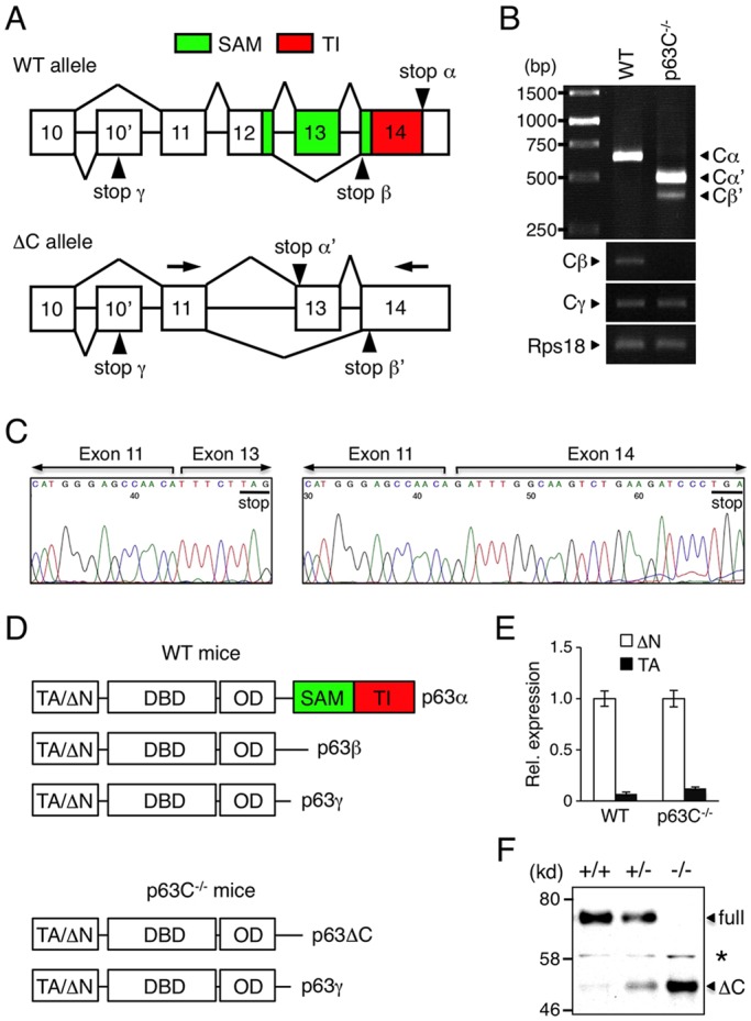
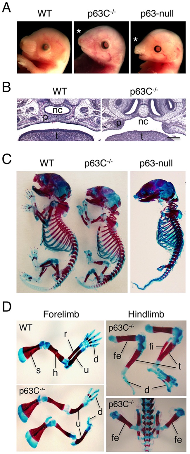
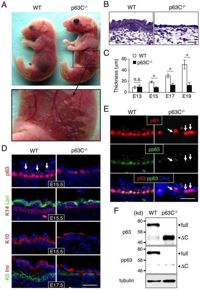
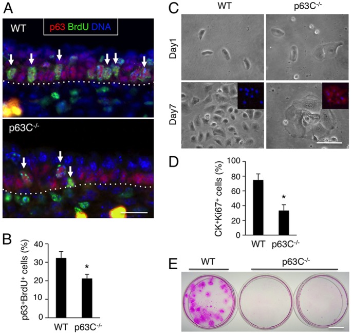
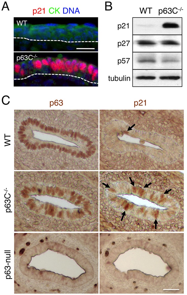
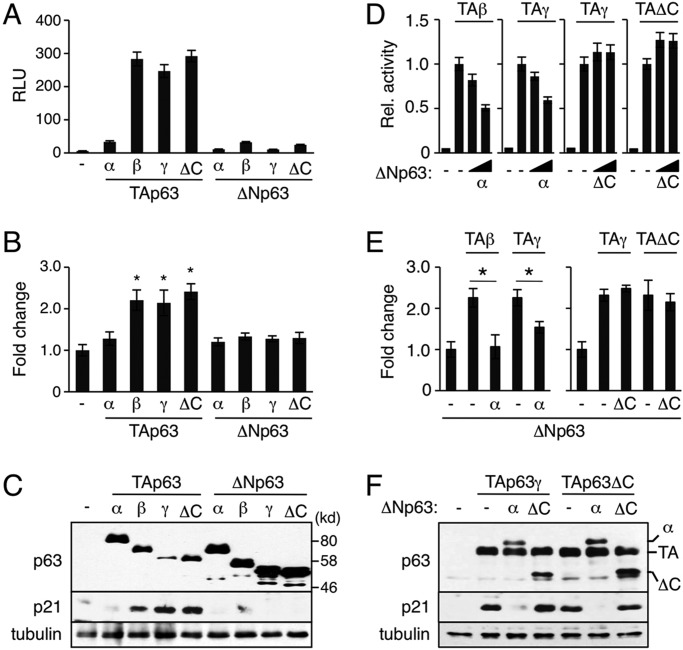
The transcription factor p63 (Trp63) plays a key role in homeostasis and regeneration of the skin. The p63 gene is transcribed from dual promoters, generating TAp63 isoforms with growth suppressive functions and dominant-negative ΔNp63 isoforms with opposing properties. p63 also encodes multiple carboxy (C)-terminal variants. Although mutations of C-terminal variants have been linked to the pathogenesis of p63-associated ectodermal disorders, the physiological role of the p63 C-terminus is poorly understood. We report here that deletion of the p63 C-terminus in mice leads to ectodermal malformation and hypoplasia, accompanied by a reduced proliferative capacity of epidermal progenitor cells. Notably, unlike the p63-null condition, we find that p63 C-terminus deficiency promotes expression of the cyclin-dependent kinase inhibitor p21(Waf1/Cip1) (Cdkn1a), a factor associated with reduced proliferative capacity of both hematopoietic and neuronal stem cells. These data suggest that the p63 C-terminus plays a key role in the cell cycle progression required to maintain the proliferative potential of stem cells of many different lineages. Mechanistically, we show that loss of Cα, the predominant C-terminal p63 variant in epithelia, promotes the transcriptional activity of TAp63 and also impairs the dominant-negative activity of ΔNp63, thereby controlling p21(Waf1/Cip1) expression. We propose that the p63 C-terminus links cell cycle control and the proliferative potential of epidermal progenitor cells via mechanisms that equilibrate TAp63 and ΔNp63 isoform function.

Keywords: Cell cycle; Epithelia; Mouse; Proliferation; Stem cells; p21; p63.


Figures

Fig. 1.
Fig. 1.
Alterative splicing at the p63 C-terminus in p63C−/− mice. (A) Structure and splicing of the p63 C-terminus in WT and ΔC p63 alleles. Arrowheads indicate stop codons in each isoform. The p63SAM and p63TI domains are illustrated. Exons are numbered. (B) Expression of p63 C-terminus variants in WT and p63C−/− epidermis as assessed by PCR. p63C−/− epidermal cells predominantly express Cα′, which lacks both the p63SAM and p63TI domains, whereas WT cells express full-length Cα. (C) Sequencing of Cα′ and Cβ′ expressed in p63C−/− mice. Stop codons are underlined. Arrows in A indicate the primer pair used to amplify the Cα′ and Cβ′ splicing variants. (D) Structure of p63 isoforms expressed in WT and p63C−/− mice. p63Cα′ and p63Cβ′ isoforms are collectively referred to as p63ΔC. DBD, DNA-binding domain; OD, oligomerization domain. (E) qPCR analysis of the N-terminus of the p63 gene in WT and p63C−/− epidermal cells. Shown is expression of TAp63 relative to ΔNp63, with the ΔNp63 level in WT being set to 1.0. Error bars indicate s.d. (F) Western blot with anti-p63 antibody of E14.5 whole epidermal cell extracts from WT, p63C−/− and p63C+/− mice. Asterisk indicates non-specific signal. Full, full-length ΔNp63α; ΔC, ΔNp63ΔC.
Fig. 2.
Fig. 2.
Craniofacial and skeletal abnormalities in p63C−/− mice. (A) Gross appearance of the head of E17.5 WT, p63C−/− and p63-null mice. Whereas p63-null mice show cleft lip, p63C−/− mice frequently lack this characteristic as indicated by asterisks. (B) H&E staining of coronal sections of E17.5 WT and p63C−/− mouse heads showing the absence and presence, respectively, of cleft of the secondary palate. nc, nasal cavity; p, palatal shelf; t, tongue. Scale bar: 250 µm. (C) Skeletal staining of E17.5 WT, p63C−/− and p63-null mice showing relatively well developed forelimbs and hindlimbs in p63C−/− mice compared with those in p63-null mice. (D) Enlarged views of forelimbs and hindlimbs of WT and p63C−/− mice. s, scapula; h, humerus; r, radius; u, ulna; d, digits; fe, femur; fi, fibula; t, tibia.
Fig. 3.
Fig. 3.
Epidermal hypoplasia in p63C−/− mice. (A) Representative E17.5 WT and p63C−/− mouse embryos (top) and an enlarged view of the p63C−/− mouse abdomen showing severe skin erosion (bottom). (B) H&E staining of E17.5 WT and p63C−/− mouse epidermis. Note that p63C−/− mouse epidermis is significantly thinner than that of WT and lacks hair follicle development. (C) Changes in epidermal thickness during development (mean±s.d.). *P<0.01; n.s., not significant. (D) Immunofluorescence of WT and p63C−/− mouse epidermis with the indicated antibodies, counterstained with Hoechst 33342 (DNA, blue). Arrows indicate p63-positive cells expanding into the suprabasal layers in WT epidermis. Lori, loricrin; Inv, involucrin. (E) Immunofluorescence of E15.5 WT and p63C−/− mouse epidermis with anti-p63 and anti-phosphorylated p63 (pp63) antibodies, counterstained with Hoechst 33342. Arrows indicate high p63-positive cells with low phosphorylation in the basal layer of the p63C−/− mouse epidermis. (F) Western blot of whole epidermal cell extracts from E15.5 WT and p63C−/− mice. The membrane was probed first with anti-pp63 antibody, stripped and then reprobed with anti-pan-p63 antibody. Full, full-length ΔNp63α; ΔC, ΔNp63ΔC. Scale bars: 50 µm.
Fig. 4.
Fig. 4.
Reduced proliferative capacity of p63C−/− epidermal cells. (A) Immunofluorescence of E15.5 WT and p63C−/− mouse epidermis with anti-BrdU and anti-p63 antibodies, counterstained with Hoechst 33342. Arrows indicate representative p63+ BrdU+ epidermal progenitor cells. Dotted lines indicate the epidermal-dermal border. (B) Quantification of A (mean±s.e.m.). *P<0.01. (C) Epidermal cells were isolated from E14.0 WT and p63C−/− mouse embryos, seeded at clonal density in Matrigel-coated dishes and cultivated for 7 days. Inset shows high expression of involucrin (red) in p63C−/− epidermal cells, which is absent in the WT counterpart. Nuclei were counterstained with Hoechst 33342. (D) Proliferation of WT and p63C−/− mouse epidermal cells at day 5 of cultivation. Shown are percentages of CK+ Ki67+ epidermal cells (mean±s.e.m.). *P<0.01. CK refers to pan-cytokeratin. (E) Staining of WT and p63C−/− mouse epidermal cells with Rhodamine B at 3 weeks of culture. No macroscopically visible clones were detected in p63C−/− cell cultures. Scale bars: 25 µm in A,C; 1 cm in E.
Fig. 5.
Fig. 5.
Upregulation of p21Waf1/Cip1 expression in p63C−/− mouse epithelia. (A) Immunofluorescence of E15.5 WT and p63C−/− mouse epidermis with anti-p21Waf1/Cip1 and anti-pan-cytokeratin (CK) antibodies, counterstained with Hoechst 33342. Dotted lines indicate epidermal-dermal border. (B) Western blot of whole epidermal cell extracts from E15.5 WT and p63C−/− mice with the antibodies to p21Waf1/Cip1, p27Kip1 and p57Kip2. Anti-Tubulin α antibody was used as a loading control. (C) Immunohistochemistry of mouse esophagus with anti-p63 (left) and anti-p21Waf1/Cip1 (right) antibodies. Data shown are representative of WT and p63C−/− mice at E15.5 and p63-null mice at E17.5. Arrows indicate epidermal cells with high p21Waf1/Cip1 expression. Scale bars: 25 µm in A; 50 µm in C.
Fig. 6.
Fig. 6.
Altered p63ΔC functions in control of p21Waf1/Cip1 expression. (A-C) Stimulation of p21Waf1/Cip1 expression by TAp63ΔC. H1299 cells were transfected with 1.0 µg plasmid expressing each p63 isoform. Samples were prepared 24 h after transfection. (A) Reporter assays using the minimal p21Waf1/Cip1 reporter (mean±s.d.). RLU, relative light unit. (B) qPCR analysis of endogenous p21Waf1/Cip1 gene expression. Values are mean±s.d. with the basal level set to 1.0. *P<0.05. (C) Western blot with anti-p63 and anti-p21Waf1/Cip1 antibodies. (D-F) Loss of dominant-negative activity of ΔNp63ΔC against TAp63-dependent transactivation of p21Waf1/Cip1. (D) Reporter assays using the minimal p21Waf1/Cip1 reporter. H1299 cells were transfected with 0.3 µg TAp63 plasmid alone or together with an increasing amount of ΔNp63 plasmid at ratios of 100:1 or 30:1 and cells were lysed 24 h after transfection. Data shown are mean±s.d. Transactivation by TAp63 alone was set to 1.0. (E) qPCR analysis of endogenous p21Waf1/Cip1 gene expression. H1299 cells were transfected with 1.0 µg TAp63 plasmid alone or together with ΔNp63 plasmid at a 50:1 ratio and RNA was extracted 24 h after transfection. Values are mean±s.d. with the basal level set to 1.0. *P<0.05. (F) Western blot analysis of p21Waf1/Cip1 expression. H1299 cells were transfected with 1.0 µg TAp63 plasmid alone or together with ΔNp63 plasmid at a 20:1 ratio and cells were lysed 36 h after transfection. The same cell extracts were used to verify expression of each p63 isoform (top). (C,F) Anti-tubulin α antibody was used as a loading control.

Similar articles

Cited by

References

    1. Ali-Khan S. E. and Hales B. F. (2003). Caspase-3 mediates retinoid-induced apoptosis in the organogenesis-stage mouse limb. Birth Defects Res. A Clin. Mol. Teratol. 67, 848-860 10.1002/bdra.10090 - DOI - PubMed
    1. Barrow L. L., van Bokhoven H., Daack-Hirsch S., Andersen T., van Beersum S. E. C., Gorlin R. and Murray J. C. (2002). Analysis of the p63 gene in classical EEC syndrome, related syndromes, and non-syndromic orofacial clefts. J. Med. Genet. 39, 559-566 10.1136/jmg.39.8.559 - DOI - PMC - PubMed
    1. Beretta C., Chiarelli A., Testoni B., Mantovani R. and Guerrini L. (2005). Regulation of the cyclin-dependent kinase inhibitor p57Kip2 expression by p63. Cell Cycle 4, 1625-1631 10.4161/cc.4.11.2135 - DOI - PubMed
    1. Bickenbach J. R., Stern M. M., Grinnell K. L., Manuel A. and Chinnathambi S. (2006). Epidermal stem cells have the potential to assist in healing damaged tissues. J. Investig. Dermatol. Symp. Proc. 11, 118-123 10.1038/sj.jidsymp.5650009 - DOI - PubMed
    1. Blanpain C. and Fuchs E. (2009). Epidermal homeostasis: a balancing act of stem cells in the skin. Nat. Rev. Mol. Cell Biol. 10, 207-217 10.1038/nrm2636 - DOI - PMC - PubMed

Publication types