

SIRT1 protects against apoptosis by promoting autophagy in degenerative human disc nucleus pulposus cells
- PMID: 25503852
- PMCID: PMC4264007
- DOI: 10.1038/srep07456
SIRT1 protects against apoptosis by promoting autophagy in degenerative human disc nucleus pulposus cells
Abstract
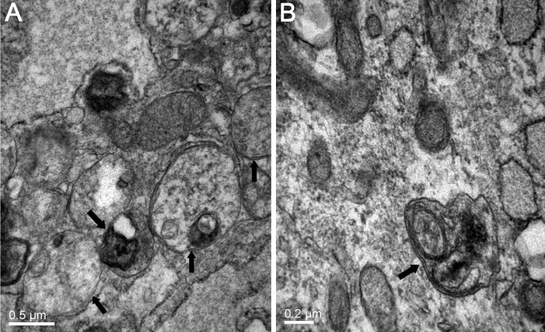
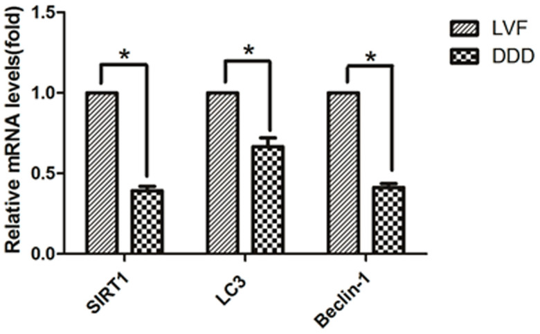
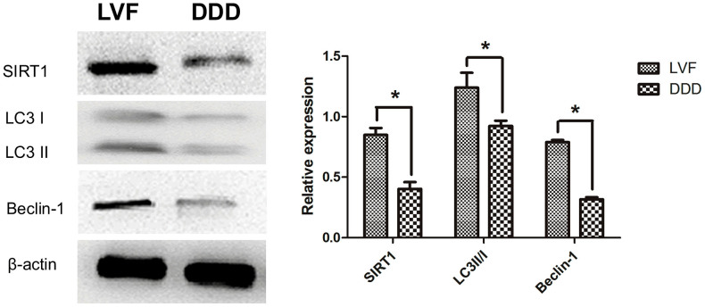
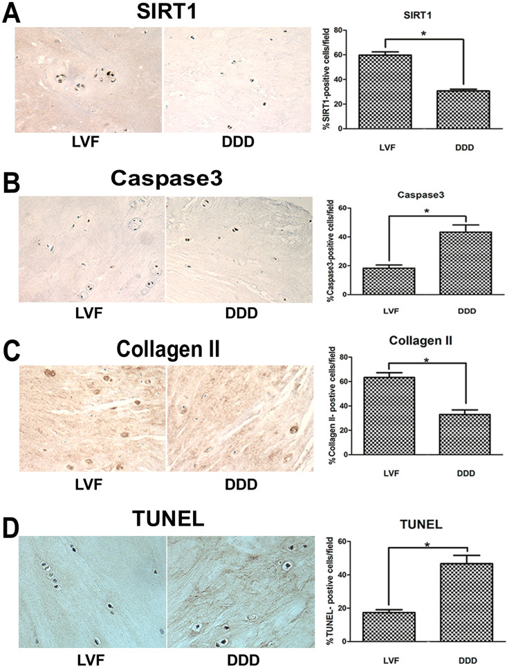
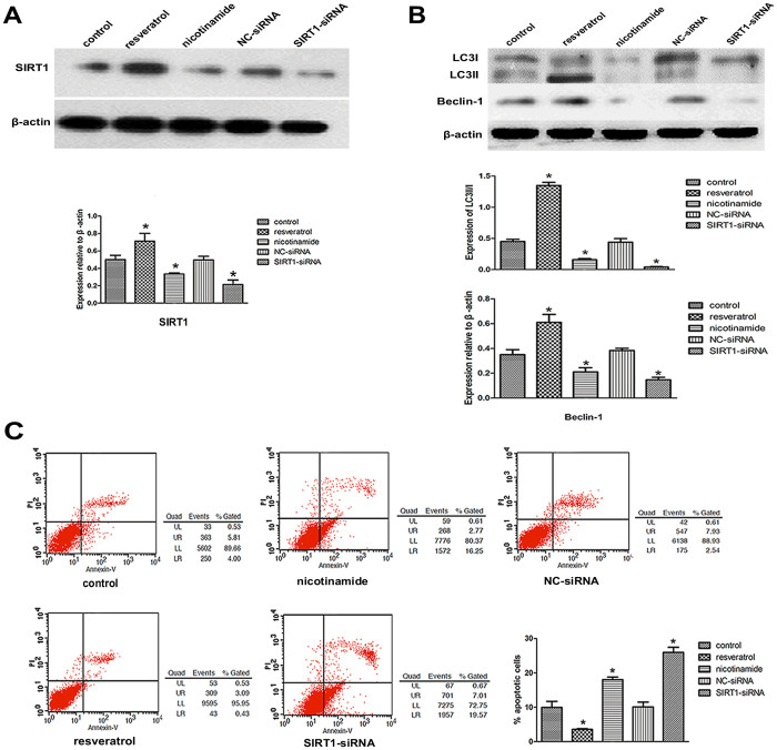
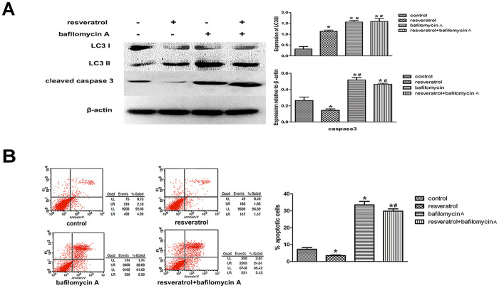
SIRT1 could protect degenerative human NP cells against apoptosis, and there were extensive and intimate connection between apoptosis and autophagy. Up to now, the role of autophagy in the process of human IVD degeneration is unclear. We sought to explore the relationship between autophagy and human IVD degeneration and to understand whether autophagy is involved in the protective effect of SIRT1 against apoptosis in NP cells. Our results showed that the autophagosomes number, the mRNA level of LC3 and Beclin-1, the protein expression of LC3-II/I and Beclin-1, decreased in NP from DDD. Resveratrol could increase the protein expression of LC3-II/I and Beclin-1, and reduce apoptosis in degenerative NP cells. In contrast, the protein levels of LC3-II/I and Beclin-1 were down-regulated and apoptosis level was significantly up-regulated in treatment with nicotinamide or SIRT1-siRNA transfection. Further analysis identified that the expression of cleaved Caspase3 and apoptosis incidence significantly increased with the pretreatment of bafilomycin A, whether resveratrol was added or not. These suggested that autophagy may play an important role in IVD degeneration, and SIRT1 protected degenerative human NP cells against apoptosis via promoting autophagy. These findings would aid in the development of novel therapeutic approaches for degenerative disc disease treatment.
Figures
Similar articles
-
Recombinant human SIRT1 protects against nutrient deprivation-induced mitochondrial apoptosis through autophagy induction in human intervertebral disc nucleus pulposus cells.Arthritis Res Ther. 2015 Sep 15;17(1):253. doi: 10.1186/s13075-015-0763-6. Arthritis Res Ther. 2015. PMID: 26373839 Free PMC article.
-
Role of Sirt1 Plays in Nucleus Pulposus Cells and Intervertebral Disc Degeneration.Spine (Phila Pa 1976). 2017 Jul 1;42(13):E757-E766. doi: 10.1097/BRS.0000000000001954. Spine (Phila Pa 1976). 2017. PMID: 27792110
-
SIRT1 inhibits apoptosis of degenerative human disc nucleus pulposus cells through activation of Akt pathway.Age (Dordr). 2013 Oct;35(5):1741-53. doi: 10.1007/s11357-012-9474-y. Epub 2012 Sep 19. Age (Dordr). 2013. PMID: 22990594 Free PMC article.
-
Cell death in intervertebral disc degeneration.Apoptosis. 2013 Jul;18(7):777-85. doi: 10.1007/s10495-013-0839-1. Apoptosis. 2013. PMID: 23512131 Review.
-
Autophagy: A double-edged sword in intervertebral disk degeneration.Clin Chim Acta. 2016 Jun 1;457:27-35. doi: 10.1016/j.cca.2016.03.016. Epub 2016 Mar 24. Clin Chim Acta. 2016. PMID: 27018178 Review.
Cited by
-
The Role of Oxidative Stress in Intervertebral Disc Degeneration.Oxid Med Cell Longev. 2022 Jan 12;2022:2166817. doi: 10.1155/2022/2166817. eCollection 2022. Oxid Med Cell Longev. 2022. PMID: 35069969 Free PMC article. Review.
-
Bushen Huoxue Formula Modulates Autophagic Flux and Inhibits Apoptosis to Protect Nucleus Pulposus Cells by Restoring the AMPK/SIRT1 Pathway.Biomed Res Int. 2022 May 27;2022:8929448. doi: 10.1155/2022/8929448. eCollection 2022. Biomed Res Int. 2022. PMID: 35669720 Free PMC article.
-
Mesenchymal Stem Cells Protect Nucleus Pulposus Cells from Compression-Induced Apoptosis by Inhibiting the Mitochondrial Pathway.Stem Cells Int. 2017;2017:9843120. doi: 10.1155/2017/9843120. Epub 2017 Dec 14. Stem Cells Int. 2017. PMID: 29387092 Free PMC article.
-
Resveratrol: Molecular Mechanisms, Health Benefits, and Potential Adverse Effects.MedComm (2020). 2025 Jun 11;6(6):e70252. doi: 10.1002/mco2.70252. eCollection 2025 Jun. MedComm (2020). 2025. PMID: 40502812 Free PMC article. Review.
-
SIRT1 Inhibits Apoptosis by Promoting Autophagic Flux in Human Nucleus Pulposus Cells in the Key Stage of Degeneration via ERK Signal Pathway.Biomed Res Int. 2021 Feb 27;2021:8818713. doi: 10.1155/2021/8818713. eCollection 2021. Biomed Res Int. 2021. PMID: 33728342 Free PMC article.
References
-
- Barrick W. T. et al. Anterior lumbar fusion improves discogenic pain at levels of prior posterolateral fusion. Spine 25, 853–857 (2000). - PubMed
-
- Kellgren J. H. The anatomical source of back pain. Rheumatol Rehabil 16, 3–12 (1977). - PubMed
-
- Kuslich S. D., Ulstrom C. L. & Michael C. J. The tissue origin of low back pain and sciatica: a report of pain response to tissue stimulation during operations on the lumbar spine using local anesthesia. Orthop Clin North Am 22, 181–187 (1991). - PubMed
Publication types
MeSH terms
Substances
LinkOut - more resources
Full Text Sources
Other Literature Sources
Research Materials
Miscellaneous
