Review
. 2015 Apr;34(2):105-118.
doi: 10.4149/gpb_2014037. Epub 2014 Dec 11.

Emerging evidence for specific neuronal functions of auxiliary calcium channel α₂δ subunits

Affiliations
Review

Emerging evidence for specific neuronal functions of auxiliary calcium channel α₂δ subunits

Stefanie Geisler et al. Gen Physiol Biophys. 2015 Apr.

Abstract

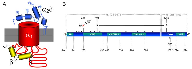
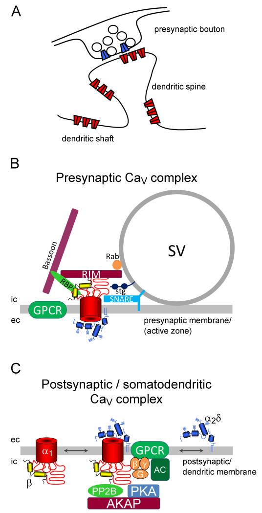
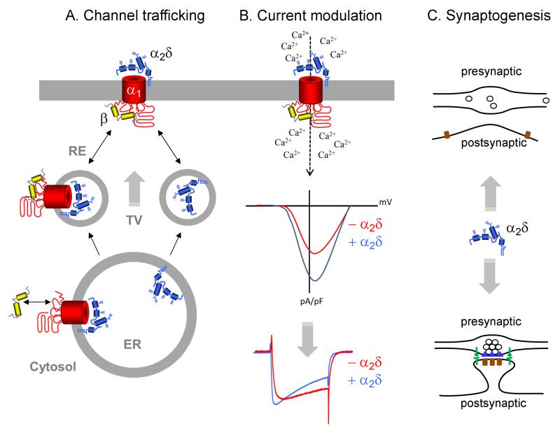
In nerve cells the ubiquitous second messenger calcium regulates a variety of vitally important functions including neurotransmitter release, gene regulation, and neuronal plasticity. The entry of calcium into cells is tightly regulated by voltage-gated calcium channels, which consist of a heteromultimeric complex of a pore forming α₁, and the auxiliary β and α₂δ subunits. Four genes (Cacna2d1-4) encode for the extracellular membrane-attached α₂δ subunits (α₂δ-1 to α₂δ-4), out of which three isoforms (α₂δ-1 to -3) are strongly expressed in the central nervous system. Over the years a wealth of studies has demonstrated the classical role of α₂δ subunits in channel trafficking and calcium current modulation. Recent studies in specialized neuronal cell systems propose roles of α₂δ subunits beyond the classical view and implicate α₂δ subunits as important regulators of synapse formation. These findings are supported by the identification of novel human disease mutations associated with α₂δ subunits and by the fact that α₂δ subunits are the target of the anti-epileptic and anti-allodynic drugs gabapentin and pregabalin. Here we review the recently emerging evidence for specific as well as redundant neuronal roles of α₂δ subunits and discuss the mechanisms for establishing and maintaining specificity.


Figures

Figure 1
Figure 1
A. The voltage-gated calcium channel complex. Voltage-gated calcium channels are composed of a transmembrane pore-forming α1 subunit (red) which forms a macromolecular complex together with extracellular α2δ (blue) and cytoplasmic β (yellow) subunits. The β subunit binds the intracellular I-II linker of α1 with high affinity. The α2δ subunit consists of posttranslationally cleaved highly glycosylated α2 and δ peptides, which are associated to each other by a disulfide bond and are most likely linked to the plasma membrane (indicated in grey) via a GPI-anchor. Note that the folding structure of the individual subunits is simplified. B. Domain structure of auxiliary α2δ subunits. Amino acid positions refer to the mouse α2δ-1 isoform [uniprot: O08532- CA2D1_MOUSE], however all isoforms have a similar topology. Amino acid residues 1—24 encode the signal peptide (SP). The α2 peptide contains a von Willebrand factor type A (VWA) domain and two sequence stretches homologous to extracellular domains of bacterial chemosensing proteins (Cache I and II). In α2δ-1 and α2δ-2 an arginine (RRR) motif proximal to the VWA domain represents the potential gabapentin (GBP) and pregabalin (PG) binding site. Two cysteine residues (AA 404 and 1059) were identified to be important for the formation of an intermolecular disulfide bond between α2 and δ. The δ peptide contains predicted ω amino acids, to which the GPI anchor can attach, and a C-terminal hydrophobic sequence (c-HS). Potential glycosylation sites are indicated by asterisks (*).
Figure 2
Figure 2
Neuronal calcium channel complexes. A. In neurons voltage-gated calcium channels are located in presynaptic boutons, where they trigger neurotransmitter release, and in postsynaptic as well as extrasynaptic positions along dendrites and dendritic spines (Obermair et al. 2004; Jenkins et al. 2010). B. In the presynaptic active zone calcium channels are organized in a dense network of presynaptic proteins (depicted are RIM, RBP, Rab, bassoon, SNARE complex) and G-protein coupled receptors (GPCR) in the vicinity of the calcium sensor synaptotagmin (stg). Together this macromolecular complex orchestrates the tight regulation of synaptic vesicle (SV) fusion and thus neurotransmitter release (reviewed in Südhof 2012). C. In the postsynaptic/somato-dendritic compartment L-type channels can be found in complexes with G-protein coupled receptors (GPCR, e.g. the β2 adrenergic receptor), an adenylyl cyclase (AC), protein kinases (PKA) and phosphatases (PP2B), and scaffolding proteins (e.g. AKAP150) (reviewed in Dai et al. 2009). Downstream signaling initiates a signaling cascade which leads to the modulation of transcription such as CREB or NFATc4. Lateral mobility of L-type calcium channels in the plasma membrane also suggests regulation via the association or dissociation with distinct signaling complexes (Di Biase et al. 2011). Considering the membrane diffusion of GPI-anchored proteins, such a mechanism may also apply to α2δ subunits. ic, intracellular; ec, extracellular.
Figure 3
Figure 3
Sketch summarizing the neuronal functions of auxiliary α2δ subunits. A. Channel trafficking: The export from the endoplasmic reticulum (ER) is mediated by β subunits; however, α2δ subunits can further enhance the trafficking. This may involve trafficking from the endoplasmic reticulum (ER) via the Golgi apparatus in transport vesicles (TV), recycling from recycling endosomes (RE), or stabilization of the channel complex at the plasma membrane. α2δ subunits can also be transported to the plasma membrane independent of the α1 subunit. B. Current modulation: Besides their role in channel trafficking α2δ subunits are important modulators of the calcium current. α2δ subunits can shift the voltage-dependence of channel activation to more negative potentials (IV curve, middle graph) and can modify the activation and inactivation kinetics (current traces, lower graph). This may be of particular relevance for somato-dendritic L-type calcium channels. C. Synaptogenesis: Accumulating evidence suggests an important role of neuronal α2δ subunits in synapse formation. Whether α2δs are necessary for the initial contact between the axon and the corresponding postsynaptic membrane (top) or for the differentiation into a fully aligned and mature synapse (bottom) remains to be answered.

Similar articles

Cited by

References

    1. Anantharaman V, Aravind L. Cache - a signaling domain common to animal Ca(2+)-channel subunits and a class of prokaryotic chemotaxis receptors. Trends Biochem. Sci. 2000;25:535–537. - PubMed
    1. Arikkath J, Campbell KP. Auxiliary subunits: essential components of the voltage-gated calcium channel complex. Curr. Opin. Neurobiol. 2003;13:298–307. - PubMed
    1. Barclay J, Balaguero N, Mione M, Ackerman SL, Letts VA, Brodbeck J, Canti C, Meir A, Page KM, Kusumi K, et al. Ducky mouse phenotype of epilepsy and ataxia is associated with mutations in the Cacna2d2 gene and decreased calcium channel current in cerebellar Purkinje cells. J. Neurosci. 2001;21:6095–6104. - PMC - PubMed
    1. Bauer CS, Nieto-Rostro M, Rahman W, Tran-Van-Minh A, Ferron L, Douglas L, Kadurin I, Sri Ranjan Y, Fernandez-Alacid L, Millar NS, et al. The increased trafficking of the calcium channel subunit alpha2delta-1 to presynaptic terminals in neuropathic pain is inhibited by the alpha2delta ligand pregabalin. J. Neurosci. 2009;29:4076–4088. - PMC - PubMed
    1. Bauer CS, Tran-Van-Minh A, Kadurin I, Dolphin AC. A new look at calcium channel alpha2delta subunits. Curr. Opin. Neurobiol. 2010;20:563–571. - PubMed

Publication types