Review
. 2015 Apr;72(7):1331-45.
doi: 10.1007/s00018-014-1800-3. Epub 2014 Dec 12.

Intracellular signaling mechanisms of the melanocortin receptors: current state of the art

Affiliations
Review

Intracellular signaling mechanisms of the melanocortin receptors: current state of the art

Adriana R Rodrigues et al. Cell Mol Life Sci. 2015 Apr.

Abstract

The melanocortin system is composed by the agonists adrenocorticotropic hormone and α, β and γ-melanocyte-stimulating hormone, and two naturally occurring antagonists, agouti and agouti-related protein. These ligands act by interaction with a family of five melanocortin receptors (MCRs), assisted by MCRs accessory proteins (MRAPs). MCRs stimulation activates different signaling pathways that mediate a diverse array of physiological processes, including pigmentation, energy metabolism, inflammation and exocrine secretion. This review focuses on the regulatory mechanisms of MCRs signaling, highlighting the differences among the five receptors. MCRs signal through G-dependent and independent mechanisms and their functional coupling to agonists at the cell surface is regulated by interacting proteins, namely MRAPs and β-arrestins. The knowledge of the distinct modulation pattern of MCRs signaling and function may be helpful for the future design of novel drugs able to combine specificity, safety and effectiveness in the course of their therapeutic use.


Conflict of interest statement

The authors have no conflict of interest.

Figures

Fig. 1
Fig. 1
Integrated mechanisms for G-protein-mediated signaling. Gs signaling is mainly conveyed through activation of AC/cAMP/PKA pathway, which inhibits C-Raf, but also leads to the activation of Rap1 that relays in B-Raf stimulation of ERK1/2 signaling. Gi inhibits AC activity blocking its inhibitory effect on C-Raf and concomitantly stimulates ERK1/2 signaling by a Ras-dependent mechanism. G12/13 strongly stimulates JNK activity but can also activate ERK1/2 through Ras or by DAG/PKC pathway. Gq can stimulate ERK1/2 via PLCβ/DAG/PKC as well as PLCβ/IP3/Ca2+-signaling mechanisms, either by direct phosphorylation of C-Raf by PKC or by a Ras-dependent manner, which may involve the recruitment of Src. Besides α-subunit, the complex βγ released from α subunit during GPCR activation is also able to promote ERK1/2 signaling through stimulation of PLCβ or PI3K. PI3K usually relays in Akt phosphorylation, but also leads to Src and/or Ras stimulation of ERK1/2. In addition, G-proteins can mediate the transactivation of RTK through its βγ-subunits. Activation of PI3K leads to Src-mediated receptor tyrosine kinase phosphorylation and subsequent recruitment of Shc, Grb2 proteins and SOS to stimulate Ras activity and ERK1/2 phosphorylation. RTK transactivation mediated by βγ-subunits may also occur through an inside-out model. This mechanism is well described for the epidermal growth factor (EGF) receptor, in which βγ activates matrix metalloprotease (MMP) proteins that cleave the ectodomains of membrane-bound growth factors (HB-EGF) to generate soluble EGF ligands that are released from the cell to activate its RTK. Whatever the mechanism of ERK1/2 activation is, these kinases are then able to phosphorylate a wide variety of cytoplasmic and nuclear targets. When translocating to the nucleus, ERK1/2 initiate gene transcription by phosphorylating several transcription factors like CREB and c-Fos
Fig. 2
Fig. 2
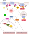
Signaling mechanisms of melanocortin 1 receptor (MC1R). MC1R couples to Gs stimulating cAMP/PKA and increasing intracellular Ca2+ levels. Ligand binding to MC1R promotes a cAMP-dependent activation of Rap1 and Ras, which leads to ERK1/2 phosphorylation through B-Raf signaling. By c-KIT transactivation, MC1R is also able to activate ERK1/2. β-arrestin 2 mediates MC1R internalization and desensitization, decreasing cAMP production but not affecting ERK1/2 activation. β-arrestin 1 competes with β-arrestin 2 for MC1R binding, which blocks β-arrestin 2-mediated cAMP inhibition. cAMP/PKA signaling increases synthesis of melanogenic pigments whereas ERK1/2 seems to have an inhibitory effect on this process. Akt/mTOR apparently increases cell survival during oxidative stress
Fig. 3
Fig. 3
Signaling mechanisms of melanocortin 2 receptor (MC2R). MC2R couples to Gs and stimulates cAMP/PKA. Activation of ERK1/2 is dependent on PKA and MC2R internalization. MC2R also leads to the phosphorylation of p38 and PKC. MC2R promotes biphasic ERK1/2 activation, with a first transient wave, dependent from receptor internalization, but independent of β-arrestins. ERK1/2 seems to be related with proliferation and differentiation processes regulating the trophic and steroidogenic properties of ACTH in adrenal glands. PKA and PKC are also mediators of adrenal steroidogenesis
Fig. 4
Fig. 4
Signaling mechanisms of melanocortin 3 receptor (MC3R). MC3R couples to Gs stimulating cAMP/PKA but is also able to couple to Gi driving to ERK1/2 pathway through activation of PI3K. MC3R receptor is also known to increase intracellular Ca2+ levels and to interact with PKC and IP3. MC3R activation induces cell proliferation through a PI3K/ERK1/2-mediated mechanism that may be related to neuronal regeneration
Fig. 5
Fig. 5
Signaling mechanisms of melanocortin 4 receptor (MC4R). MC4R couples to Gs stimulating cAMP/PKA and to Gq activating PKC dependently from PLCβ and IP3. It also activates ERK1/2 via Gi or through mechanisms dependent from Ca2+, PKA, PKC or PI3K. A PKA-dependent ERK1/2 signaling is associated with food intake suppression, whereas a PKA-independent ERK1/2 seems to decrease cell apoptosis upon neuronal damage. MC4R also inhibits JNK activity, possibly to increase insulin-stimulated glucose uptake
Fig. 6
Fig. 6
Signaling mechanisms of melanocortin 5 receptor (MC5R). MC5R activates two parallel signaling pathways, Gs/cAMP/PKA and Gi/PI3K/ERK1/2 that promote CREB phosphorylation and c-fos expression, respectively. MC5R is also known to increase intracellular Ca2+ levels and promote JAK/STAT activation. ERK1/2 signaling occurs through a biphasic fashion with an early transient peak dependent from Gi protein and a second one, more sustained in time, regulated by Gi and β-arrestins. MC5R internalizes independently from β-arrestins but these scaffold molecules seem to retain ERK1/2 signaling in the cytoplasm to mediate MC5R-dependent decrease of fatty acid re-esterification in adipocytes. MC5R-dependent increase of lipolysis rate is regulated by PKA activation

Similar articles

Cited by

References

    1. Cooray SN, Clark AJ. Melanocortin receptors and their accessory proteins. Mol Cell Endocrinol. 2011;331:215–221. - PubMed
    1. Smith AI, Funder JW Proopiomelanocortin processing in the pituitary, central nervous system, and peripheral tissues. Endocr Rev. 1988;9:159–179. - PubMed
    1. Bultman SJ, Michaud EJ, Woychik RP. Molecular characterization of the mouse agouti locus. Cell. 1992;71:1195–1204. - PubMed
    1. Wilson BD, Ollmann MM, Kang L, Stoffel M, Bell GI, Barsh GS. Structure and function of ASP, the human homolog of the mouse agouti gene. Hum Mol Genet. 1995;4:223–230. - PubMed
    1. Voisey J, Kelly G, Van Daal A. Agouti signal protein regulation in human melanoma cells. Pigment Cell Res. 2003;16:65–71. - PubMed

Publication types

LinkOut - more resources