. 2015 Feb;8(1):193-202.
doi: 10.1161/CIRCEP.114.002049. Epub 2014 Dec 10.

Phospholamban as a crucial determinant of the inotropic response of human pluripotent stem cell-derived ventricular cardiomyocytes and engineered 3-dimensional tissue constructs

Affiliations

Phospholamban as a crucial determinant of the inotropic response of human pluripotent stem cell-derived ventricular cardiomyocytes and engineered 3-dimensional tissue constructs

Gaopeng Chen et al. Circ Arrhythm Electrophysiol. 2015 Feb.

Abstract

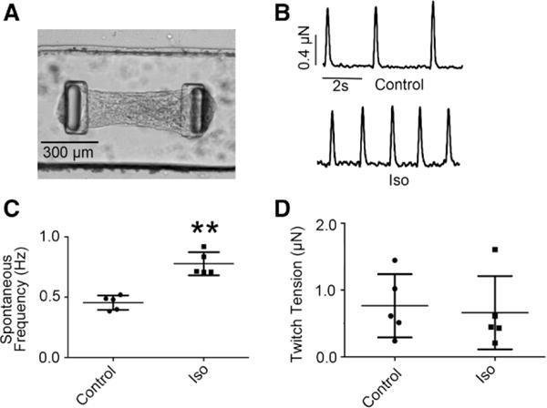
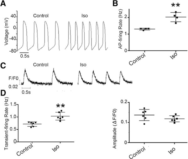
Background: Human (h) embryonic stem cells (ESCs) and induced pluripotent stem cells (iPSCs) serve as a potential unlimited ex vivo source of cardiomyocytes (CMs). However, a well-accepted roadblock has been their immature phenotype. hESC/iPSC-derived ventricular (v) CMs and their engineered cardiac microtissues (hvCMTs) similarly displayed positive chronotropic but null inotropic responses to β-adrenergic stimulation. Given that phospholamban (PLB) is robustly present in adult but poorly expressed in hESC/iPSC-vCMs and its defined biological role in β-adrenergic signaling, we investigated the functional consequences of PLB expression in hESC/iPSC-vCMs and hvCMTs.

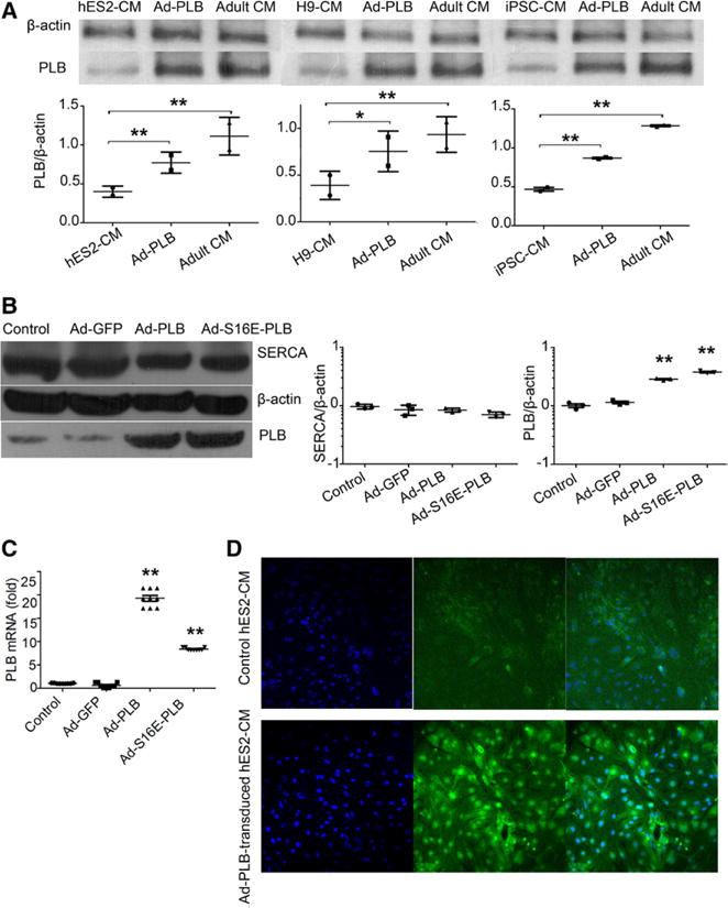
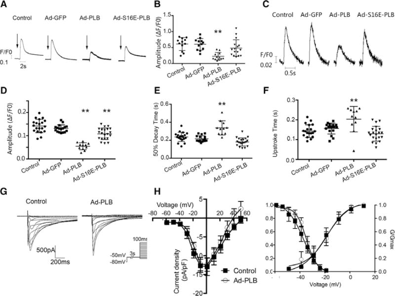
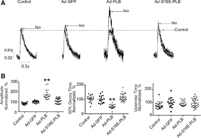
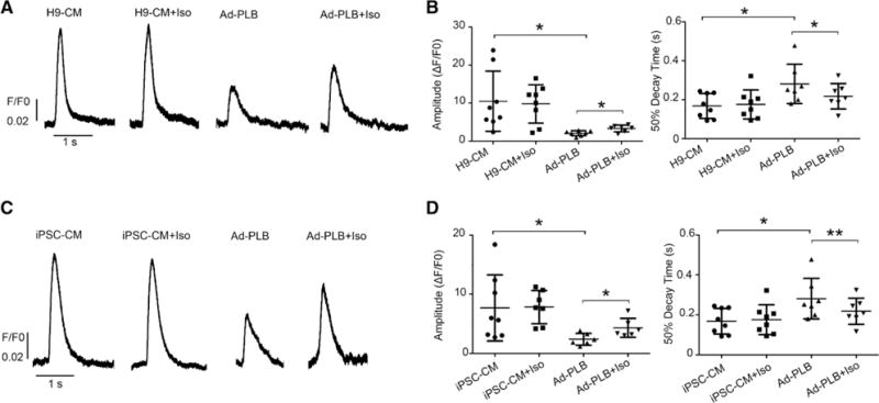
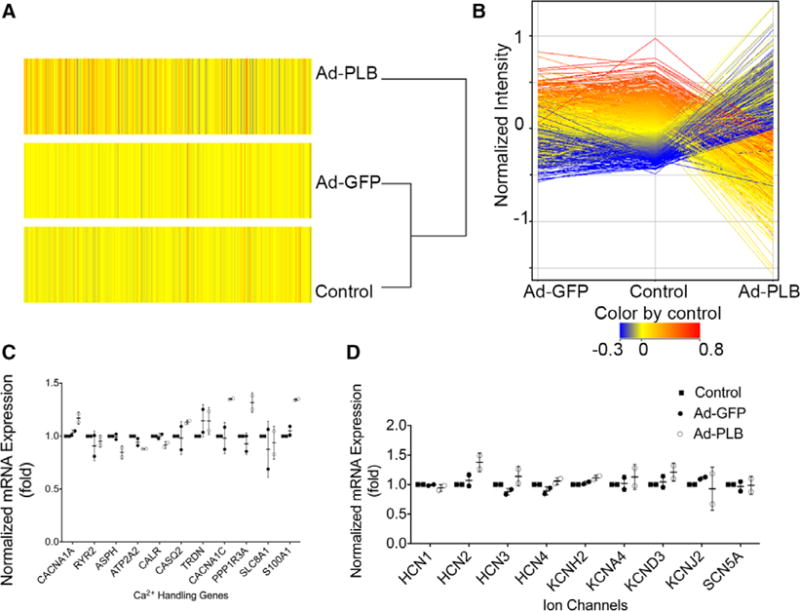
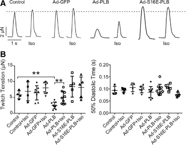
Methods and results: First, we confirmed that PLB protein was differentially expressed in hESC (HES2, H9)- and iPSC-derived and adult vCMs. We then transduced hES2-vCMs with the recombinant adenoviruses (Ad) Ad-PLB or Ad-S16E-PLB to overexpress wild-type PLB or the pseudophosphorylated point-mutated variant, respectively. As anticipated from the inhibitory effect of unphosphorylated PLB on sarco/endoplasmic reticulum Ca2+-ATPase, Ad-PLB transduction significantly attenuated electrically evoked Ca2+ transient amplitude and prolonged the 50% decay time. Importantly, Ad-PLB-transduced hES2-vCMs uniquely responded to isoproterenol. Ad-S16E-PLB-transduced hES2-vCMs displayed an intermediate phenotype. The same trends were observed with H9- and iPSC-vCMs. Directionally, similar results were also seen with Ad-PLB-transduced and Ad-S16E-transduced hvCMTs. However, Ad-PLB altered neither the global transcriptome nor ICa,L, implicating a PLB-specific effect.

Conclusions: Engineered upregulation of PLB expression in hESC/iPSC-vCMs restores a positive inotropic response to β-adrenergic stimulation. These results not only provide a better mechanistic understanding of the immaturity of hESC/iPSC-vCMs but will also lead to improved disease models and transplantable prototypes with adult-like physiological responses.

Keywords: adrenergic effects; phospholamban; pluripotent stem cells; tissues.


Figures

Figure 1
Figure 1
Positive chronotropic but null inotropic responses of hES2-derived ventricular microtissues (hvCMTs) to β-adrenergic stimulation. A, Image of a typical hvCMT on a microfabricated tissue gauge device. B, Representative raw tracings of hvCMT at the baseline (upper) and after 10 μmol/L isoproterenol (Iso) treatment (down). C, Iso increased the spontaneous twitch frequency (n=5; **P<0.01). D, Iso did not affect the developed twitch tensions (P=0.729).
Figure 2
Figure 2
Isoproterenol (Iso) increased the spontaneous action potential (AP) and Ca2+ transient firings (chronotropy) of single hES2-ventricular microtissues (vCMs) but not the transient amplitude. A, Representative AP tracings of single hES2-vCMs before and after Iso. B, The corresponding AP firing frequencies (n=4; **P=0.008). C, Representative recordings of spontaneous Ca2+ transients. D, Transient rate (**P=0.005) and amplitude (P=0.182) of Ca2+ transients (n=6).
Figure 3
Figure 3
Effects of adenovirus (Ad)-green fluorescence protein (GFP), Ad-phospholamban (PLB), and Ad-S16E-PLB transduction on the expression levels of PLB, sarco/endoplasmic reticulum Ca2+-ATPase (SERCA). A, Western blot showing Ad-PLB transduction increased PLB protein expression in hES2-ventricular cardiomyocytes (vCMs; left), H9-vCMs (middle), and induced pluripotent stem cell (iPSC)-vCMs (right) but did not reach the adult level (n=2; P=0.226, 0.644, and 0.001 for hES2-vCMs, H9-vCMs, and iPSC-vCMs). B, Western blot analysis shows that the PLB protein levels of the Ad-PLB and Ad-S16E-PLB groups were significantly increased, but those for SERCA were not different from the untransduced and Ad-GFP–transduced groups (n=3). C, PLB transcript levels as gauged by quantitative polymerase chain reaction (n=3). D, Immunostaing images show that Ad-PLB–transduced hES2-vCMs were 100% positive: cell nucleus indicated by DAPI (left), PLB staining (middle), and merged image (right).
Figure 4
Figure 4
Effects of adenovirus (Ad)-green fluorescence protein (GFP), Ad-phospholamban (PLB), and Ad-S16E-PLB transduction on the Ca2+ transient properties, SR load, and ICa,L of hES2-ventricular cardiomyocytes (vCMs). A, Representative raw tracings of caffeine-induced Ca2+ transients (arrows indicate the time of caffeine application). B, Bar graphs summarizing the amplitudes of the same groups as indicated. Ad-PLB transductions significantly decreased the sarcoplasmic reticulum (SR) load (n=11, 11, 11, and 15 for control, Ad-GFP, Ad-PLB, and Ad-S16E-PLB, respectively). Untransduced vs Ad-S16E-PLB–tranduced: P=0.17. C, Representative raw tracings of electrically induced Ca2+ transients of untransduced, Ad-GFP–transduced, Ad-PLB–transduced, and Ad-S16E-PLB–transduced hES2-vCMs paced at 1 Hz. D–F, Bar graphs summarizing the transient parameters of the same groups from C. Ad-PLB transduction significantly decreased the transient amplitude (D) and prolonged the 50% decay (E) and upstroke (F). Ad-S16E-PLB transduction produced an intermediate phenotype (n=20, 17, 11, and 21 for control, Ad-GFP, Ad-PLB, and Ad-S16E-PLB. Untransduced vs Ad-S16E-PLB–tranduced: P=0.29 for upstroke time, P=0.14 for 50% decay time, and **P<0.01, compared with control. G and H, Representative ICa,L tracings of nontransduced and Ad-PLB–transduced hES2-vCMs. Electrophysiological protocol is given in the inset. Current density (I)–voltage (V) plots and steady-state activation curves revealed no significant differences. n=7 and 5 for control and Ad-PLB–transduced CMs, respectively.
Figure 5
Figure 5
Restoration of a positive inotropic response of hES2-ventricular cardiomyocytes (vCMs) by phospholamban (PLB) overexpression. A, Representative raw tracings of electrically induced Ca2+ transients of untransduced, adenovirus (Ad)-green fluorescence protein (GFP)–transduced, Ad-PLB–transduced, and Ad-S16E-PLB–transduced human embryonic stem cell (hESC)–derived vCMs paced at 1 Hz recorded in the absence and presence of isoproterenol (Iso) as indicated. Peaks recorded under control drug–free condition of all groups have been normalized for comparison. B, Bar graphs summarizing the transient parameters of the same groups from (A). Ad-PLB–transduced hESC-vCMs uniquely responded to Iso (n=15, 15, 15, and 25 for control, Ad-GFP, Ad-PLB, and Ad-S16E-PLB respectively; **P<0.01, compared with parameters under drug-free condition).
Figure 6
Figure 6
Restoration of a positive inotropic response of human (H)9-ventricular cardiomyocytes (vCMs) and induced pluripotent stem cell (iPSC)–derived vCMs by phospholamban (PLB) overexpression. A, Representative raw tracings of electrically induced Ca2+ transients of untransduced or adenovirus (Ad)-PLB–transduced H9-CMs paced at 0.5 Hz with/without isoproterenol (Iso) application. B, Bar graphs summarizing amplitude and 50% decay time of the same groups from A (n=8 and 7 for untransduced or Ad-PLB–transduced H9-CMs). C, Representative raw tracings of electrically induced Ca2+ transients of untransduced or Ad-PLB–transduced iPSC-CMs paced at 0.5 Hz with/without Iso. D, Bar graphs summarizing amplitude and 50% decay time of the same groups from C (n=7 and 6 for untransduced or Ad-PLB–transduced iPSC-CMs; *P<0.05 and **P<0.01).
Figure 7
Figure 7
Transcriptomic analysis. A, Heat maps of microarray transcriptomic profiling of untransduced, adenovirus (Ad)-green fluorescence protein (GFP)–transduced, and Ad-phospholamban (PLB)–transduced human embryonic stem cell–derived ventricular cardiomyocytes. B, No significant changes in the global gene expression or those of (C) calcium-handling genes and (D) or ion channels were detected (n=2).
Figure 8
Figure 8
Restoration of a positive inotropic response in engineered cardiac microtissues (hvCMTs) by phospholamban (PLB) overexpression. Adenovirus (Ad)-PLB transduction uniquely attenuated the developed tensions of hvCMTs but restored a positive inotropic response to isoproterenol (Iso). A, Raw tracings of untransduced, Ad-green fluorescence protein (GFP)–transduced, Ad-PLB–transduced, and Ad-S16E-PLB–transduced hvCMTs recorded paced at 0.5 Hz in the absence and presence of Iso as indicated. The dotted line shows the peak of untransduced hvCMT recorded under Iso-free condition. B, Bar graph summarizing the twitch tension (the dotted line shows twitch tension of untransduced hvCMTs recorded under Iso-free condition) and 50% diastolic time from the same groups (n=4, 8, 9, and 5 for control, Ad-GFP, Ad-PLB, and Ad-S16E-PLB, respectively; **P<0.01).

References

    1. Thomson JA, Itskovitz-Eldor J, Shapiro SS, Waknitz MA, Swiergiel JJ, Marshall VS, Jones JM. Embryonic stem cell lines derived from human blastocysts. Science. 1998;282:1145–1147. - PubMed
    1. Takahashi K, Tanabe K, Ohnuki M, Narita M, Ichisaka T, Tomoda K, Yamanaka S. Induction of pluripotent stem cells from adult human fibroblasts by defined factors. Cell. 2007;131:861–872. doi: 10.1016/j.cell.2007.11.019. - DOI - PubMed
    1. Yang L, Soonpaa MH, Adler ED, Roepke TK, Kattman SJ, Kennedy M, Henckaerts E, Bonham K, Abbott GW, Linden RM, Field LJ, Keller GM. Human cardiovascular progenitor cells develop from a kdr plus embryonic-stem-cell-derived population. Nature. 2008;453:524–528. - PubMed
    1. Kehat I, Kenyagin-Karsenti D, Snir M, Segev H, Amit M, Gepstein A, Livne E, Binah O, Itskovitz-Eldor J, Gepstein L. Human embryonic stem cells can differentiate into myocytes with structural and functional properties of cardiomyocytes. J Clin Invest. 2001;108:407–414. doi: 10.1172/JCI12131. - DOI - PMC - PubMed
    1. Weng Z, Kong CW, Ren L, Karakikes I, Geng L, He J, Chow MZ, Mok CF, Keung W, Chow H, Leung AY, Hajjar RJ, Li RA, Chan CW. A simple, cost-effective but highly efficient system for deriving ventricular cardiomyocytes from human pluripotent stem cells. Stem Cells Dev. 2014;23:1704–1716. doi: 10.1089/scd.2013.0509. - DOI - PMC - PubMed

Publication types

MeSH terms

Substances