Comparative Study
. 2014 Dec 12;346(6215):1256846.
doi: 10.1126/science.1256846.

Convergent transcriptional specializations in the brains of humans and song-learning birds

Affiliations
Comparative Study

Convergent transcriptional specializations in the brains of humans and song-learning birds

Andreas R Pfenning et al. Science. .

Abstract

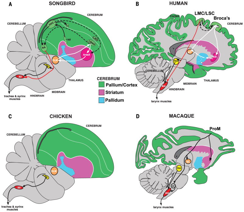
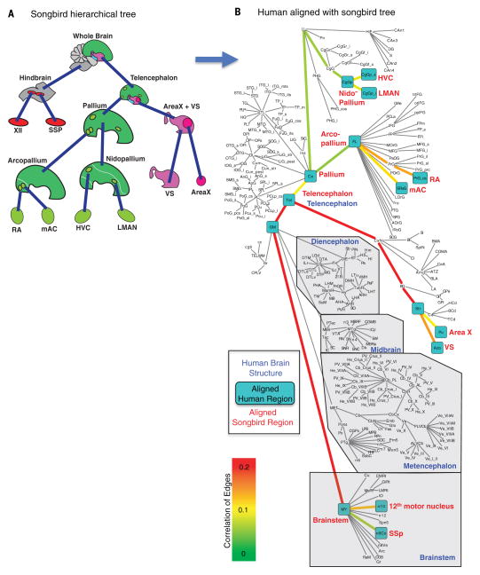
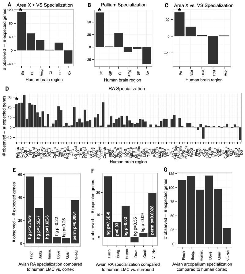
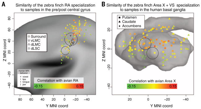
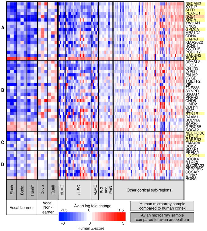
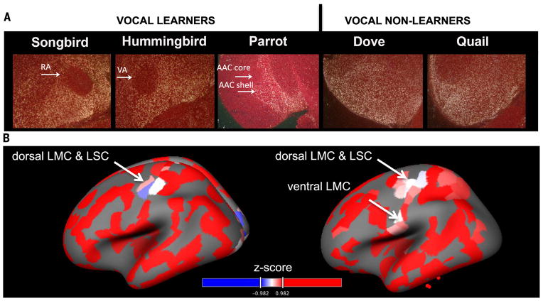
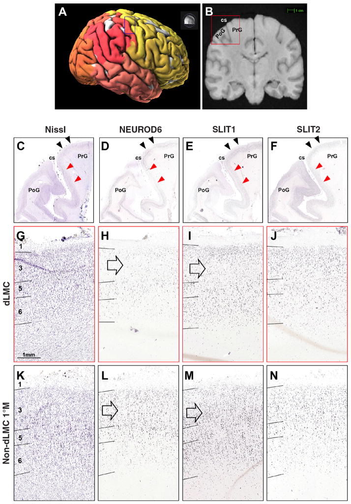
Song-learning birds and humans share independently evolved similarities in brain pathways for vocal learning that are essential for song and speech and are not found in most other species. Comparisons of brain transcriptomes of song-learning birds and humans relative to vocal nonlearners identified convergent gene expression specializations in specific song and speech brain regions of avian vocal learners and humans. The strongest shared profiles relate bird motor and striatal song-learning nuclei, respectively, with human laryngeal motor cortex and parts of the striatum that control speech production and learning. Most of the associated genes function in motor control and brain connectivity. Thus, convergent behavior and neural connectivity for a complex trait are associated with convergent specialized expression of multiple genes.


Figures

Fig. 1
Fig. 1. Comparative brain relationships, connectivity, and cell types among vocal learners and nonlearners
(A) Drawing of a zebra finch male brain section showing profiled song nuclei: Area X, HVC, LMAN, RA, and the 12th motor nucleus (XII). (B) Drawing of a human brain section showing proposed vocal pathway connectivity including LMC/LSC in the precentral gyrus. Black arrows, connections and regions of the posterior vocal motor pathway; white arrows, connections and regions of the anterior vocal pathway; dashed arrows, connections between the two pathways. The thick blue arrows show the analogous brain regions predicted by this study across finch and human. Red arrows show the direct projections found only in vocal learners, from vocal motor cortex regions to brainstem vocal motor neurons. (C) Known connectivity of a vocal-nonlearning bird showing absence of forebrain song nuclei. (D) Known connectivity of vocal-nonlearning primates (i.e., macaque) showing presence of forebrain regions that have an indirect projection to nucleus ambiguus (Amb) but have no known role in production of vocalizations.
Fig. 2
Fig. 2. Optimal alignment of human and zebra finch brain hierarchies
(A) Tree representing zebra finch brain regions based on hierarchical expression of gene expression profiles. (B) Tree representing the hierarchy of the human brain based on the current knowledge of mammalian brain organization (http://human.brain-map.org). Each node (name) in the tree is a brain region. The daughters of a node are the subregions found within a brain region. Each edge (line) is a specialization of a subregion relative from the brain region that encompasses it. Turquoise boxes are human regions (black text) where the zebra finch brain regions (red text) optimally aligned. The blue font highlights larger human brain structures. Edges are colored by the correlation value (0 to 0.2) of the aligned avian and human specializations. Human region abbreviations are annotated (table S1A).
Fig. 3
Fig. 3. Relative number of genes with significantly shared specialized expression between avian and human brain regions
Each panel shows a plot of the number of genes significantly specialized (P < 0.05; hypergeometric test) in common between the avian and human samples relative to the number of genes expected to be specialized by chance. (A) Finch Area X + VS specialization compared to all subregions of the human telencephalon. (B) Finch pallial region (RA, neighboring arcopallium, HVC, and LMAN combined) specialization compared to all subregions of the human telencephalon. (C) Finch Area X specialization compared to all subregions of the human striatum. (D) Finch RA specialization compared to the specialization of every subregion from the human cortex, which optimally aligned to the zebra finch pallium. (E) Avian RA analogs (vocal learners) and mAC (nonlearners) relative to the adjacent arcopallium compared to human LMC/dLSC relative to cortex. (F) Avian RA analogs (vocal learners) and mAC (nonlearners) relative to the arcopallium compared to human LMC/dLSC relative to PrG/PoG. (G) Avian arcopallium versus whole brain specialized genes compared to human cortex versus whole brain specialized genes. In (A) to (D), asterisks denote the human specializations determined to be similar to the avian specialization on the basis of the optimal alignment and correlation. In (E) to (G), P values less than 0.05 indicate that the number of specialized genes is greater than chance according to a hypergeometric test.
Fig. 4
Fig. 4. Overlap of regions with convergent specialized expression and speech activation
Shown are the coordinates in MNI (Montreal Neurological Institute) space of each human brain microarray sample from the striatum (A) and the precentral/postcentral gyrus (B). The different subregions within the striatum and precentral/postcentral gyrus are labeled by differently shaped symbols. These points are placed on top of a representative image of the cortical surface map from the Allen Human Brain Atlas (38). Only the left hemisphere, which has higher sample density, is shown. In (A), each point is colored on the basis of the correlation between the specialization of that sample relative to the entire human striatum, and of Area X relative to Area X and VS together. Circles represent high-confidence regions of speech activation from multiple coordinates in multiple studies (–62). In (B), each point is colored on the basis of the correlation between the specialization of that sample relative to the entire human PrG/PoG, and of zebra finch’s RA specialization. A similar plot with the average of all vocal learners is shown in fig. S4C.
Fig. 5
Fig. 5. Heat map of gene expression specialization in avian RA analogs versus the arcopallium and human LMC/dLSC regions versus other cortical regions
(A to D) Each row represents a gene (table S4), sorted according to whether or not they are significant in hummingbird RA analog [(A) and (B)], significant in individual human LMC/LSC regions relative to the entire PrG and PoG [(A) and (C)], or all LMC/dLSC regions (dLMC, vLMC, LSC) combined (D). Samples within each section are ordered on the basis of estimated log fold difference in LMC/LSC versus PrG and PoG surround. Each column is a microarray sample from an avian species (dark gray) or human (light gray) as listed at the bottom. For the avian species samples, each entry in the heat map shows the log fold change between each microarray sample and median gene expression value for the entire finch arcopallium (needed one species and microarray platform to normalize). For the human samples, each entry is the Z-score specialization relative to the entire cortex for the human sample or brain region. Red, higher expression; blue, lower expression; white, no difference between the compared regions. In the hummingbird microarray data set, one animal was an outlier for some genes [(C) and (D); third column], which we believe is due to an error in the laser capture dissection for a subset of sections (fig. S17); in situ hybridization data validated the hummingbird profiles of one of these genes as an example (fig. S11). Yellow highlights show validated genes; orange highlights were not able to be validated.
Fig. 6
Fig. 6. Convergent differential regulation of SLIT1 in the RA analog and human LMC
(A) In situ hybridization of SLIT1 in the RA analog and arcopallium of vocal-learning and vocal-nonlearning avian species. Shown are frontal sections; dorsal is up, right is midline. White, SLIT1 mRNA detected by 35S riboprobe in dark-field view; red, cresyl violet stain of brain cells. (B) Cortical surface map of SLIT1 relative gene expression levels (Z-score) in the human brain measured by microarrays (http://human.brain-map.org/static/brainexplorer). Red, higher expression; blue, lower expression. Two example persons are shown (left hemisphere), one with both dLMC/LSC and vLMC. Dorsal is up, front is left.
Fig. 7
Fig. 7. In situ hybridization localization of the putative dLMC/dLSC in the human brain
(A) Surface image of a human brain showing the different cortical lobes (colors) and the region dissected for the in situ hybridization analyses (box). (B) Magnetic resonance image showing the location of the region dissected for in situ hybridization analyses in the right hemisphere (box). (C) Nissl stain of the examined region. (D) NEUROD6 down-regulation in a distinct region of the PrG, and in the upper layers of the adjacent PoG. (E) SLIT1 down-regulation in the same PrG dLMC region (see I versus M), as well as in the adjacent PoG. (F) SLIT2 control showing no noticeable difference. (G to N) Red arrows correspond to the boundaries of the regions represented in the higher-power images of (G) to (J); black arrows correspond to (K) to (N). Down-regulation in dLMC is strongest in layer 3 (open arrow), but also in layer 5 for NEUROD6.

References

    1. Petkov CI, Jarvis ED. Birds, primates, and spoken language origins: Behavioral phenotypes and neurobiological substrates. Front Evol Neurosci. 2012;4:12. doi: 10.3389/fnevo.2012.00012. - DOI - PMC - PubMed
    1. Jürgens U. The neural control of vocalization in mammals: A review. J Voice. 2009;23:1–10. doi: 10.1016/j.jvoice.2007.07.005;. - DOI - PubMed
    1. Egnor SE, Hauser MD. A paradox in the evolution of primate vocal learning. Trends Neurosci. 2004;27:649–654. doi: 10.1016/j.tins.2004.08.009. - DOI - PubMed
    1. Doupe AJ, Kuhl PK. Birdsong and human speech: Common themes and mechanisms. Annu Rev Neurosci. 1999;22:567–631. doi: 10.1146/annurev.neuro.22.1.567. - DOI - PubMed
    1. Bolhuis JJ, Okanoya K, Scharff C. Twitter evolution: Converging mechanisms in birdsong and human speech. Nat Rev Neurosci. 2010;11:747–759. doi: 10.1038/nrn2931. - DOI - PubMed

Publication types

MeSH terms

LinkOut - more resources