

In vivo measurement of localized tibiofemoral cartilage strains in response to dynamic activity
- PMID: 25504809
- PMCID: PMC4315145
- DOI: 10.1177/0363546514559821
In vivo measurement of localized tibiofemoral cartilage strains in response to dynamic activity
Abstract
Background: Altered local mechanical loading may disrupt normal cartilage homeostasis and play a role in the progression of osteoarthritis. Currently, there are limited data quantifying local cartilage strains in response to dynamic activity in normal or injured knees.
Purpose/hypothesis: To directly measure local tibiofemoral cartilage strains in response to a dynamic hopping activity in normal healthy knees. We hypothesized that local regions of cartilage will exhibit significant compressive strains in response to hopping, while overall compartmental averages may not.
Study design: Controlled laboratory study.
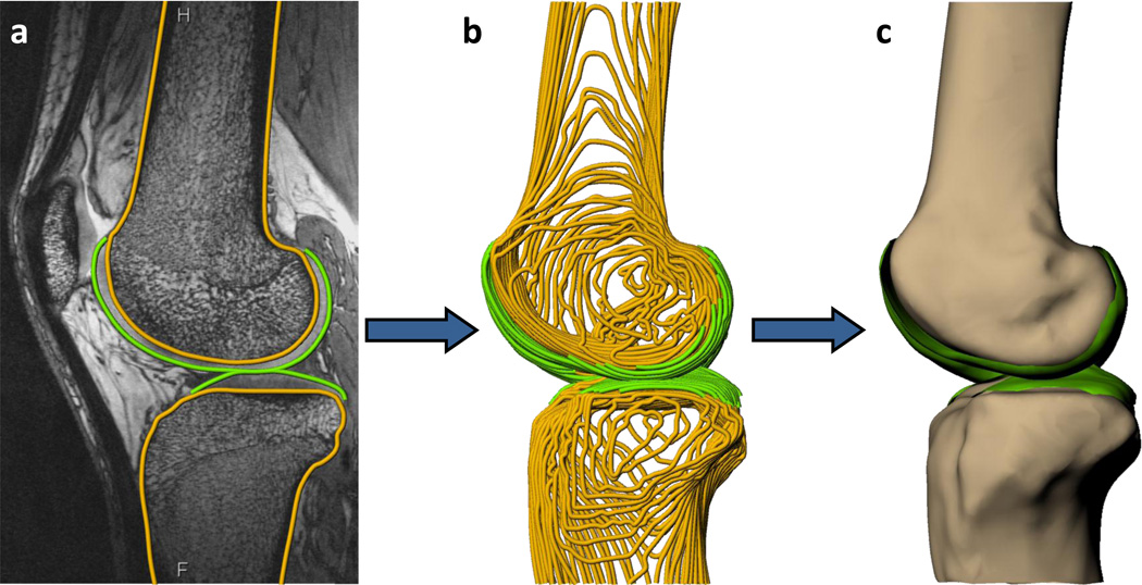
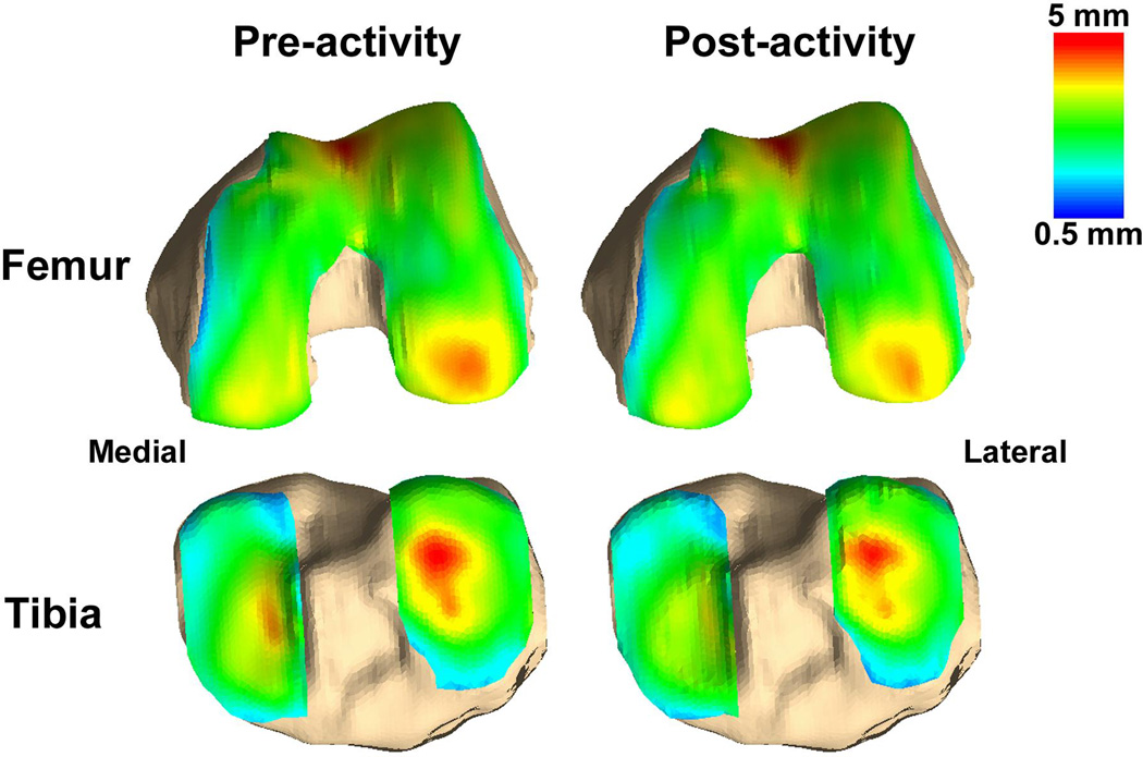
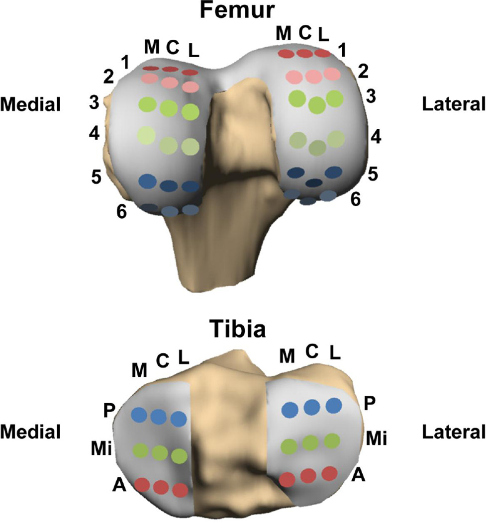
Methods: Both knees of 8 healthy subjects underwent magnetic resonance imaging before and immediately after a dynamic hopping activity. Images were segmented and then used to create 3-dimensional surface models of bone and cartilage. These pre- and postactivity models were then registered using an iterative closest point technique to enable site-specific measurements of cartilage strain (defined as the normalized change in cartilage thickness before and after activity) on the femur and tibia.
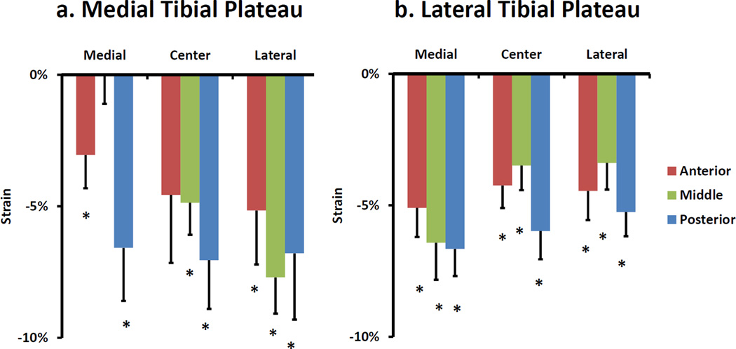
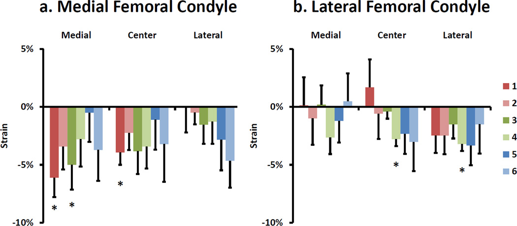
Results: Significant strains were observed in both the medial and lateral tibial cartilage, with each compartment averaging a decrease of 5%. However, these strains varied with location within each compartment, reaching a maximum compressive strain of 8% on the medial plateau and 7% on the lateral plateau. No significant averaged compartmental strains were observed in the medial or lateral femoral cartilage. However, local regions of the medial and lateral femoral cartilage experienced significant compressive strains, reaching maximums of 6% and 3%, respectively.
Conclusion: Local regions of both the femur and tibia experienced significant cartilage strains as a result of dynamic activity. An understanding of changes in cartilage strain distributions may help to elucidate the biomechanical factors contributing to cartilage degeneration after joint injury.
Clinical relevance: Site-specific measurements of in vivo cartilage strains are important because altered loading is believed to be a factor contributing to the development and progression of osteoarthritis. Specifically, this methodology and data could be used to evaluate the effects of soft tissue injuries (such as ligament or meniscus tears) on cartilage strains in response to dynamic activities of daily living.
Keywords: biomechanics; cartilage; hopping; jumping; knee; magnetic resonance imaging; osteoarthritis; strain; stress test; tibiofemoral.
© 2014 The Author(s).
Figures
Similar articles
-
Effects of Anterior Cruciate Ligament Deficiency on Tibiofemoral Cartilage Thickness and Strains in Response to Hopping.Am J Sports Med. 2019 Jan;47(1):96-103. doi: 10.1177/0363546518802225. Epub 2018 Oct 26. Am J Sports Med. 2019. PMID: 30365903 Free PMC article.
-
In Vivo Tibial Cartilage Strains in Regions of Cartilage-to-Cartilage Contact and Cartilage-to-Meniscus Contact in Response to Walking.Am J Sports Med. 2017 Oct;45(12):2817-2823. doi: 10.1177/0363546517712506. Epub 2017 Jul 3. Am J Sports Med. 2017. PMID: 28671850 Free PMC article.
-
Effect of normal gait on in vivo tibiofemoral cartilage strains.J Biomech. 2016 Sep 6;49(13):2870-2876. doi: 10.1016/j.jbiomech.2016.06.025. Epub 2016 Jun 27. J Biomech. 2016. PMID: 27421206 Free PMC article.
-
Mechanobiology of the meniscus.J Biomech. 2015 Jun 1;48(8):1469-78. doi: 10.1016/j.jbiomech.2015.02.008. Epub 2015 Feb 9. J Biomech. 2015. PMID: 25731738 Free PMC article. Review.
-
Effects of and Response to Mechanical Loading on the Knee.Sports Med. 2022 Feb;52(2):201-235. doi: 10.1007/s40279-021-01579-7. Epub 2021 Oct 20. Sports Med. 2022. PMID: 34669175 Review.
Cited by
-
Acromial and glenoid morphology in glenohumeral osteoarthritis: a three-dimensional analysis.JSES Int. 2021 Mar 21;5(3):398-405. doi: 10.1016/j.jseint.2021.01.006. eCollection 2021 May. JSES Int. 2021. PMID: 34136846 Free PMC article.
-
Morphological evaluation of the sagittal plane femoral load-bearing surface in computer-simulated virtual total knee arthroplasty implantation at different flexion angles.Knee Surg Sports Traumatol Arthrosc. 2017 Sep;25(9):2880-2886. doi: 10.1007/s00167-016-3997-1. Epub 2016 Jan 25. Knee Surg Sports Traumatol Arthrosc. 2017. PMID: 26811034
-
The Characteristic Recovery Time as a Novel, Noninvasive Metric for Assessing In Vivo Cartilage Mechanical Function.Ann Biomed Eng. 2020 Dec;48(12):2901-2910. doi: 10.1007/s10439-020-02558-1. Epub 2020 Jul 14. Ann Biomed Eng. 2020. PMID: 32666421 Free PMC article.
-
In Vivo Assessment of Exercise-Induced Glenohumeral Cartilage Strain.Orthop J Sports Med. 2018 Jul 13;6(7):2325967118784518. doi: 10.1177/2325967118784518. eCollection 2018 Jul. Orthop J Sports Med. 2018. PMID: 30023404 Free PMC article.
-
Obesity impacts the mechanical response and biochemical composition of patellofemoral cartilage: An in vivo, MRI-based investigation.J Biomech. 2022 Mar;134:110991. doi: 10.1016/j.jbiomech.2022.110991. Epub 2022 Feb 7. J Biomech. 2022. PMID: 35176590 Free PMC article.
References
-
- Andriacchi TP, Mundermann A, Smith RL, Alexander EJ, Dyrby CO, Koo S. A framework for the in vivo pathomechanics of osteoarthritis at the knee. Ann Biomed Eng. 2004;32(3):447–457. - PubMed
-
- Beaupre GS, Stevens SS, Carter DR. Mechanobiology in the development, maintenance, and degeneration of articular cartilage. J Rehabil Res Dev. 2000;37(2):145–151. - PubMed
-
- Bedi A, Kelly N, Baad M, Fox AJ, Ma Y, Warren RF, Maher SA. Dynamic contact mechanics of radial tears of the lateral meniscus: implications for treatment. Arthroscopy. 2012;28(3):372–381. - PubMed
Publication types
MeSH terms
Grants and funding
LinkOut - more resources
Full Text Sources
Other Literature Sources
