. 2015 Sep;42(3):763-70.
doi: 10.1002/jmri.24822. Epub 2014 Dec 15.

MRI findings of radiation-associated angiosarcoma of the breast (RAS)

Affiliations

MRI findings of radiation-associated angiosarcoma of the breast (RAS)

Sona A Chikarmane et al. J Magn Reson Imaging. 2015 Sep.

Abstract

Purpose: To describe the magnetic resonance imaging (MRI) characteristics of radiation-associated breast angiosarcomas (RAS).

Materials and methods: In this Institutional Review board (IRB)-approved retrospective study, 57 women were diagnosed with pathologically confirmed RAS during the study period (January 1999 to May 2013). Seventeen women underwent pretreatment breast MRI (prior to surgical resection or chemotherapy), of which 16 studies were available for review. Imaging features, including all available mammograms, ultrasounds, and breast MRIs, of these patients were evaluated by two radiologists independently and correlated with clinical management and outcomes.

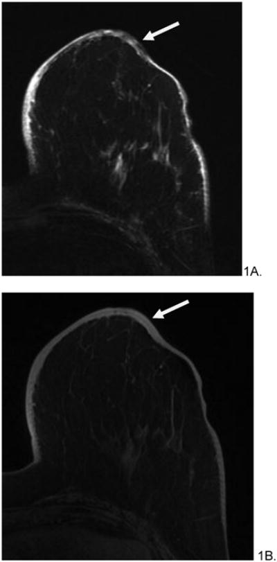
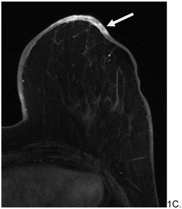
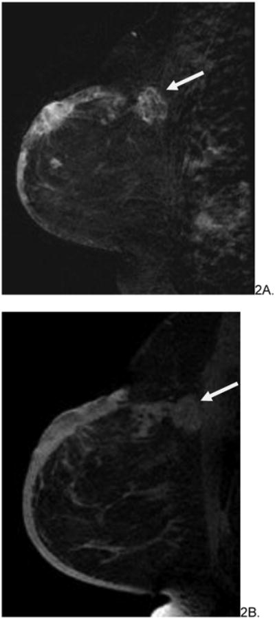
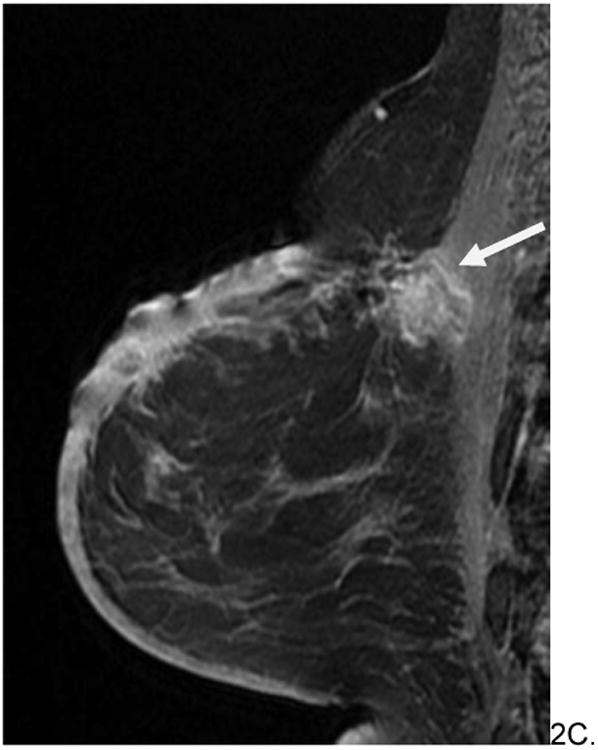
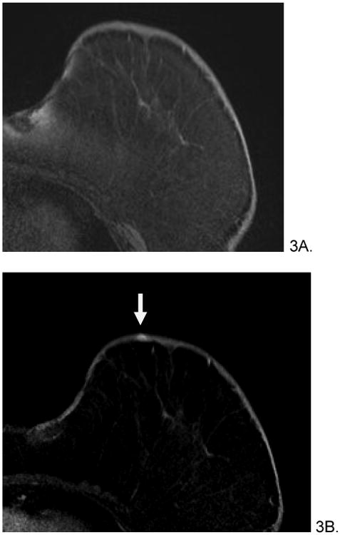
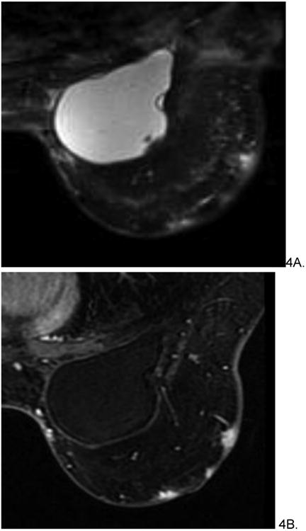
Results: The median age of patients at original breast cancer diagnosis was 69.3 years (range 42-84 years), with average time from initial radiation therapy to diagnosis of RAS of 7.3 years (range 5.1-9.5 years). Nine women had mammograms (9/16, 56%) and six had breast ultrasound (US) (6/16, 38%) prior to MRI, which demonstrated nonsuspicious findings in 5/9 mammograms and 3/6 ultrasounds. Four patients had distinct intraparenchymal masses on mammogram and MRI. MRI findings included diffuse T2 high signal skin thickening (16/16, 100%). Nearly half (7/16, 44%) of patients had T2 low signal intensity lesions; all lesions rapidly enhanced on postcontrast T1 -weighted imaging. All women underwent surgical resection, with 8/16 (50%) receiving neoadjuvant chemotherapy. Four women died during the study period.

Conclusion: Clinical, mammographic, and sonographic findings of RAS are nonspecific and may be occult on conventional breast imaging; MRI findings of RAS include rapidly enhancing dermal and intraparenchymal lesions, some of which are low signal on T2 weighted imaging.

Keywords: angiosarcoma; breast magnetic resonance imaging; radiation-associated.


Figures

Figure 1
Figure 1
82 year old female with history of left breast invasive ductal carcinoma, status post lumpectomy and radiation treatment who presented with bruising around her nipple five years after radiation treatment. Initial mammogram (not shown) was reported as normal. Skin punch biopsy revealed postradiation angiosarcoma. Pre-surgical MRI revealed diffuse, non-mass like T2 hypointensity within skin (A, arrow), which appeared T1 isointense (B), and rapidly enhanced on post-contrast imaging (C). Subsequent mastectomy was performed and the patient has been disease free for 25 months after treatment.
Figure 1
Figure 1
82 year old female with history of left breast invasive ductal carcinoma, status post lumpectomy and radiation treatment who presented with bruising around her nipple five years after radiation treatment. Initial mammogram (not shown) was reported as normal. Skin punch biopsy revealed postradiation angiosarcoma. Pre-surgical MRI revealed diffuse, non-mass like T2 hypointensity within skin (A, arrow), which appeared T1 isointense (B), and rapidly enhanced on post-contrast imaging (C). Subsequent mastectomy was performed and the patient has been disease free for 25 months after treatment.
Figure 2
Figure 2
60 year old woman with history of left breast invasive ductal carcinoma status post lumpectomy and radiation eight years prior who presented with increased edema around surgical skin scar. No mammograms or ultrasounds were available for review. Skin bunch biopsy revealed post-radiation angiosarcoma. Pre-surgical MRI revealed predominately T2 hypointense non-mass skin thickening with an associated T2 hypointense posterior irregular mass (arrows, A), which were T1 hypointense (B) and enhanced rapidly on post contrast imaging (C). The patient underwent pre-surgical chemotherapy with paclitaxil and gemcitabine before radical mastectomy and chest wall resection. She is currently disease free.
Figure 2
Figure 2
60 year old woman with history of left breast invasive ductal carcinoma status post lumpectomy and radiation eight years prior who presented with increased edema around surgical skin scar. No mammograms or ultrasounds were available for review. Skin bunch biopsy revealed post-radiation angiosarcoma. Pre-surgical MRI revealed predominately T2 hypointense non-mass skin thickening with an associated T2 hypointense posterior irregular mass (arrows, A), which were T1 hypointense (B) and enhanced rapidly on post contrast imaging (C). The patient underwent pre-surgical chemotherapy with paclitaxil and gemcitabine before radical mastectomy and chest wall resection. She is currently disease free.
Figure 3
Figure 3
66 year old female with history of invasive ductal and lobular carcinoma of left breast, status post lumpectomy and radiation therapy 7 years prior who presented with left breast violaceous skin lesion. Skin punch biopsy revealed angiosarcoma. Pre-treatment breast MRI revealed T1 isointense (A), T2 heterogenous (not shown), focus of rapid enhancement (B, arrow). The patient underwent pre-surgical chemotherapy with taxol and gemcitabine before mastectomy. She is currently disease free.
Figure 4
Figure 4
54 year old female with history of invasive ductal carcinoma, status post neoadjuvant chemotherapy, lumpectomy and radiation treatment 7 years prior who presented with bruising in left breast. Skin punch biopsy revealed post-radiation angiosarcoma involving dermis and subcutaneous tissue. Pre-treatment MRI demonstrated multifocal T2 hyperintense (A), rapidly enhancing small masses (B) without diffuse skin enhancement. The patient underwent pre-surgical chemotherapy with taxol before radical mastectomy and chest wall resection. She is currently disease free.

References

    1. Huang J, Mackillop WJ. Increased risk of soft tissue sarcoma after radiotherapy in women with breast carcinoma. Cancer. 2001;92:172–180. - PubMed
    1. Glazebrook KN, Magut MJ, Reynolds C. Angiosarcoma of the breast. AJR American journal of roentgenology. 2008;190:533–538. - PubMed
    1. Cabete J, Lencastre A, Fidalgo A, Lobo L, Joao A, Serrao V. Postradiation cutaneous angiosarcoma of the breast: a diagnosis to keep in mind. The breast journal. 2014;20:89–90. - PubMed
    1. Morgan EA, Kozono DE, Wang Q, et al. Cutaneous radiation-associated angiosarcoma of the breast: poor prognosis in a rare secondary malignancy. Annals of surgical oncology. 2012;19:3801–3808. - PubMed
    1. Abbott R, Palmieri C. Angiosarcoma of the breast following surgery and radiotherapy for breast cancer. Nature clinical practice Oncology. 2008;5:727–736. - PubMed

MeSH terms

Supplementary concepts