. 2015 Feb;89(4):2253-67.
doi: 10.1128/JVI.02716-14. Epub 2014 Dec 10.

Identification and functional comparison of seven-transmembrane G-protein-coupled BILF1 receptors in recently discovered nonhuman primate lymphocryptoviruses

Affiliations

Identification and functional comparison of seven-transmembrane G-protein-coupled BILF1 receptors in recently discovered nonhuman primate lymphocryptoviruses

Katja Spiess et al. J Virol. 2015 Feb.

Abstract

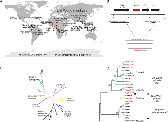
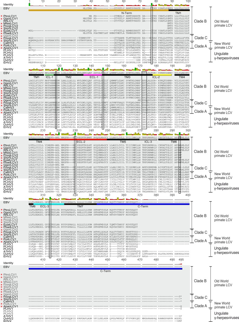
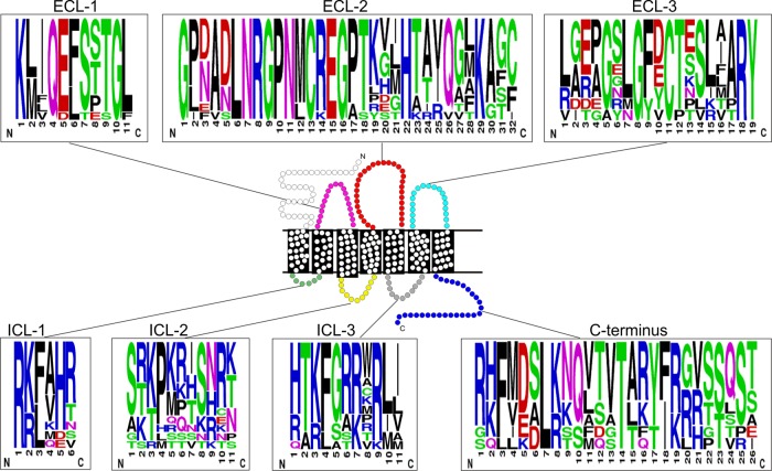
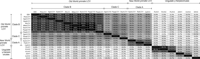
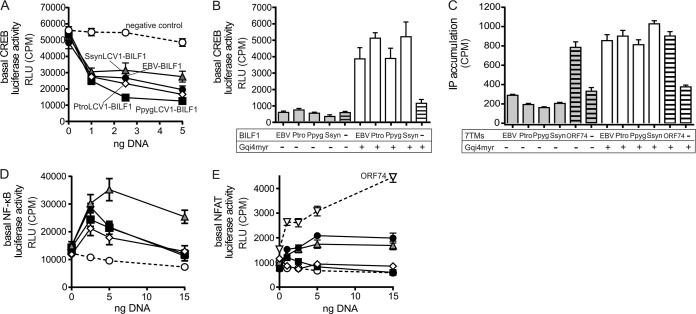
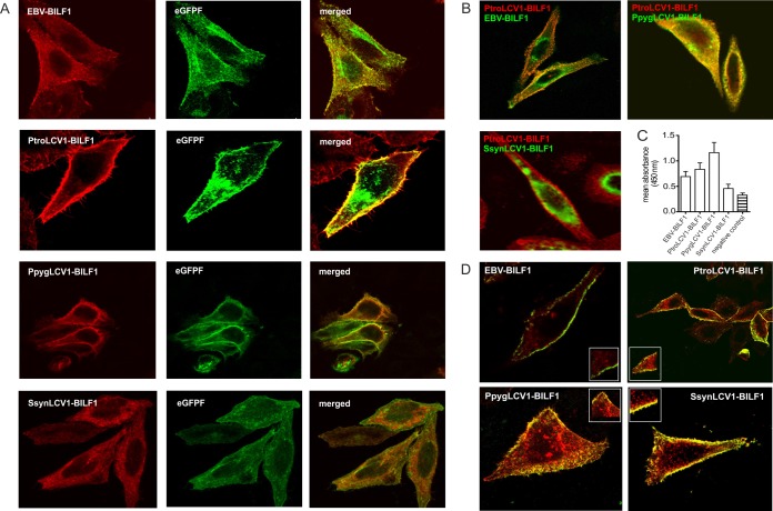
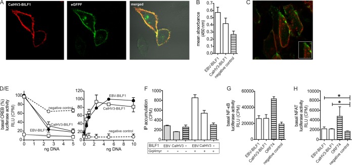
Coevolution of herpesviruses with their respective host has resulted in a delicate balance between virus-encoded immune evasion mechanisms and host antiviral immunity. BILF1 encoded by human Epstein-Barr virus (EBV) is a 7-transmembrane (7TM) G-protein-coupled receptor (GPCR) with multiple immunomodulatory functions, including attenuation of PKR phosphorylation, activation of G-protein signaling, and downregulation of major histocompatibility complex (MHC) class I surface expression. In this study, we explored the evolutionary and functional relationships between BILF1 receptor family members from EBV and 12 previously uncharacterized nonhuman primate (NHP) lymphocryptoviruses (LCVs). Phylogenetic analysis defined 3 BILF1 clades, corresponding to LCVs of New World monkeys (clade A) or Old World monkeys and great apes (clades B and C). Common functional properties were suggested by a high degree of sequence conservation in functionally important regions of the BILF1 molecules. A subset of BILF1 receptors from EBV and LCVs from NHPs (chimpanzee, orangutan, marmoset, and siamang) were selected for multifunctional analysis. All receptors exhibited constitutive signaling activity via G protein Gαi and induced activation of the NF-κB transcription factor. In contrast, only 3 of 5 were able to activate NFAT (nuclear factor of activated T cells); chimpanzee and orangutan BILF1 molecules were unable to activate NFAT. Similarly, although all receptors were internalized, BILF1 from the chimpanzee and orangutan displayed an altered cellular localization pattern with predominant cell surface expression. This study shows how biochemical characterization of functionally important orthologous viral proteins can be used to complement phylogenetic analysis to provide further insight into diverse microbial evolutionary relationships and immune evasion function.

Importance: Epstein-Barr virus (EBV), known as an oncovirus, is the only human herpesvirus in the genus Lymphocryptovirus (LCV). EBV uses multiple strategies to hijack infected host cells, establish persistent infection in B cells, and evade antiviral immune responses. As part of EBV's immune evasion strategy, the virus encodes a multifunctional 7-transmembrane (7TM) G-protein-coupled receptor (GPCR), EBV BILF1. In addition to multiple immune evasion-associated functions, EBV BILF1 has transforming properties, which are linked to its high constitutive activity. We identified BILF1 receptor orthologues in 12 previously uncharacterized LCVs from nonhuman primates (NHPs) of Old and New World origin. As 7TM receptors are excellent drug targets, our unique insight into the molecular mechanism of action of the BILF1 family and into the evolution of primate LCVs may enable validation of EBV BILF1 as a drug target for EBV-mediated diseases, as well as facilitating the design of drugs targeting EBV BILF1.


Figures

FIG 1
FIG 1
Identification of novel BILF1 receptor sequences from uncharacterized LCVs of nonhuman primates and phylogenetic studies. (A) World map of primate hosts harboring novel and known LCVs. The viruses are indicated in red and gray circles (novel and known LCVs, respectively); names of viruses and their hosts are written in bold blac (novel viruses) and italic gray (known viruses). Three ape hosts (gorilla, siamang, and orangutan) harbor 2 distinct LCVs. (B) Map of amplified genes and diagram of PCR strategy. EBV ORF BALF5 (DPOL), BILF1, LF1, and LF2 are symbolized as arrows (BILF1 ORF in red). A scale (in kb) oriented on the EBV genome is given below. The right side shows degenerate nested primers (light gray squares) used to amplify part of the LF2 gene. A short solid black line represents the amplified fragment. The left side represents published sequence information for the DNA polymerase gene of the investigated LCV (solid black line). Based on both DPOL and LF2 sequences, specific primers (gray squares) were selected and long-distance PCR was performed. A final contiguous sequence of approximately 3.5 kb was obtained, including the BILF1 ORF (red). (C) Phylogenetic tree of viral and endogenous 7TM receptors. The amino acid sequences were aligned with MAFFT and an unrooted tree was constructed using the neighbor-joining algorithm. The published U12/UL33 (purple), US28 (yellow), poxvirus-encoded receptor (blue), human chemokine receptor (black), ORF74 (red), and UL78/U51 (orange branches) sequences were aligned with the published and newly detected BILF1 sequences (green branches) and subjected to phylogenetic analyses. (D) Phylogenetic tree of BILF1 receptor family. BILF1 amino acid sequences were aligned with MAFFT and a rooted tree was constructed, using the phyml/bootstrap algorithm and the KSHV ORF74 sequence as an outgroup. The BILF1 sequences of New World primate LCVs (clade A; bright green branches) separate from those of Old World primate LCVs (clades B and C; dark green branches). The BILF1-homologous sequences from gammaherpesviruses of ungulates (gray branches) cluster separately from the primate BILF1 sequences. Newly discovered BILF1 sequences are highlighted in red, and BILF1 receptors used for functional studies are marked with a star.
FIG 2
FIG 2
Multiple-sequence alignment of primate BILF1 receptors. An alignment of 21 BILF1 amino acid sequences was performed using MAFFT (Geneious 6.16). TM1 to TM7 of the BILF1 receptors are indicated with black bars. The intracellular loop regions (ICL-1, -2, and -3) are indicated with green, yellow, and gray bars, and the extracellular loop regions (ECL-1, ECL-2, and ECL-3) are indicated with bars in magenta, red and light blue. N and C termini are highlighted with bars in brown and dark blue, respectively. The alternative DRY motif and important conserved amino acid residues are marked with rectangles. The primate BILF1 amino acid sequences are highlighted with gray boxes and newly detected BILF1 amino acid sequences with red stars.
FIG 3
FIG 3
Sequence logo of the ICL and ECL domains as well as from the C termini of the BILF1 receptors. In the schematic picture of a 7TM receptor, the ECL and the ICL regions are depicted with the same colors as in Fig. 2. In the sequence logo, the chemical properties of the amino acids are represented in color (polar, green; neutral, purple; basic, blue; acidic, red; and hydrophobic, black). The figure was created using the web-based program WebLogo (http://weblogo.berkeley.edu).
FIG 4
FIG 4
BILF1 amino acid sequence identities. Amino acid sequence identity was estimated based on an alignment of 21 BILF1 sequences and is displayed as a heat map.
FIG 5
FIG 5
CREB-mediated transcription regulation and activation of the transcription factors NF-κB and NFAT by BILF1 receptors. (A) Inhibition of forskolin (10 μM)-induced CREB activation with increasing doses of BILF1 receptor DNA in transiently transfected HEK-293 cells. The negative control is displayed in white circles, SsynLCV1 BILF1 in gray triangles, EBV-BILF1 in black circles, PtroLCV1 BILF1 in white squares, and PpygLCV1 BILF1 in black squares. (B) CREB activation by the BILF1 receptors in the absence (gray bars) and presence (white bars) of the promiscuous chimeric G protein GαΔ6qi4myr. The negative controls are shown as bars with black horizontal lines. (C) IP3 turnover in HEK-293 cells transfected with receptor DNA and in the absence (gray) or presence (white columns) of GαΔ6qi4myr. Positive controls (KSHV-ORF74) and negative controls (empty vector) are shown as bars with black lines. (D/E) Activation of the NF-κB (D) and the NFAT (E) transcription factors in transiently transfected HEK-293 cells by the different BILF1 receptors, with negative (vector) and positive (KSHV ORF74) controls. The controls and the BILF1 receptors are displayed as described for Fig. 3A (results of all experiments are shown as means ± SEM; n = 3).
FIG 6
FIG 6
Distinct cellular localization patterns of BILF1 receptors. (A) HEK-293 cells were cotransfected with EBV BILF1, PtroLCV1 BILF1, PpygLCV1 BILF1, and SsynLCV1 BILF1 and the farnesylated enhanced green fluorescent protein (eGFPF). The representative pictures show the localization of the BILF1 receptors detected with a primary HA antibody against the HA tag expressed at the N termini of the receptors (red signal, left side), the signal of eGFPF (green signal, middle), and the merge signals of the BILF1 receptors with eGFPF (orange signal, right side). (B) Representative pictures of EBV BILF1- and PpygLCV1 BILF1-expressing HEK-293 cells (top) and SsynLCV1 BILF1-expressing HEK-293 cells (bottom), cotransfected with PtroLCV1 BILF1. EBV, PpygLCV1, and SsynLCV1 BILF1 molecules were detected with an anti-HA FITC-conjugated antibody using an N-terminal HA tag (green signal). PtroLCV1-BILF1was detected with a primary antibody against the c-myc tag (N terminal) and a secondary Cy5-conjugated antibody (red signal). (C) The graph shows cell surface expression levels of the BILF1 receptors as estimated in ELISA using an N-terminal HA tag (means ± SEM; n = 3). (D) Representative pictures of the constitutive internalization of the BILF1 receptors determined by antibody uptake studies. HEK-293 cells were transfected with HA-tagged EBV BILF1, PtroLCV1 BILF1, PpygLCV1 BILF1, and SsynLCV1 BILF1. Forty-eight hours after the transfection, receptors present at the cell surface were stained with a hemagglutinin antibody for 1 h at 4°C. Subsequently, cells were incubated at 37°C for 30 min to induce internalization and fixed afterwards. Labeled receptors still residing at the cell surface were detected with a FITC-conjugated antibody (green signal) prior to permeabilization, while internalized receptors were detected with a rhodamine-conjugated antibody (red signal).
FIG 7
FIG 7
Functional studies of CalHV3 BILF1. (A and B) To estimate cell surface expression levels of CalHV3 BILF1 in comparison to EBV BILF1, confocal microscopy (A) and ELISA (B) studies were performed as described for Fig. 6A and C. (C) Internalization studies were performed for CalHV3 BILF1 as described for Fig. 6D. (D/E) As shown in Fig. 5A and B for the ape and Old World monkey BILF1 receptors, CalHV3 BILF1-mediated CREB activity was estimated by cotransfection of CalHV3 BILF1 with GαΔ6qi4myr and in the presence of forskolin (D), as well as by cotransfection of CalHV3 BILF1 with GαΔ6qi4myr (E). EBV BILF1 was included in both experiments as a positive control. (F) IP3 turnover for CalHV3 BILF1 and EBV BILF1 is shown in the presence and in the absence of. GαΔ6qi4myr as described for Fig. 5C. (G and H) NF-κB (G) and NFAT (H) activation by CalHV3 BILF1 and EBV BILF1 was estimated by cotransfecting HEK-293 cells with receptor and reporter plasmid DNA. The significance of the NFAT activity is proven by Student's 2-tailed unpaired t test (for EBV BILF1, P < 0.011, and for CalHV3 BILF1, P < 0.021; significance is indicated with an asterisk for both). Representative pictures from the confocal microscopy studies (A and C) are shown, and the ELISA (B) and signaling (D to H) studies were performed 3 times (values are means ± SEM).

Similar articles

Cited by

References

    1. Küppers R. 2003. B cells under influence: transformation of B cells by Epstein-Barr virus. Nat Rev Immunol 3:801–812. doi:10.1038/nri1201. - DOI - PubMed
    1. Henle G, Henle W, Diehl V. 1968. Relation of Burkitt's tumor-associated herpes-ytpe virus to infectious mononucleosis. Proc Natl Acad Sci U S A 59:94–101. doi:10.1073/pnas.59.1.94. - DOI - PMC - PubMed
    1. Kutok JL, Wang F. 2006. Spectrum of Epstein-Barr virus-associated diseases. Annu Rev Pathol 1:375–404. doi:10.1146/annurev.pathol.1.110304.100209. - DOI - PubMed
    1. Hislop AD, Ressing ME, van Leeuwen D, Pudney VA, Horst D, Koppers-Lalic D, Croft NP, Neefjes JJ, Rickinson AB, Wiertz EJHJ. 2007. A CD8+ T cell immune evasion protein specific to Epstein-Barr virus and its close relatives in Old World primates. J Exp Med 204:1863–1873. doi:10.1084/jem.20070256. - DOI - PMC - PubMed
    1. Crawford DH. 2001. Biology and disease associations of Epstein-Barr virus. Philos Trans R Soc Lond B Biol Sci 356:461–473. doi:10.1098/rstb.2000.0783. - DOI - PMC - PubMed

Publication types

LinkOut - more resources