. 2015 Jan 23;290(4):1936-51.
doi: 10.1074/jbc.M114.598698. Epub 2014 Dec 10.

Generation and characterization of ATP analog-specific protein kinase Cδ

Affiliations

Generation and characterization of ATP analog-specific protein kinase Cδ

Varun Kumar et al. J Biol Chem. .

Abstract

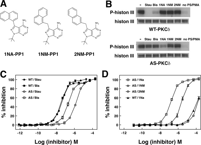
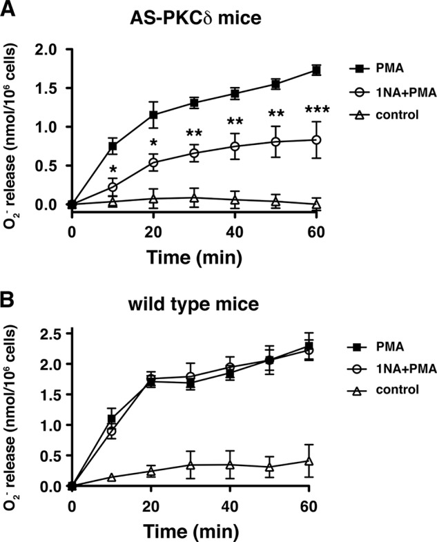
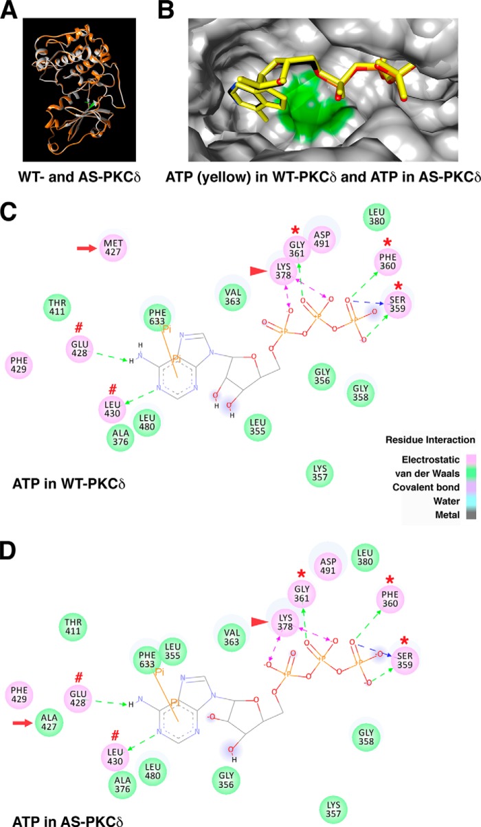
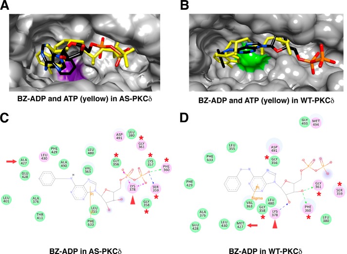
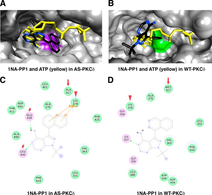
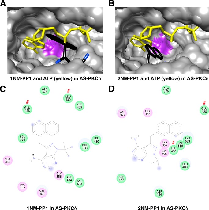
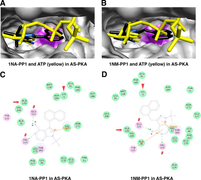
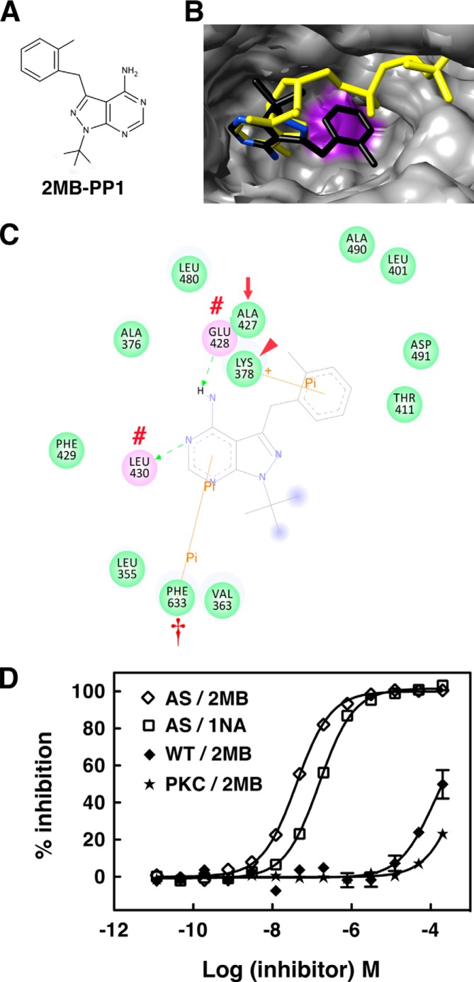
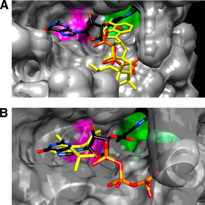
To better study the role of PKCδ in normal function and disease, we developed an ATP analog-specific (AS) PKCδ that is sensitive to specific kinase inhibitors and can be used to identify PKCδ substrates. AS PKCδ showed nearly 200 times higher affinity (Km) and 150 times higher efficiency (kcat/Km) than wild type (WT) PKCδ toward N(6)-(benzyl)-ATP. AS PKCδ was uniquely inhibited by 1-(tert-butyl)-3-(1-naphthyl)-1H-pyrazolo[3,4-d]pyrimidin-4-amine (1NA-PP1) and 1-(tert-butyl)-3-(2-methylbenzyl)-1H-pyrazolo[3,4-d]pyrimidin-4-amine (2MB-PP1) but not by other 4-amino-5-(4-methylphenyl)-7-(t-butyl)pyrazolo[3,4-d]pyrimidine (PP1) analogs tested, whereas WT PKCδ was insensitive to all PP1 analogs. To understand the mechanisms for specificity and affinity of these analogs, we created in silico WT and AS PKCδ homology models based on the crystal structure of PKCι. N(6)-(Benzyl)-ATP and ATP showed similar positioning within the purine binding pocket of AS PKCδ, whereas N(6)-(benzyl)-ATP was displaced from the pocket of WT PKCδ and was unable to interact with the glycine-rich loop that is required for phosphoryl transfer. The adenine rings of 1NA-PP1 and 2MB-PP1 matched the adenine ring of ATP when docked in AS PKCδ, and this interaction prevented the potential interaction of ATP with Lys-378, Glu-428, Leu-430, and Phe-633 residues. 1NA-PP1 failed to effectively dock within WT PKCδ. Other PP1 analogs failed to interact with either AS PKCδ or WT PKCδ. These results provide a structural basis for the ability of AS PKCδ to efficiently and specifically utilize N(6)-(benzyl)-ATP as a phosphate donor and for its selective inhibition by 1NA-PP1 and 2MB-PP1. Such homology modeling could prove useful in designing molecules to target PKCδ and other kinases to understand their function in cell signaling and to identify unique substrates.

Keywords: ATP; Chemical Biology; Protein Kinase C (PKC); Protein Phosphorylation; Stroke.


Figures

FIGURE 1.
FIGURE 1.
Design and expression of AS PKCδ. A, alignment of the gatekeeper (boldface type) and its surrounding residues within the nucleotide-binding domain of human PKC isozymes and PKA. Glu and Val/Leu residues are underlined. B and C, expression of WT and AS PKCδ in COS-7 (B) and Neuro2A cells (C) was detected by Western blot analysis using anti-PKCδ antibody. Transfection with the pcDNA3 vector (V) was used as a control.
FIGURE 2.
FIGURE 2.
AS PKCδ, but not WT PKCδ, utilized N6-(benzyl)-ATP (BZ-ATP) to phosphorylate substrates. A, chemical structure of an ATP analog. R, site of N6 modification. B, phosphorylation of histone 3 by WT and AS PKCδ in the presence of [γ-32P]ATP or N6-(benzyl)-[γ-32P]ATP. Top, autoradiogram of phosphorylated histone 3; bottom, Coomassie-stained histone 3. C, mouse neutrophil lysates (100 μg) were labeled with AS PKCδ and N6-(benzyl)-ATPγS. After para-nitrobenzyl mesylate (PNBM) alkylation, the labeled substrates were detected by Western blot analysis using an antibody that detects thiophosphate esters. Immunoreactivity was increased in samples treated with the PKC activator PMA. D, the same amounts of neutrophil lysates as in D were loaded and visualized by silver staining.
FIGURE 3.
FIGURE 3.
The kinase activity of AS PKCδ is specifically inhibited by 1NA-PP1. A, structure of PP1 analogs. B, WT or AS PKCδ was incubated with [γ-32P]ATP, histone 3, and a 1 μm concentration of staurosporine (Stau), bisindolylmaleimide I (Bis), or PP1 analogs. Top, autoradiogram showing phosphorylated histone 3; bottom, histone 3 in a silver-stained gel. Inhibition of WT and AS PKCδ by staurosporine and bisindolylmaleimide I (C) and by PP1 analogs (D) was measured by fluorescence polarization assays (n = 3). Error bars, S.E.
FIGURE 4.
FIGURE 4.
Generation of AS PKCδ knock-in mice. A, organization of the mouse Prkcd gene, targeting constructs, and M425A mutant allele resulting from homologous recombination. Filled boxes, exons 7–18. Met-425 is in exon 13. Arrowheads, loxP sequences. The targeting vector was composed of a 3.1-kb short arm carrying the M425A mutation, a Neo expression cassette flanked by two loxP sites, a 5.4-kb-long arm, and a diphtheria toxin A (DTA) gene. B, verification of genotypes by PCR using the G8-PCR-F and G8–35-R primers. The wild type allele (+) generates a 0.47-kb product, and the M425A mutant allele (A) generates a 0.6-kb product. C, Western blot showing similar PKCδ immunoreactivity in neutrophils from WT and AS PKCδ knock-in (KI) mice.
FIGURE 5.
FIGURE 5.
Pattern of PKCδ immunoreactivity in neutrophils of WT PKCδ (A and C) and AS PKCδ (B and D) mice. PKCδ (red) was detected diffusely in the cytoplasm at baseline (A and B) and at the plasma membrane after incubation with 200 nm PMA for 2 min (C and D). Nuclei (blue) were detected with DAPI in the bottom panels. Scale bar, 25 μm.
FIGURE 6.
FIGURE 6.
1NA-PP1 inhibits superoxide anion (O2) production from PMA-stimulated neutrophils of AS PKCδ (A) but not WT (B) mice. A, PMA-stimulated (100 nm) O2 production was significantly reduced by 5 μm 1NA-PP1 (Ftreatment (1,24) = 18.1, p = 0.0132; Ftime (6,24) = 57.5, p < 0.0001; Finteraction (6,24) = 5.7, p = 0.0009). *, p < 0.05; **, p < 0.01; ***, p < 0.001 compared with PMA-treated neutrophils (Bonferroni test). B, PMA-stimulated (100 nm) O2 production from WT neutrophils was not reduced by 5 μm 1NA-PP1 (Ftreatment (1,24) = 0.00, p = 0.9822; Ftime (6,24) = 142.1, p < 0.0001; Finteraction (6,24) = 0.67, p = 0.6748). Error bars, S.E.
FIGURE 7.
FIGURE 7.
Comparison of ATP interactions in the nucleotide-binding pocket of WT and AS PKCδ. A, the homology model of WT PKCδ (gray) was superimposed with the AS PKCδ model (orange). The gatekeeper residue Met-427 in WT PKCδ is highlighted in green. B, ATP (yellow) docked in WT PKCδ was superimposed with ATP (colored by atom type: nitrogen (blue), carbon (black), oxygen (red), and phosphorus (orange)) docked in AS PKCδ. Shown are the ATP-interacting residues in the nucleotide-binding pocket of WT (C) and AS PKCδ (D). Residues involved in electrostatic (hydrogen bond, charge, or polar), van der Waals, covalent bond, water, and metal interactions with the ligand are shaded in pink, green, magenta, aquamarine, and dark gray, respectively. Hydrogen bond interactions with amino acid main chain and amino acid side chain residues are represented by green and blue dashed arrows directed toward the electron donor. Charged interaction is shown by a pink dashed arrow with heads on both sides. Pi interaction is represented by an orange line with, π indicating the interaction. In both WT and AS PKCδ, ATP forms hydrogen bonds with Glu-428 and Leu-430 (red number symbols) near the gatekeeper (red arrow) and Ser-359, Phe-360, and Gly-361 in the glycine-rich loop (red asterisks) and charged interactions with invariant Lys-378 (red arrowhead). The π-π interaction of ATP with Phe-633 is shown as an orange line in WT and AS PKCδ.
FIGURE 8.
FIGURE 8.
Comparison of N6-(benzyl)-ADP (BZ-ADP) interactions in the nucleotide-binding pocket of AS and WT PKCδ. Shown are the superimposed structures of BZ-ADP (colored by atom type) and ATP (yellow) in AS (A) and WT PKCδ (B). Ala-427 is highlighted in purple in AS PKCδ, and Met-427 is highlighted in green in WT PKCδ. Shown are the BZ-ADP-interacting residues in the nucleotide-binding pocket of AS (C) and WT PKCδ (D). ATP interacts with the residues in the glycine-rich loop (red asterisks) and the invariant Lys-378 (red arrowheads). The gatekeeper is indicated by a red arrow.
FIGURE 9.
FIGURE 9.
Comparison of 1NA-PP1 interactions in the nucleotide-binding pocket of AS and WT PKCδ. A and B, superimposed structure of 1NA-PP1 (colored by atom type) and ATP (yellow) in AS (A) and WT PKCδ (B). Ala-427 is highlighted in purple in AS PKCδ, and Met-427 is highlighted in green in WT PKCδ. C and D, 1NA-PP1-interacting residues in the nucleotide-binding pocket of AS (C) and WT PKCδ (D). 1NA-PP1 forms hydrogen bonds with Glu-428 and Leu-430 (red number symbols), and π-σ interaction with Lys-378 (red arrowheads). The gatekeeper is indicated by a red arrow.
FIGURE 10.
FIGURE 10.
Interactions of 1NM-PP1 and 2NM-PP1 with AS PKCδ. A, superimposed structures of 1NM-PP1 (colored by atom type) and ATP (yellow) in AS PKCδ. B, superimposed structures of 2NM-PP1 (colored by atom type) and ATP (yellow) in AS PKCδ. The mutated gatekeeper (Ala-427) in AS PKCδ is highlighted in purple. C and D, 1NM-PP1- (C) and 2NM-PP1-interacting (D) residues in the nucleotide-binding pocket of AS PKCδ. Glu-428 and Leu-430 are indicated by red number symbols.
FIGURE 11.
FIGURE 11.
Interactions of 1NA-PP1 and 1NM-PP1 with AS PKA. A, superimposed structures of 1NA-PP1 (colored by atom type) and ATP (yellow) in AS PKA. B, superimposed structures of 1NM-PP1 (colored by atom type) and ATP (yellow) in AS PKA. The mutated gatekeeper (Ala-120) in AS PKA is highlighted in purple. C and D, 1NA-PP1- (C) and 1NM-PP1-interacting (D) residues in the nucleotide-binding pocket of AS PKA. The gatekeeper, invariant Lys, and residue forming hydrogen bonds with ligand are indicated by red arrows, arrowheads, and red number symbols, respectively.
FIGURE 12.
FIGURE 12.
AS PKCδ is more sensitive to inhibition by 2MB-PP1 than 1NA-PP1. A, chemical structure of 2MB-PP1. B and C, interactions of 2MB-PP1 in the nucleotide-binding pocket of AS PKCδ. B, shown are the superimposed structure of 2MB-PP1 (colored by atom type) and ATP (yellow) in AS PKCδ. Ala-427 is highlighted in purple in AS PKCδ. C, 2MB-PP1-interacting residues in the nucleotide-binding pocket of AS PKCδ. 2MB-PP1 forms hydrogen bonds with Glu-428 and Leu-430 (red number symbols), π-σ interaction with Lys-378 (red arrowhead), and π-π interaction with Phe-633 (red dagger). The gatekeeper is indicated by a red arrow. D, inhibition of AS PKCδ, WT PKCδ, and a commercial mixture of PKC isozymes (PKC) (Calbiochem) by 2MB-PP1 and 1NA-PP1 was measured using fluorescence polarization assays (n = 3). Error bars, S.E.
FIGURE 13.
FIGURE 13.
Interactions of N6-(benzyl)-ATP and 1NA-PP1 with mAC. A, superimposed structures of N6-(benzyl)-ATP (yellow) and MANT-GTP (colored by atom type) in the catalytic site of mAC. The residues (Thr-408, Ala-409, Gln-410, and Glu-411) in the hydrophobic pocket of mAC facing the MANT group are highlighted in green. The Trp-1020 residue close to the guanine ring of MANT-GTP is highlighted in magenta. B, superimposed structures of 1NA-PP1 (yellow) and MANT-GTP (colored by atom type) in the catalytic site of mAC. The naphthyl ring of 1NA-PP1 forms a steric clash with one of hydrophobic residues (Trp-1020) highlighted in magenta.

References

    1. Steinberg S. F. (2008) Structural basis of protein kinase C isoform function. Physiol. Rev. 88, 1341–1378 - PMC - PubMed
    1. Newton A. C. (2010) Protein kinase C: poised to signal. Am. J. Physiol. Endocrinol. Metab. 298, E395–E402 - PMC - PubMed
    1. Choi D. S., Wei W., Deitchman J. K., Kharazia V. N., Lesscher H. M., McMahon T., Wang D., Qi Z. H., Sieghart W., Zhang C., Shokat K. M., Mody I., Messing R. O. (2008) Protein kinase Cδ regulates ethanol intoxication and enhancement of GABA-stimulated tonic current. J. Neurosci. 28, 11890–11899 - PMC - PubMed
    1. Chou W. H., Choi D. S., Zhang H., Mu D., McMahon T., Kharazia V. N., Lowell C. A., Ferriero D. M., Messing R. O. (2004) Neutrophil protein kinase Cδ as a mediator of stroke-reperfusion injury. J. Clin. Invest. 114, 49–56 - PMC - PubMed
    1. Bishop A. C., Buzko O., Shokat K. M. (2001) Magic bullets for protein kinases. Trends Cell Biol. 11, 167–172 - PubMed

Publication types

MeSH terms