. 2015 Feb 6;290(6):3680-92.
doi: 10.1074/jbc.M114.611210. Epub 2014 Dec 10.

Activity of plasma membrane V-ATPases is critical for the invasion of MDA-MB231 breast cancer cells

Affiliations

Activity of plasma membrane V-ATPases is critical for the invasion of MDA-MB231 breast cancer cells

Kristina Cotter et al. J Biol Chem. .

Abstract

The vacuolar (H(+))-ATPases (V-ATPases) are a family of ATP-driven proton pumps that couple ATP hydrolysis with translocation of protons across membranes. Previous studies have implicated V-ATPases in cancer cell invasion. It has been proposed that V-ATPases participate in invasion by localizing to the plasma membrane and causing acidification of the extracellular space. To test this hypothesis, we utilized two separate approaches to specifically inhibit plasma membrane V-ATPases. First, we stably transfected highly invasive MDA-MB231 cells with a V5-tagged construct of the membrane-embedded c subunit of the V-ATPase, allowing for extracellular expression of the V5 epitope. We evaluated the effect of addition of a monoclonal antibody directed against the V5 epitope on both V-ATPase-mediated proton translocation across the plasma membrane and invasion using an in vitro Matrigel assay. The addition of anti-V5 antibody resulted in acidification of the cytosol and a decrease in V-ATPase-dependent proton flux across the plasma membrane in transfected but not control (untransfected) cells. These results demonstrate that the anti-V5 antibody inhibits activity of plasma membrane V-ATPases in transfected cells. Addition of the anti-V5 antibody also inhibited in vitro invasion of transfected (but not untransfected) cells. Second, we utilized a biotin-conjugated form of the specific V-ATPase inhibitor bafilomycin. When bound to streptavidin, this compound cannot cross the plasma membrane. Addition of this compound to MDA-MB231 cells also inhibited in vitro invasion. These studies suggest that plasma membrane V-ATPases play an important role in invasion of breast cancer cells.

Keywords: Acidification; Breast Cancer; Invasion; Migration; Proton Transport; Vacuolar ATPase.


Figures

FIGURE 1.
FIGURE 1.
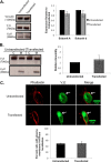
c-V5-transfected cells express V5 both intracellularly and at the plasma membrane. A, cell lysates were prepared from untransfected cells and cells transfected with the V5-tagged construct of subunit c. For each preparation, 20 μg of protein was separated by SDS-PAGE using 4–15% gradient acrylamide gels, and proteins were transferred to nitrocellulose. Immunoblotting was conducted using monoclonal antibodies against V5 and α-tubulin as described under “Experimental Procedures.” The blot displayed is representative of data obtained from two separate experiments. B, c-V5-transfected cells were grown as a monolayer on coverslips in 6-well plates. Permeabilized cells (Perm) were treated with 0.1% Triton X-100 for 5 min, whereas nonpermeabilized cells (Non-Perm) were not. Cells were immunostained using a monoclonal antibody against V5 and Alexa Fluor® 594 phalloidin to stain actin followed by incubation with secondary antibodies as described under “Experimental Procedures.” Images were taken with identical exposure times and antibody concentrations. The top panels show permeabilized cells, whereas the bottom panels show nonpermeabilized cells. The left panels display phalloidin staining, the middle panels display V5 staining, and the right panels display the merged images. The results shown are representative of three independent trials.
FIGURE 2.
FIGURE 2.
c-V5-transfected cells experience a reduction in cytosolic pH following incubation in the presence of an anti-V5 antibody. Untransfected control or c-V5-transfected cells were treated with or without a monoclonal antibody against V5 and then loaded with the pH-sensitive fluorescence probe SNARF-1. Cytosolic pH was measured as described under “Experimental Procedures.” The values represent the mean cytosolic pH and error bars indicate S.D. (n = 9). *, p < 0.05 compared with untreated untransfected cells.
FIGURE 3.
FIGURE 3.
The more alkaline cytosolic pH of c-V5-transfected cells is not a result of enhanced V-ATPase subunit expression, an alteration in pump assembly, or plasma membrane localization. A, cell lysates from untransfected control cells and cells transfected with c-V5 were prepared. 10 μg of protein for each sample was separated by SDS-PAGE using 4–15% gradient acrylamide gels, and proteins were transferred to nitrocellulose. Immunoblotting was conducted using monoclonal antibodies against subunit A, subunit d1, and vinculin. The blot displayed is representative of data obtained from three separate experiments. Western blots from each experiment were quantitated, and the ratio of the expression of subunits A and d in transfected cells versus untransfected cells was calculated. B, untransfected control and c-V5-transfected cells were harvested, and cytosolic (lanes C) and membrane (lanes M) fractions were prepared as explained under “Experimental Procedures.” 19 μg of protein from each fraction was then separated by SDS-PAGE using 4–15% gradient acrylamide gels, and proteins were transferred to nitrocellulose. Immunoblotting was conducted using monoclonal antibodies against subunit A, subunit d1, and vinculin (not pictured). The blot displayed is representative from three separate experiments. The ratio of subunit A in the membrane versus the cytosolic fraction was calculated as a measure of V-ATPase assembly. The graphed data represent the average degrees of assembly for transfected cells relative to untransfected cells. C, untransfected control and c-V5-transfected cells were grown as a monolayer on coverslips in 6-well plates. Permeabilized cells were immunostained using an antibodies against the V1E subunit of the V-ATPase and Alexa Fluor® 568 phalloidin to stain actin followed by incubation with secondary antibodies as described under “Experimental Procedures.” Images were taken with identical exposure times and antibody concentrations. The three panels show representative staining for phalloidin (left panel), V1E (middle panel), and the merged images (right panel) for both untransfected and transfected cells. White arrows indicate plasma membrane V-ATPase expression. To quantitate plasma membrane V-ATPase expression, 90 cells from thee separate experiments were counted, and the number of cells showing plasma membrane V-ATPase localization was determined. The graphed data represent the percentage of untransfected cells and c-V5-transfected cells that exhibit plasma membrane V-ATPase localization. All error bars indicate S.E.
FIGURE 4.
FIGURE 4.
Treatment with anti-V5 antibody reduces proton flux across the plasma membrane of c-V5-transfected cells without altering V-ATPase assembly. A, untransfected control and c-V5-transfected cells were incubated in the absence or presence of a monoclonal antibody against V5 and then loaded with SNARF-1. Cells were perfused with 50 mm K+-acetate, which causes a rapid intracellular acidification followed by a pHcyt recovery. The rate of proton extrusion (JH+) during pHcyt recovery was measured as described under “Experimental Procedures.” The values are the mean of nine experiments and error bars indicate S.D. *, p < 0.05 compared with untreated untransfected cells. B, untransfected control and c-V5-transfected cells were treated in the presence or absence of a monoclonal antibody against V5 for 8 h. Cells were then harvested, and cytosolic (lanes C) and membrane (lanes M) fractions were prepared as explained under “Experimental Procedures.” 13 μg of protein from each fraction was then separated by SDS-PAGE using 4–15% gradient acrylamide gels, and proteins were transferred to nitrocellulose. Immunoblotting was conducted using monoclonal antibodies against subunit A, subunit d1, and vinculin (not pictured). The blot displayed is representative from three separate experiments. The ratio of subunit A in the membrane versus the cytosolic fraction was calculated as a measure of V-ATPase assembly. The graphed data represent the average degrees of assembly relative to untransfected, untreated cells, and error bars indicate S.E.
FIGURE 5.
FIGURE 5.
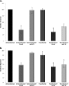
In vitro invasion and migration of c-V5-transfected cells is reduced after treatment with an anti-V5 antibody. A and B, untransfected control cells or c-V5-transfected cells were treated in the presence or absence of the V-ATPase specific inhibitor concanamycin A (ConA) or a monoclonal antibody against V5 and then plated on MatrigelTM-coated FluoroBlokTM inserts (A, invasion) or uncoated FluoroBlokTM inserts (B, migration) and allowed to migrate toward a chemoattractant on the trans-side of the well for 8 h. Cells were then stained with calcein AM, and the number of cells that had invaded or migrated to the trans-side was counted, with three wells analyzed per sample and an average of 10 images analyzed per well. The values for both assays are the means of at least three independent experiments expressed relative to untransfected, untreated cells. All error bars indicate S.E. *, p < 0.05 compared with untransfected, untreated cells.
FIGURE 6.
FIGURE 6.
Streptavidin-bound biotin-bafilomycin is membrane-impermeable and inhibits in vitro invasion and migration of MB231 cells. A, vacuolar membranes were isolated from the yeast strain YPH500, and ATP-dependent proton transport activity was measured as described under “Experimental Procedures.” The values are the means of two independent experiments set relative to vacuoles treated with DMSO alone. B and C, MB231 cells were treated in the presence or absence of the V-ATPase specific inhibitor concanamycin A (ConA) or streptavidin-bound, biotin-conjugated bafilomycin and then plated on MatrigelTM-coated FluoroBlokTM inserts (B, invasion) or uncoated FluoroBlokTM inserts (C, migration) and allowed to migrate toward a chemoattractant on the trans-side of the well for 8 h. The cells were then stained with calcein AM, and the number of cells that had invaded or migrated to the trans-side was counted, with three wells analyzed per sample and an average of 10 images analyzed per well. The values for both assays are the means of at least three independent experiments expressed relative to cells treated with DMSO and streptavidin alone. All error bars indicate S.E. *, p < 0.05 compared with cells treated with DMSO and streptavidin alone.

References

    1. Nguyen D. X., Bos P. D., Massagué J. (2009) Metastasis: from dissemination to organ-specific colonization. Nat. Rev. Cancer 9, 274–284 - PubMed
    1. Spano D., Zollo M. (2012) Tumor microenvironment: a main actor in the metastasis process. Clin. Exp. Metastasis 29, 381–395 - PubMed
    1. Gupta G. P., Massagué J. (2006) Cancer metastasis: building a framework. Cell 127, 679–695 - PubMed
    1. Webb B. A., Chimenti M., Jacobson M. P., Barber D. L. (2011) Dysregulated pH: a perfect storm for cancer progression. Nat. Rev. Cancer 11, 671–677 - PubMed
    1. Martínez-Zaguilán R., Seftor E. A., Seftor R. E., Chu Y. W., Gillies R. J., Hendrix M. J. (1996) Acidic pH enhances the invasive behavior of human melanoma cells. Clin. Exp. Metastasis 14, 176–186 - PubMed

Publication types