

MicroRNA-146a and microRNA-146b regulate human dendritic cell apoptosis and cytokine production by targeting TRAF6 and IRAK1 proteins
- PMID: 25505246
- PMCID: PMC4317016
- DOI: 10.1074/jbc.M114.591420
MicroRNA-146a and microRNA-146b regulate human dendritic cell apoptosis and cytokine production by targeting TRAF6 and IRAK1 proteins
Abstract
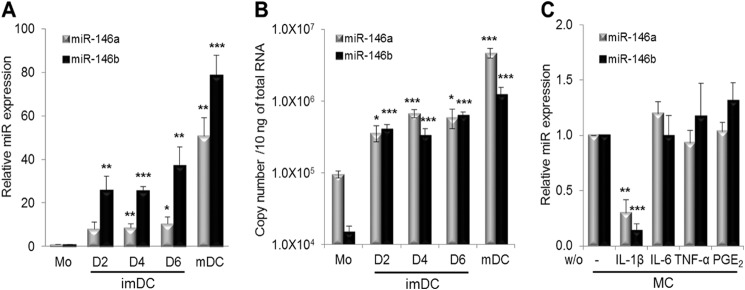
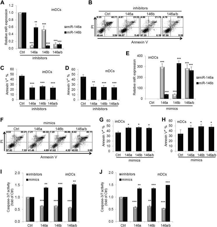
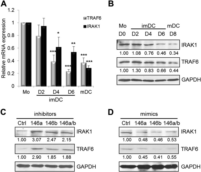
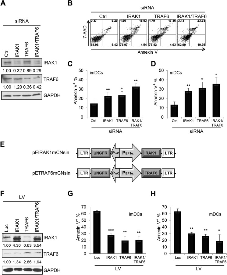
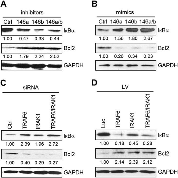
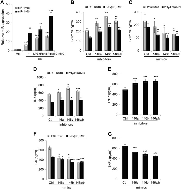
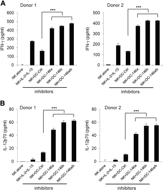
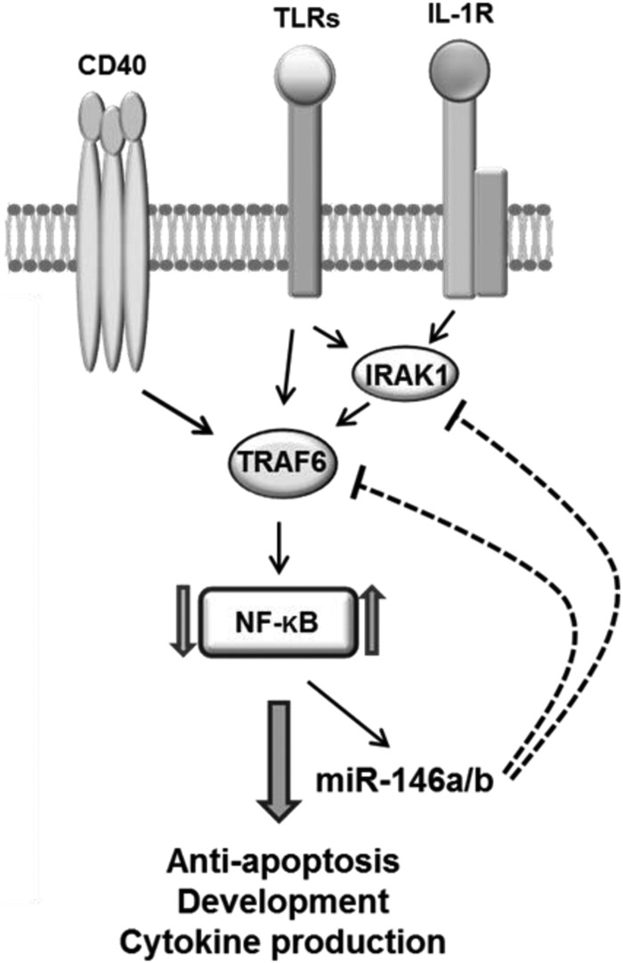
We have previously reported 27 differentially expressed microRNAs (miRNAs) during human monocyte differentiation into immature dendritic cells (imDCs) and mature DCs (mDCs). However, their roles in DC differentiation and function remain largely elusive. Here, we report that microRNA (miR)-146a and miR-146b modulate DC apoptosis and cytokine production. Expression of miR-146a and miR-146b was significantly increased upon monocyte differentiation into imDCs and mDCs. Silencing of miR-146a and/or miR-146b in imDCs and mDCs significantly prevented DC apoptosis, whereas overexpressing miR-146a and/or miR-146b increased DC apoptosis. miR-146a and miR-146b expression in imDCs and mDCs was inversely correlated with TRAF6 and IRAK1 expression. Furthermore, siRNA silencing of TRAF6 and/or IRAK1 in imDCs and mDCs enhanced DC apoptosis. By contrast, lentivirus overexpression of TRAF6 and/or IRAK1 promoted DC survival. Moreover, silencing of miR-146a and miR-146b expression had little effect on DC maturation but enhanced IL-12p70, IL-6, and TNF-α production as well as IFN-γ production by IL-12p70-mediated activation of natural killer cells, whereas miR-146a and miR-146b overexpression in mDCs reduced cytokine production. Silencing of miR-146a and miR-146b in DCs also down-regulated NF-κB inhibitor IκBα and increased Bcl-2 expression. Our results identify a new negative feedback mechanism involving the miR-146a/b-TRAF6/IRAK1-NF-κB axis in promoting DC apoptosis.
Keywords: Apoptosis; Cytokine; Dendritic Cell; Differentiation; IL-1 Receptor-associated Kinases (IRAK); MicroRNA (miRNA); NF-kappa B (NF-κB); TNF Receptor-associated Factor (TRAF); miR-146.
© 2015 by The American Society for Biochemistry and Molecular Biology, Inc.
Figures
Similar articles
-
Dexmedetomidine exerts cardioprotective effect through miR-146a-3p targeting IRAK1 and TRAF6 via inhibition of the NF-κB pathway.Biomed Pharmacother. 2021 Jan;133:110993. doi: 10.1016/j.biopha.2020.110993. Epub 2020 Nov 18. Biomed Pharmacother. 2021. PMID: 33220608
-
Overexpression of miR-146b-5p Ameliorates Neonatal Hypoxic Ischemic Encephalopathy by Inhibiting IRAK1/TRAF6/TAK1/NF-αB Signaling.Yonsei Med J. 2020 Aug;61(8):660-669. doi: 10.3349/ymj.2020.61.8.660. Yonsei Med J. 2020. PMID: 32734729 Free PMC article.
-
Differential regulation of microRNA-146a and microRNA-146b-5p in human retinal pigment epithelial cells by interleukin-1β, tumor necrosis factor-α, and interferon-γ.Mol Vis. 2013 Apr 3;19:737-50. Print 2013. Mol Vis. 2013. PMID: 23592910 Free PMC article.
-
MicroRNA in TLR signaling and endotoxin tolerance.Cell Mol Immunol. 2011 Sep;8(5):388-403. doi: 10.1038/cmi.2011.26. Epub 2011 Aug 8. Cell Mol Immunol. 2011. PMID: 21822296 Free PMC article. Review.
-
Unveiling the therapeutic potential of miR-146a: Targeting innate inflammation in atherosclerosis.J Cell Mol Med. 2024 Oct;28(19):e70121. doi: 10.1111/jcmm.70121. J Cell Mol Med. 2024. PMID: 39392102 Free PMC article. Review.
Cited by
-
Cell death pathways and viruses: Role of microRNAs.Mol Ther Nucleic Acids. 2021 Mar 19;24:487-511. doi: 10.1016/j.omtn.2021.03.011. eCollection 2021 Jun 4. Mol Ther Nucleic Acids. 2021. PMID: 33898103 Free PMC article. Review.
-
Inflammation related miRNAs as an important player between obesity and cancers.J Diabetes Metab Disord. 2019 Nov 26;18(2):675-692. doi: 10.1007/s40200-019-00459-2. eCollection 2019 Dec. J Diabetes Metab Disord. 2019. PMID: 31890692 Free PMC article. Review.
-
Immunological Molecular Responses of Human Retinal Pigment Epithelial Cells to Infection With Toxoplasma gondii.Front Immunol. 2019 May 1;10:708. doi: 10.3389/fimmu.2019.00708. eCollection 2019. Front Immunol. 2019. PMID: 31118929 Free PMC article.
-
Osteoarthritis and Toll-Like Receptors: When Innate Immunity Meets Chondrocyte Apoptosis.Biology (Basel). 2020 Mar 30;9(4):65. doi: 10.3390/biology9040065. Biology (Basel). 2020. PMID: 32235418 Free PMC article. Review.
-
A Review of Macrophage MicroRNAs' Role in Human Asthma.Cells. 2019 May 8;8(5):420. doi: 10.3390/cells8050420. Cells. 2019. PMID: 31071965 Free PMC article. Review.
References
-
- Lanzavecchia A., Sallusto F. (2004) Lead and follow: the dance of the dendritic cell and T cell. Nat. Immunol. 5, 1201–1202 - PubMed
-
- Banchereau J., Briere F., Caux C., Davoust J., Lebecque S., Liu Y. J., Pulendran B., Palucka K. (2000) Immunobiology of dendritic cells. Annu. Rev. Immunol. 18, 767–811 - PubMed
-
- Steinman R. M., Hawiger D., Nussenzweig M. C. (2003) Tolerogenic dendritic cells. Annu. Rev. Immunol. 21, 685–711 - PubMed
-
- Jiang H., Van De Ven C., Satwani P., Baxi L. V., Cairo M. S. (2004) Differential gene expression patterns by oligonucleotide microarray of basal versus lipopolysaccharide-activated monocytes from cord blood versus adult peripheral blood. J. Immunol. 172, 5870–5879 - PubMed
-
- Jiang H., van de Ven C., Baxi L., Satwani P., Cairo M. S. (2009) Differential gene expression signatures of adult peripheral blood vs cord blood monocyte-derived immature and mature dendritic cells. Exp. Hematol. 37, 1201–1215 - PubMed
Publication types
MeSH terms
Substances
Grants and funding
LinkOut - more resources
Full Text Sources
Other Literature Sources
