. 2015 Feb 6;290(6):3293-307.
doi: 10.1074/jbc.M114.614578. Epub 2014 Dec 12.

Topological analysis of Hedgehog acyltransferase, a multipalmitoylated transmembrane protein

Affiliations

Topological analysis of Hedgehog acyltransferase, a multipalmitoylated transmembrane protein

Antonio D Konitsiotis et al. J Biol Chem. .

Abstract

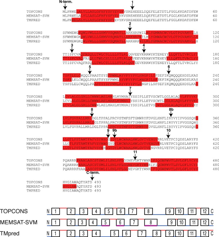
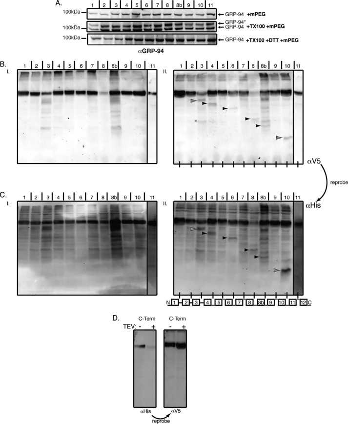
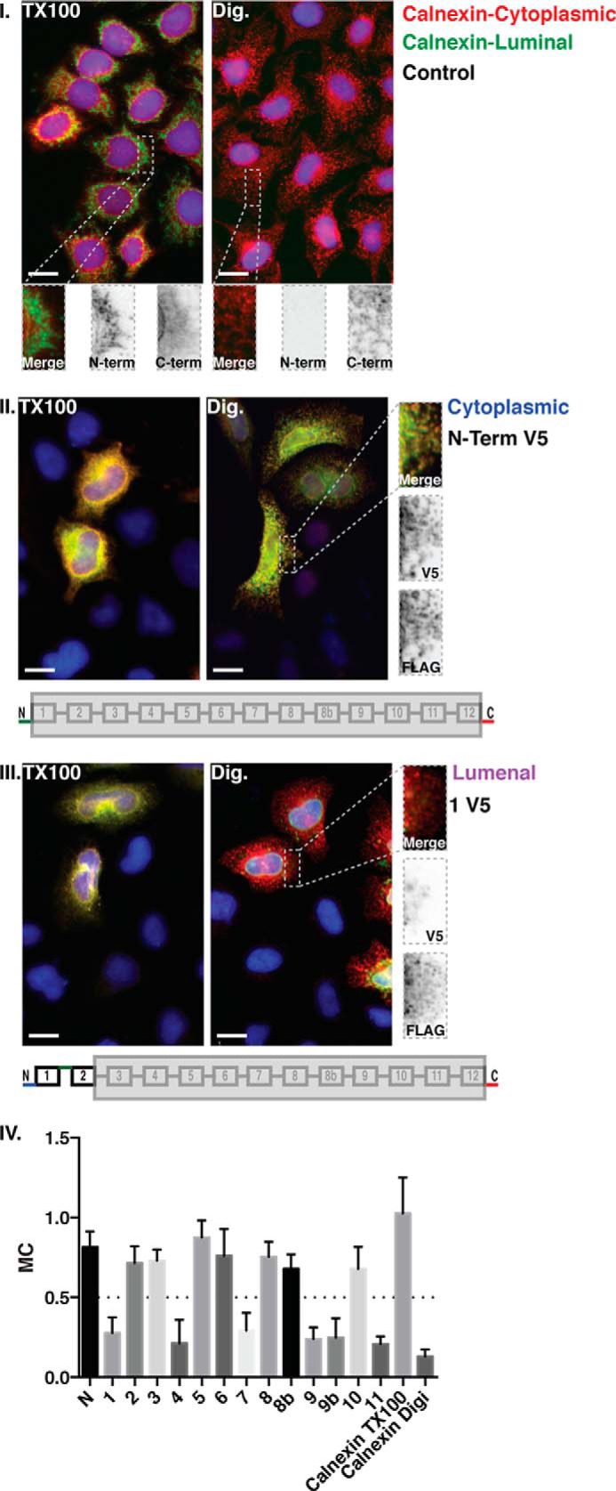
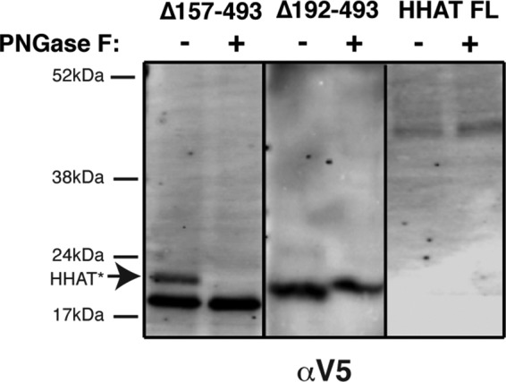
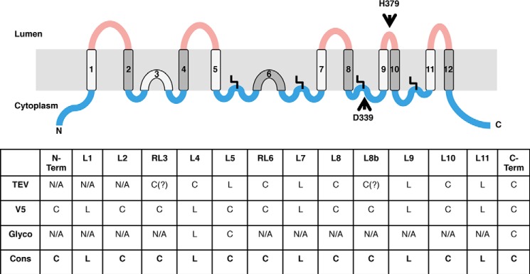
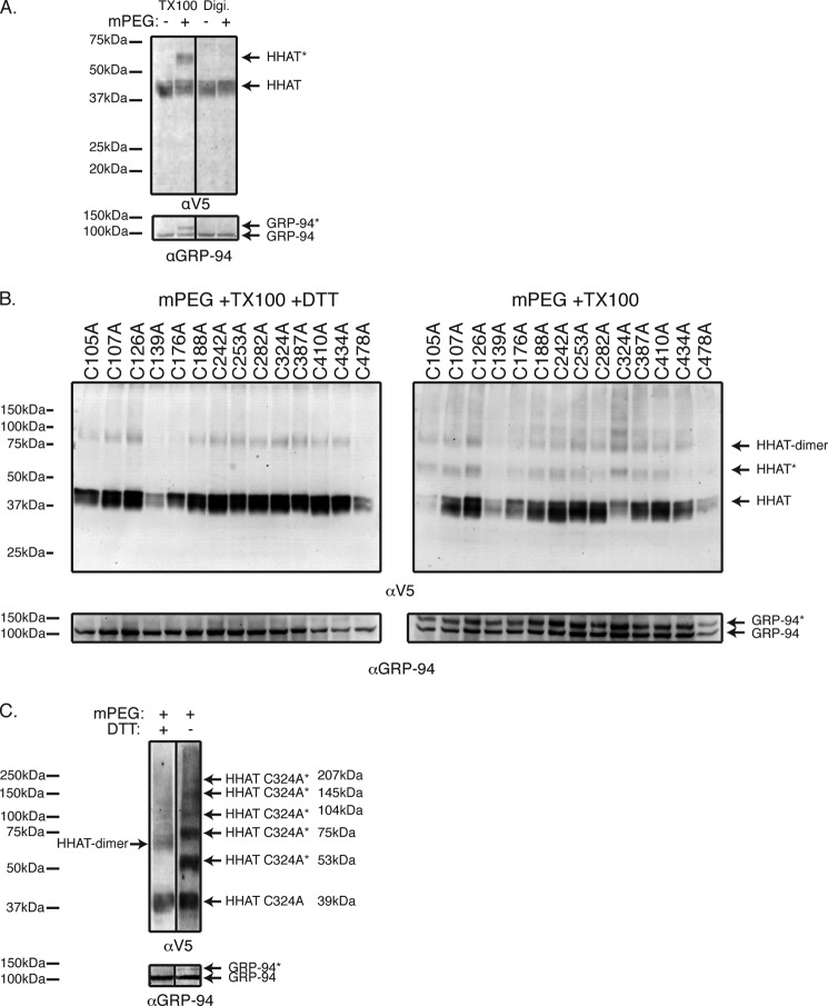
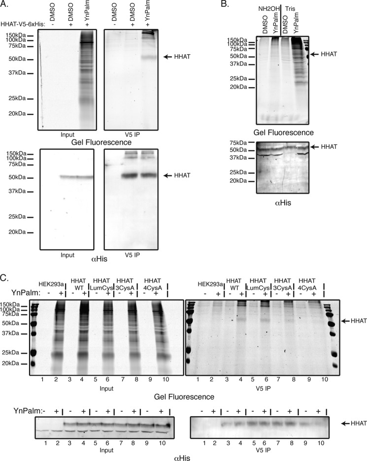
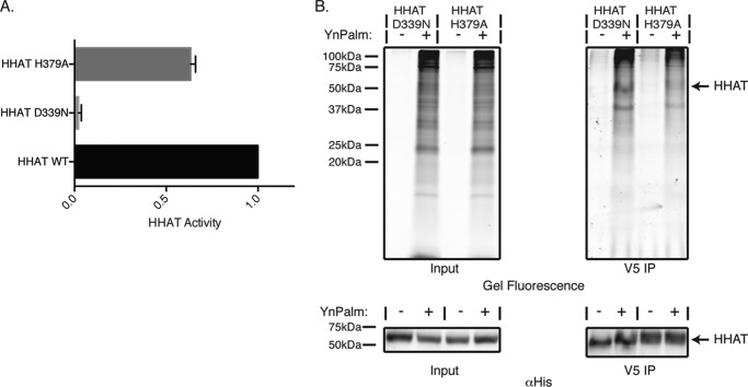
Hedgehog proteins are secreted morphogens that play critical roles in development and disease. During maturation of the proteins through the secretory pathway, they are modified by the addition of N-terminal palmitic acid and C-terminal cholesterol moieties, both of which are critical for their correct function and localization. Hedgehog acyltransferase (HHAT) is the enzyme in the endoplasmic reticulum that palmitoylates Hedgehog proteins, is a member of a small subfamily of membrane-bound O-acyltransferase proteins that acylate secreted proteins, and is an important drug target in cancer. However, little is known about HHAT structure and mode of function. We show that HHAT is comprised of ten transmembrane domains and two reentrant loops with the critical His and Asp residues on opposite sides of the endoplasmic reticulum membrane. We further show that HHAT is palmitoylated on multiple cytosolic cysteines that maintain protein structure within the membrane. Finally, we provide evidence that mutation of the conserved His residue in the hypothesized catalytic domain results in a complete loss of HHAT palmitoylation, providing novel insights into how the protein may function in vivo.

Keywords: Acyltransferase; Cancer; Click Chemistry; HHAT; Hedgehog Signaling Pathway; MBOAT; Protein Palmitoylation; Topology; Transmembrane Domain.


Figures

FIGURE 1.
FIGURE 1.
Bioinformatic prediction analysis of HHAT sequences identifies a maximum of 13 predicted transmembrane domains. Clustal Omega sequence alignment of human HHAT sequence with the TM predictions from three separate bioinformatic algorithms: TOPCONS, MEMSAT-SVM, and TMpred. Predicted TMs are highlighted in red, and loops are numbered in accordance with the epitope insertion analysis from our experiments. The TMpred algorithm predicted a helix within loop 8, and so mutants were made at two places in the loop between predicted TMs 8 and 9 to accommodate this prediction. These mutants were numbered 8 and 8b. In all models, the critical His-379 residue is predicted to be part of TM 9. Positions selected for epitope and TEV tag insertions used in this study are indicated by arrows. The bottom panel depicts the predicted topologies from each of the algorithms along with the predicted loop location. The black boxes depict the predicted TMs, whereas the lines depict the loop regions. Loops are colored red and blue depending on whether they were predicted to be luminal or cytosolic, respectively. In the MEMSAT-SVM diagram, TMs 5, 6, and 8 were predicted to be pore-lining helices, which are depicted in magenta. Loops and TMs are not drawn exactly to scale but do indicate relative size differences. N-term., N-terminal; C-term., C-terminal.
FIGURE 2.
FIGURE 2.
Mapping of HHAT topology by TEV cleavage of microsomal preparations. HHAT constructs containing a C-terminal V5-His6 epitope tag and an internal TEV protease tag at the indicated loops were transfected into HEK293a cells, and intact microsomal preparations were prepared by ultracentrifugation. A, to show that the microsomal preparations have intact ER membranes, mPEG, a hydrophilic reagent of 5 kDa that reacts with -SH groups, did not react with the non-disulfide-bonded cysteine in the ER luminal protein GRP94 when it was added to 10 μg of the microsomal preparations (top panel). After treatment of the microsomes with 0.2% Triton X-100 (TX100, center panel), GRP94 is modified by mPEG, and the reaction could be blocked by the reducing agent DTT (bottom panel). GRP94* indicates the position of the modified proteins. B, microsome preparations were treated with TEV buffer (I) or Pro-TEV protease (see “Experimental Procedures”) (II) for 4 h at 25 °C with agitation. The reaction was then stopped with sample buffer, and samples were analyzed by SDS-PAGE and probed first with anti-V5 antibody and then reprobed with anti-His6 antibody. Because the microsomal preparations are intact, any TEV cleavage sites on luminal loops will be inaccessible to the Pro-TEV protease. However, any cytosolic sites will be accessible to the protease, and so the protein will be cleaved and produce a shorter C-terminal fragment that will be identified by antibody labeling. C, using this method, the HHAT topology model (II, bottom panel) was produced. Red lines indicate that the TEV site was inaccessible and, hence, luminal. Blue lines indicate that the TEV site was accessible and, hence, cytoplasmic. Cleaved bands are indicated by black arrowheads, whereas gray arrowheads indicate potential cleaved products. It was unclear whether mutants 1/2 and 2/3 produced any cleavage product. Immunoblots shown are representative results from five independent experiments. D, a construct containing the TEV protease tag in the linker sequence between the C-terminal (C-term.) V5 epitope and the His6 epitope tag was used to examine the topology of the C terminus of HHAT. Specifically, if the C terminus is cytosolic and accessible to TEV protease cleavage, then reactivity of the protein with a His6 antibody would be lost after treatment with Pro-TEV, whereas reactivity with the V5 antibody should be retained. This is what was observed in the experiments with this construct, indicating that the C terminus is cytosolic.
FIGURE 3.
FIGURE 3.
Mapping of HHAT topology by V5 epitope indirect immunofluorescence in selectively permeabilized cells. HHAT constructs containing a C-terminal FLAG epitope tag and an internal V5 epitope tag at the indicated loops were transfected into HeLa cells plated on 96-well imaging plates. Cell membranes were either permeabilized with 0.2% Triton X-100 (TX100) or with 0.04% digitonin (Dig). Permeabilization with digitonin maintains the ER membrane intact, whereas permeabilization with Triton X-100 fully permeabilizes all membranes of the cell. FLAG and V5 epitopes were stained with specific primary antibodies, followed by secondary antibodies with 555-nm (red) or 488-nm (green) excitation wavelengths, respectively. To control for the selective permeabilization of the ER membrane, untransfected cells were selectively permeabilized and stained for Calnexin using two different antibodies, one that binds to an epitope on the luminal N terminus of the protein and another that bind to an epitope on the cytosolic C terminus of the protein (I). Only two conditions are shown, for the V5 epitope in the N terminus of the protein (II) and in predicted loop 1 (III). The N and C termini of HHAT were both cytosolic (II), therefore the FLAG epitope will always be stained regardless of the detergent used for permeabilization. Images were then analyzed using Volocity image analysis software to determine the colocalization coefficient of the two channels. The insets show a higher magnification of the indicated regions. The histogram (IV) shows mean Manders' coefficients (MC) for colocalization of the red (555-nm) channel with the green (488-nm) channel for different HHAT mutants and for the Calnexin control (n > 40 cells, data are mean ± S.D.). High Manders' coefficients indicate better colocalization of FLAG-tagged proteins with V5-tagged proteins. Values less than 0.5 indicate low colocalization. Identical exposures and image normalization for both permeabilizations ensure a fair side-by-side comparison. The bottom panels of II and III illustrate the positions of the stained epitopes. Images are examples of each condition from at least three independent experiments. Scale bars = 15 μm.
FIGURE 4.
FIGURE 4.
N-glycosylation analysis of full-length HHAT, HHAT-Δ157–493, and HHAT-Δ192–493 truncation mutants containing a C-terminal N-glycosylation motif. In vitro translation/translocation of HHAT truncations ending in predicted loop regions 4 (Δ157–493 mutant) and 5 (Δ192–493 mutant) or full-length (FL) HHAT cDNA C-terminally tagged with an N-glycosylation site and a V5 epitope. Luminal N-glycosylation sites become glycosylated, and the covalent attachment to the protein can be detected by a shift in molecular weight on an SDS-PAGE gel. Following expression, lysates were treated with PNGase F, an endoglycosidase that cleaves N-glycans between the innermost sugar moiety and Asn, removing the shift in molecular weight of glycosylated proteins, or left untreated. Lysates were analyzed by SDS-PAGE on 7.5% gels, followed by immunoblotting with anti-V5 antibody. HHAT*, glycosylated HHAT protein.
FIGURE 5.
FIGURE 5.
HHAT topology experimental consensus model indicating ten TMs and two RLs. Shown is the consensus HHAT topology model, including palmitoylation modifications, on the basis of the data shown in the bottom panel which summarizes the results from the three different experiments, indicating whether a loop is luminal (L) or cytoplasmic (C). The consensus (Cons.) model is indicated in the bottom row. Our data indicate that predicted TM 3 and TM 6 are likely to be RLs. The cytosolic loops containing cysteines that are palmitoylated display the fatty acid modification as a bent line. Relative loop length is approximated by the size of the loops but not precisely scaled. Cytosolic loops are colored blue, and luminal loops are red. The positions of the two conserved MBOAT residues, Asp-339 and His-379, are indicated by arrows.
FIGURE 6.
FIGURE 6.
HHAT cysteine mapping in selectively permeabilized cells. A, HEK293a cells expressing HHAT-V5-His6 were permeabilized with 0.04% digitonin (Digi.) or 1% Triton X-100 (TX100) as indicated. Modification was carried out for 30 min at 4 °C with 1 mm mPEG. Samples were analyzed by SDS-PAGE on 7.5% gels, followed by immunoblotting with antibodies to V5 (top panel) or GRP94 (bottom panel). B, HEK293a cells expressing the indicated HHAT single cysteine mutants were permeabilized with 1% Triton X-100. Modification was carried out for 30 min at 4 °C with 1 mm mPEG. Where indicated, DTT was present during the reaction. Samples were analyzed by SDS-PAGE on 7.5% gels, followed by immunoblotting with antibodies to V5 (top panels) or GRP94 (bottom panels). The immunoblots shown are representative results from four independent experiments. C, HEK293a cells expressing HHAT C324A mutant were permeabilized with 1% Triton X-100. Modification was carried out for 30 min at 4 °C with 1 mm mPEG. Where indicated, DTT was present during the reaction. Samples were analyzed by SDS-PAGE on 7.5% gels, followed by immunoblotting with antibodies to V5 (top panel) or GRP94 (bottom panel). HHAT* and GRP94* indicate the positions of the modified proteins.
FIGURE 7.
FIGURE 7.
HHAT is palmitoylated on multiple cysteines via thioester linkage. A, HEK293a cells were transfected with a construct expressing the WT human HHAT cDNA C-terminally tagged with V5 and His6 epitopes. Cells were then fed overnight with 50 μm palmitic acid analog with a clickable alkyne moiety (YnPalm) or with vehicle (0.1% DMSO). Cells were lysed, and HHAT was immunoprecipitated (IP) using anti-V5 antibody. Immunoprecipitated proteins or lysates were ligated by CuAAC to AzTB. Tagged proteins were separated on 15% SDS-PAGE gels and analyzed by in-gel fluorescence (top panels) and immunoblotting with antibody against His6 (bottom panels). B, lysates were prepared as in the previous experiment. However, before immunoprecipitation, lysates were either treated with 1 m NH2OH (pH 7.5) or 1 m Tris (pH 7.5) for 5 h at room temperature, as described under “Experimental Procedures.” Lysates were then precipitated by chloroform/methanol precipitation, resolubilized in 0.2% Triton X-100 in PBS, immunoprecipitated with αV5 antibody, ligated by CuAAC to AzTB, and analyzed by SDS-PAGE in-gel fluorescence (top panel) and anti-His6 immunoblotting (bottom panel). C, HEK293a cells were transfected with constructs expressing WT human HHAT cDNA, the luminal cysteine mutant HHAT-LumCys, or the cytosolic cysteine mutants HHAT-3CysA or HHAT-4CysA. Cells were then fed overnight with 50 μm YnPalm or 0.1% DMSO. Cells were then lysed, immunoprecipitated (IP) with anti-V5 antibody, ligated by CuAAC to AzTB, and analyzed by SDS-PAGE in-gel fluorescence (top panels) and anti-His6 immunoblotting (bottom panels).
FIGURE 8.
FIGURE 8.
His-379 is implicated in HHAT palmitoylation. A, HHAT activity for the D339N and H379A mutants was determined by measuring the ability of the purified and solubilized HHAT mutant proteins to palmitoylate a Shh peptide using an in-house developed in vitro click chemistry-based palmitoylation assay. HHAT-D339N-V5-His6 showed only ∼3% activity compared with the WT, whereas the HHAT-H379A-V5-His6 mutant had ∼63% activity compared with the WT. Data are presented as mean ± S.D. and normalized to HHAT-WT-V5-His6. B, HEK293a cells were transiently transfected with HHAT-D339N-V5-His6 or HHAT-H379A-V5-His6 mutant constructs. Cells were then fed overnight with 50 μm YnPalm or with 0.1% DMSO. Cells were lysed and HHAT was immunoprecipitated (IP) using a V5 antibody. Immunoprecipitated proteins or lysates were ligated by CuAAC to AzTB. Tagged proteins were separated on 15% SDS-PAGE gels and analyzed by in-gel fluorescence (top panel) and immunoblotting with antibody against the His tag (bottom panel). Gels and immunoblots shown are representative results from two independent experiments.

Similar articles

Cited by

References

    1. McMahon A. P., Ingham P. W., Tabin C. J. (2003) Developmental roles and clinical significance of hedgehog signaling. Curr. Top. Dev. Biol. 53, 1–114 - PubMed
    1. Ingham P. W., McMahon A. P. (2001) Hedgehog signaling in animal development: paradigms and principles. Genes Dev. 15, 3059–3087 - PubMed
    1. Barakat M. T., Humke E. W., Scott M. P. (2010) Learning from Jekyll to control Hyde: Hedgehog signaling in development and cancer. Trends Mol. Med. 16, 337–348 - PMC - PubMed
    1. Scales S. J., de Sauvage F. J. (2009) Mechanisms of Hedgehog pathway activation in cancer and implications for therapy. Trends Pharmacol. Sci. 30, 303–312 - PubMed
    1. Mann R. K., Beachy P. A. (2004) Novel lipid modifications of secreted protein signals. Annu. Rev. Biochem. 73, 891–923 - PubMed

Publication types