. 2014 Dec 10;34(50):16594-604.
doi: 10.1523/JNEUROSCI.2585-14.2014.

Neuronal transgene expression in dominant-negative SNARE mice

Affiliations

Neuronal transgene expression in dominant-negative SNARE mice

Takumi Fujita et al. J Neurosci. .

Erratum in

  • J Neurosci. 2016 Apr 6;36(14):4136-7

Abstract

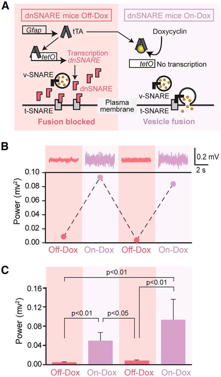
Experimental advances in the study of neuroglia signaling have been greatly accelerated by the generation of transgenic mouse models. In particular, an elegant manipulation that interferes with astrocyte vesicular release of gliotransmitters via overexpression of a dominant-negative domain of vesicular SNARE (dnSNARE) has led to documented astrocytic involvement in processes that were traditionally considered strictly neuronal, including the sleep-wake cycle, LTP, cognition, cortical slow waves, depression, and pain. A key premise leading to these conclusions was that expression of the dnSNARE was specific to astrocytes. Inconsistent with this premise, we report here widespread expression of the dnSNARE transgene in cortical neurons. We further demonstrate that the activity of cortical neurons is reversibly suppressed in dnSNARE mice. These findings highlight the need for independent validation of astrocytic functions identified in dnSNARE mice and thus question critical evidence that astrocytes contribute to neurotransmission through SNARE-dependent vesicular release of gliotransmitters.

Keywords: EEG; GFAP; SNARE; adenosine; sleep.


Figures

Figure 1.
Figure 1.
Induction of dnSNARE by removal of doxycycline suppresses cortical EEG power reversibly in dnSNARE mice. A, Schematic outlining the tTA system used in dnSNARE mice. Dox administration via drinking water prevents transcription activation. Removal of Dox induces redundant expression of dnSNARE, which is a cytosolic portion (lacking transmembrane region) of vesicular SNARE (v-SNARE) and thus formation of the SNARE complex between vesicles expressing v-SNARE and the plasma membrane expressing target SNARE (t-SNARE) is inhibited. DnSNARE mice that are positive for both transactivator (GFAP-tTA) and tet operators (tetO-dnSNARE) were treated with (On-Dox) or without (Off-Dox) Dox for 2 weeks before each EEG collection. B, Top, Representative EEG trace collected from a dnSNARE mouse under On-Dox or Off-Dox condition. Bottom, Representative EEG power showing a reversible decrease in EEG power during Off-Dox periods in dnSNARE mouse. C, Quantification of EEG power in dnSNARE mice under On-Dox or Off-Dox condition. DnSNARE mice during On-Dox periods showed significantly higher EEG power compared with those during Off-Dox periods. One-way ANOVA with post hoc Tukey-Kramer test (n = 4, first Off-Dox; n = 5, first On-Dox and second Off-Dox; n = 3, second On-Dox). Error bars indicate SEM.
Figure 2.
Figure 2.
Neuronal expression of dnSNARE and 2 reporter genes LacZ and EGFP in dnSNARE mice. A, Top, Schematic outlining a collection of cortical neurons from 8-d old C57BL/6 WT or dnSNARE mice under Off-Dox condition using density gradient-based dissociation method and FACS. Neurons were enriched and separated from myelin debris by collecting Fraction 2 and 3 before FACS. Bottom, Under Off-Dox condition, human GFAP promoter drives the expression of 3 reporter units, dnSNARE, LacZ, and EGFP, in dnSNARE mice. B, qPCR analysis of markers expression in purified mouse PSA-NCAM+ population. Neuronal markers; RNA binding protein fox-1 homolog 3 (Rbfox3), Tubulin, beta 3 (Tubb3), Neurofilament, light polypeptide (Nefl), Neurofilament, medium polypeptide (Nefm), Glutamic acid decarboxylase 1 (Gad1), Glutamic acid decarboxylase 2 (Gad2), Choline acetyltransferase (Chat) and Solute carrier family 6, member 4 (Slc6a4). Astrocyte markers; Aquaporin 4 (Aqp4), Glial fibrillary acidic protein (Gfap), Excitatory amino acid transporter 2 (Slc1a2) and aldehyde dehydrogenase family 1, member L1 (Aldh1l1). Oligodendrocyte precursor marker, platelet derived growth factor receptor, α polypeptide (Pdgfra). Oligodendrocyte marker, myelin basic protein (Mbp). Microglial marker, Integrin α M (Itgam). n = 7 biological replicates. C, Immunolabeling analysis of neuronal marker expression in purified mouse PSA-NCAM+ population. Neuronal marker NeuN is highly enriched in cortical PSA-NCAM+ populations compared with PSA-NCAM populations in which the astrocyte marker Aqp4 is highly expressed. Scale bar, 100 μm. D, qPCR analysis of PSA-NCAM+ and PSA-NCAM populations showed neuronal expression of dnSNARE and reporter transgenes (LacZ and EGFP) in dnSNARE mice (n = 4 biological samples) and no expression in both populations isolated from WT mice (n = 3 biological samples). E, Expression of the tTA transgene in PSA-NCAM+ and PSA-NCAM populations in dnSNARE mice but not in WT mice (n = 4 each). F, Relative expression of transgenes (dnSNARE, LacZ, and EGFP) in cerebral cortex in adult Off-Dox dnSNARE mice compared with On-Dox condition (n = 4). G, Enrichment of neuronal marker gene Rbfox3 expression in neurons isolated from adult dnSNARE mice compared with tissue (n = 3). H, Neuronal expression of transgenes (dnSNARE, LacZ, and EGFP) in adult dnSNARE mice under Off-Dox condition compared with On-Dox condition (n = 3). Error bars indicate SEM in B and DH.
Figure 3.
Figure 3.
Expression of EGFP and β-galactosidase driven by GFAP promoter in widespread neuronal population and basal leakiness of “Tet-Off” system in dnSNARE mice. A, Immunohistochemical analysis of the cellular expression of VAMP2 in cerebral cortex (VAMP2, green; MAP2, orange; DAPI, blue). White arrows indicate VAMP2- and MAP2-positive neurons. Scale bars, 100 μm (left), 20 μm (right), 10 μm (inset). B, Analysis of VAMP2 expression in cortical astrocytic and cortical neuronal cultures. Immunocytochemistry for VAMP2 (green) together with MAP2 (orange) or GFAP (yellow). Red arrows indicate VAMP2- and MAP2-expressing cells. Scale bar, 100 μm. C, Neuronal EGFP expression in cortex and hippocampus. Unstained sagittal sections of dnSNARE Off-Dox, On-Dox, GFAP-tTA negative and wild-type C57BL/6J (WT) mice. Although scattered bright EGFP signals indicate a bushy astrocyte appearance in hippocampus and to a minor extend in cortex, NeuN-positive neurons (purple) also exhibit low to moderate level of EGFP expression throughout cortex and hippocampus in dnSNARE mice under the Off-Dox condition. EGFP expression is suppressed in On-Dox dnSNARE mice compared with that in the Off-Dox condition, but broad basal expression was observed. EGFP expression was also found in GFAP-tTA negative mice but was absent in WT mice. Insets are magnified images. Scale bars, 200 μm (main images); 40 μm (insets). D, X-gal staining (blue) and NeuN immunohistochemistry (brown) shows that several neurons express β-galactosidase (β-gal) in both cortex and hippocampus in dnSNARE Off-Dox mice. No β-gal positive cells were evident in WT mice. Scale bar, 100 μm. E, qPCR analysis of transgenes expressions (dnSNARE, LacZ, and EGFP) in cerebral cortex showing the leaky expressions of all 3 transgenes in adult GFAP-tTA-negative mice (n = 4, one-way ANOVA followed by Tukey–Kramer test). Error bars indicate SEM. F, GFAP promoter induces neuronal expression of Gi-coupled RASSL (receptor activated solely by synthetic ligand) Ro1. Representative images collected from a double-transgenic mice line that expresses Ro1 under the control of GFAP promoter using “Tet-Off” system. Ro1 expression was examined in brain sections prepared from Ro1 mice by immunostaining against FLAG (the epitope tag fused to Ro1). Majority of neurons express FLAG in both cortex and hippocampus. Scale bar, 50 μm.
Figure 4.
Figure 4.
EEG activity pattern and adenosine follow a circadian rhythm in dnSNARE mice. A, Schematic diagram depicting how ECoG recordings and microdialysis samples were collected in freely moving mice. B, Comparison of the raw power spectrum of dnSNARE mice under Off-Dox and On-Dox conditions (n = 5). C, Representative power spectrum analysis show the shift in the percentage prevalence of delta waves between dark and light phase in On-Dox (left) and Off-Dox (right) conditions. D, Comparison of the extracellular concentration of adenosine in wild-type C57BL/6J (WT) and Off-Dox dnSNARE mice during the dark phase or light phase in basal forebrain (top, WT, n = 7; Off-Dox dnSNARE, n = 9) or in hippocampus (bottom, WT, n = 9; Off-Dox dnSNARE, n = 6; p > 0.05, Student's t test compared with WT). E, Adenosine measurements over 48 h in microdialysates collected before, during and after sleep deprivation from basal forebrain (top) and hippocampus (bottom) in WT (basal forebrain, n = 5; hippocampus, n = 7) and in Off-Dox dnSNARE mice (basal forebrain, n = 6; hippocampus, n = 4). F, Adenosine concentrations during sleep deprivation and subsequent recovery sleep periods. No significant difference in adenosine concentrations was observed between WT and Off-Dox dnSNARE mice in either basal forebrain (top left, sleep deprivation period; top right, recovery sleep period; n = 5–6) or hippocampus (bottom left, sleep deprivation period; bottom right, recovery sleep period; n = 5–8). Error bars indicate SEM in B and DF. n.s., Not significant.

Comment in

Similar articles

Cited by

References

    1. Agha-Mohammadi S, O'Malley M, Etemad A, Wang Z, Xiao X, Lotze MT. Second-generation tetracycline-regulatable promoter: repositioned tet operator elements optimize transactivator synergy while shorter minimal promoter offers tight basal leakiness. J Gene Med. 2004;6:817–828. doi: 10.1002/jgm.566. - DOI - PubMed
    1. Agulhon C, Petravicz J, McMullen AB, Sweger EJ, Minton SK, Taves SR, Casper KB, Fiacco TA, McCarthy KD. What is the role of astrocyte calcium in neurophysiology? Neuron. 2008;59:932–946. doi: 10.1016/j.neuron.2008.09.004. - DOI - PMC - PubMed
    1. Araque A, Carmignoto G, Haydon PG, Oliet SH, Robitaille R, Volterra A. Gliotransmitters travel in time and space. Neuron. 2014;81:728–739. doi: 10.1016/j.neuron.2014.02.007. - DOI - PMC - PubMed
    1. Barker JM, Torregrossa MM, Taylor JR. Low prefrontal PSA-NCAM confers risk for alcoholism-related behavior. Nat Neurosci. 2012;15:1356–1358. doi: 10.1038/nn.3194. - DOI - PMC - PubMed
    1. Barres BA. What is a glial cell? Glia. 2003;43:4–5. doi: 10.1002/glia.10252. - DOI - PubMed

Publication types

LinkOut - more resources