. 2014 Nov 18;11(1):82.
doi: 10.1186/s12983-014-0082-9. eCollection 2014.

Sensing deep extreme environments: the receptor cell types, brain centers, and multi-layer neural packaging of hydrothermal vent endemic worms

Affiliations

Sensing deep extreme environments: the receptor cell types, brain centers, and multi-layer neural packaging of hydrothermal vent endemic worms

Shuichi Shigeno et al. Front Zool. .

Abstract

Introduction: Deep-sea alvinellid worm species endemic to hydrothermal vents, such as Alvinella and Paralvinella, are considered to be among the most thermotolerant animals known with their adaptability to toxic heavy metals, and tolerance of highly reductive and oxidative stressful environments. Despite the number of recent studies focused on their overall transcriptomic, proteomic, and metabolic stabilities, little is known regarding their sensory receptor cells and electrically active neuro-processing centers, and how these can tolerate and function in such harsh conditions.

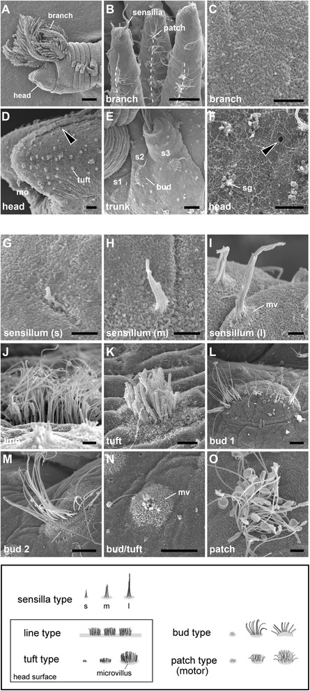
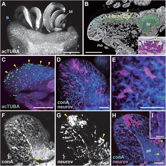
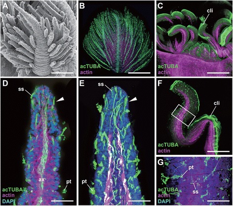
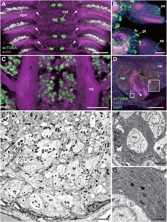
Results: We examined the extra- and intracellular organizations of the epidermal ciliated sensory cells and their higher centers in the central nervous system through immunocytochemical, ultrastructural, and neurotracing analyses. We observed that these cells were rich in mitochondria and possessed many electron-dense granules, and identified specialized glial cells and serial myelin-like repeats in the head sensory systems of Paralvinella hessleri. Additionally, we identified the major epidermal sensory pathways, in which a pair of distinct mushroom bodies-like or small interneuron clusters was observed. These sensory learning and memory systems are commonly found in insects and annelids, but the alvinellid inputs are unlikely derived from the sensory ciliary cells of the dorsal head regions.

Conclusions: Our evidence provides insight into the cellular and system-wide adaptive structure used to sense, process, and combat the deep-sea hydrothermal vent environment. The alvinellid sensory cells exhibit characteristics of annelid ciliary types, and among the most unique features were the head sensory inputs and structure of the neural cell bodies of the brain, which were surrounded by multiple membranes. We speculated that such enhanced protection is required for the production of normal electrical signals, and to avoid the breakdown of the membrane surrounding metabolically fragile neurons from oxidative stress. Such pivotal acquisition is not broadly found in the all body parts, suggesting the head sensory inputs are specific, and these heterogenetic protection mechanisms may be present in alvinellid worms.

Keywords: Annelids; Brain; Deep-sea; Evolution; Glia; Nervous system; Sensory cells.


Figures

Figure 1
Figure 1
Images of the vent endemic alvinellid worm, Paralvinella hessleri . A: Live worms viewed on board. B: A paraformaldehyde-fixed sample viewed from the lateral side. Scale bar, 1 mm.
Figure 2
Figure 2
Schemes of body plans of Paralvinella hessleri : the sensory and neural structure. A: Lateral view of the general body plan. Most of the parapodia setae are omitted. B: The epidermal sensory systems (green), major epidermal cilia. The patch type cilia on the branchial crown are represented as lines, and most of the cilia on trunk are omitted for clarity. C: The central nervous system and some peripheral nerves. The cell bodies (purple), major neural axons (red lines), and sensory organs (green patches). Divisions between the subesophageal ganglia and ventral nerve cords are not clear. Below left: The enlargement of rostral parts of the body to show the details of neural projections and epidermal ciliary sensory cells, side view. Below right: Dorsal view of the bi-lobed prostomium and the brain. On the left-hand side, the repeated features of glial and ciliary sensory cells (green) are located inside of the nuchal groove. The position of small globuli cell clusters or the mushroom body and distributed sensory cells are shown with axonal projection to the brain.
Figure 3
Figure 3
Scanning electron microscopy images of epidermal ciliary sensory cells. The cell types are summarized below for each body part. A: An overall side view of the rostral part. B: Branchial crown and the dotted lines show lines of patch type cilia along the lateral sides. C: Enlargement of a branch to show the no-pore condition of the epidermis. D: The head part and nuchal groove (arrowhead). E: The setigerous segments to show the distribution of the bud type cilia. F: The pores (arrowhead) and granules on the epidermis of the head. G - O: The epidermal cilia. mo, mouth opening; mv, microvilli; s1-3, setigerous segment 1–3; sg, secretory granules. Scale bar in A: 100 μm; B, D, E: 20 μm; M-O, 5 μm; C, F, G-L: 2 μm.
Figure 4
Figure 4
A primary sensilla cell type and the intracellular organella. Transmission electron microscopy (TEM) images. A: Cross section of the single layer epidermis of the branchial crown. B: Enlarged view of the same cell to show the organella. Inset: a cross-section showing the microtubules of cilia outside of the cuticle. ax, axon; cu, cuticle; gry; yellow-color granule; mf, muscle fiber; mt, mitochondrion; nu, nucleus; sc, supporting cells; ve, vesicle. Scale bar in A, B: 5 μm; Inset, 200 nm.
Figure 5
Figure 5
The brain, ciliary sensory cells, and glial repeats of the head or prostomium: whole-mount laser confocal microscopy images. A: Dorsal view of the prostomium with buccal tentacles. B: A semi-sagittal histological section cut as shown in A, showing a negative image of the hematoxylin and eosin stained section. Pseudocolors were used to enhance the position of line type cilia (light blue), axonal bundles and the brain (green), and secretory cells in the nuchal groove (yellow). Inset: granules stained with eosin. The arrowheads indicate the same position. C: A side view of the prostomium showing the distribution of acetylated alpha-tubulin (acTUBA) positive ciliary cells. The arrowheads indicate the line type cilia. The concanavalin A (conA, blue) and phalloidin (actin, red) cell membrane or muscle markers used for counterstaining. D, E: The sensory cell bodies and their axons coupled with dye (neurov: NeuroVue®Red) and repeated myelin-like glia stained with conA membrane marker (blue): a dorsal view of whole-mount prostomium and the enlargement. F-G: The sensory cells and the axonal projection patterns in the image series of conA, neurov, and merge. The numbers indicate the glial repeats and the arrowheads are positions filled with dye, and some labeled axons in the brain. I: Enlarged view showing the axons in the glial repeats. br, brain; bt, buccal tentacle; mo, mouth opening; pil, neuropil. Scale bar in A-C: 100 μm; D-H: 40 μm; I: 20 μm.
Figure 6
Figure 6
The myelin-like glial wrappers. The transverse sections, TEM images. A: The axonal bundles cut with sensory cell level, and B: the glial inter-repeat region with two different axonal types. The arrowhead indicates possible glial cell body. C: The detailed structure of glial membrane. D: Two axonal bundles. The type A and B run along the ventral and dorsal sides, respectively. E: Type B axonal bundles showing details of the membrane and mitochondria. ax, axonal fibers; co, collagen fibers; gc, glial cell wrappers; se, line type sensory cell. Scale bar in A, B, D: 2 μm; C, 300 nm; E: 1 μm.
Figure 7
Figure 7
The brains and higher sensory centers. A: A horizontal confocal optical section of the whole head region. B: A sagittal histological section of the head to show the brain subdivisions. The white dotted line covered with pseudo-green color indicates the position of the mushroom body-like structure in a section stained with hematoxylin-eosin. The dotted lines indicate optical cutting sites for confocal microscopy. C: The direct sensory pathways from tuft type cilia to the mushroom body, a sagittal section of the head. D-F: The nuclei distribution and neuropil structure. Horizontal optical sections viewed from the dorsal side at the level of the neuropil of the mushroom body (encircled with dotted red line). G-I: The highly mingled axons from small mushroom body neurons (G), the brain neuropil (H), and more ventral region (I). Nuclei stained with DAPI and tubulin positive fibers to show the distinct neuropils and axonal bundle patterns. The horizontal optical section series of confocal microscopy viewed from the dorsal (globuli cluster level) to ventral side. Arrowheads indicate the axonal fiber patterns running from dorsal to ventral, and to connectives. The phalloidin (actin) was used for counterstaining of neural fibers and muscles. br, brain; cen, central neuropil; co 1–5, connective to ventral nerve cords; ent, sensory axon entry region of the neuropil; pt, patch type cilia; mbc, mushroom body cells; np, neuropil; tt, tuft type cilia. Scale bar in A, D-I: 100 μm; B: 50 μm; C: 20 μm.
Figure 8
Figure 8
The mushroom body-like structure and the cell body wrappers. A schematic figure and TEM images. A: Schematic figure showing the brain organization and associated nerves, dorsal view. The thick longitudinal line indicates the cutting site for Figure 8B. B: TEM images of small interneurons and a large neighbor neuron. The arrowhead indicates granule-rich cell. C, D: The small or large interneurons covered with glia (an arrowhead). E: The granule-rich axons from the small cells. F: The neuropil of mushroom body. G: Details of the mushroom body neuropil. H: The enlarged view of glial membranes of small cells. I: Granules in the neurons. ax, axons; ent, sensory axon entry region of the neuropil; gr, granules; gry, yellow color granules; lt, line type sensory neurons; mt, mitochondrion; neu, a large neighbor neuron; np, neuropil; nu, nucleus; tt1 an tt2, tuft type cell 1 or 2; mbc, mushroom body cells. Scale bar in B, D, G: 4 μm; C, E, F: 1 μm, H, I: 0.1 μm.
Figure 9
Figure 9
The ciliary epidermal cells of the branchial crown and buccal tentacles. A: Scanning electron microcopy image of a branch. B-G: Laser confocal microscopy images. B: The whole view of the single branch with many patch type cilia. C: The buccal tentacles with dense patch type cilia arranged along each ventral side. D: An optical section of the leaflet showing the sensillia type cells (arrowhead) and axonal bundles. E: The 3D reconstruction images of optical sections showing the pathways from the sensilla type cells. F, G: A buccal tentacle and enlarged view to show the distribution of cilia. ax, axon; cli, ciliary line; pt, patch type cilia; ss, sensilla type cell. Scale bar in A-C, F: 100 μm; D, E: 10 μm; G: 25 μm.
Figure 10
Figure 10
The trunk nervous system with the sensory cilia and ventral nerve cord. A-D: The laser scanning microscopy views. A: Distribution of ventral patch type cilia with smaller cilia allayed along the ventro-lateral sides (arrowheads) with autofluorescence of the neuropodia. B: Notopodia and sensilla distributed at the tips. The axonal projections are seen from sensilla of the tips (ss), but such projections are not seen from patch type cilia (pt). C: Cellular distribution and neuropils of the ventral nerve cord. D: A cross-section of the ventral nerve cord to show the position of tubulinergic and serotonergic cells (arrowheads) within the cell body layers and axons in the neuropils. The two rectangles indicate the position of analysis for Figures E and F. The TEM images. E: Neuropil without obvious wrapping structure. The large axons are possibly motor neurons. F: The wrapped cell body of the ventral nerve cords. G: An enlarged view of glial membranes. ax, a relatively giant axonal bundle; ce, cell body layer; ss, sensilla type cell; pt, patch type cell; mt, mitochondrion; np, neuropil; nu, nucleus; npo, neruopodium. Scale bar in A-D: 50 μm; E, F: 2 μm; G: 0.1μm.
Figure 11
Figure 11
Schemes of the sensory pathways and the differentially specialized protection systems. In contrast to the primary sensory cells situated on the epidermis, the sensory neurons are multiply covered by glial wrappers. The myelin-like repeats are only formed in the head ciliary lines and not in the ventral nerve cords. Insets: the schemes show two distinct structural types of primary sensory receptor cells of epidermis and neuronal cells of the brain and ventral nerve cords. ax, axonal fiber; ct, cuticle; gc, glial cell membranes; gry, yellow color granule; lt, line type cell; mt, mitochondrion; ss, sensilla type cell; tt1 an tt2, tuft type cell 1 or 2.
Figure 12
Figure 12
Comparative schemes of the sensory inputs and motor outputs to emphasize the functional subdivisions. The tuft type sensory cells and their inputs are characterized for alvinellid worms. The two distinct tuft type cells on the head or prostomium are identified. Note that the branchial crown of Terebellida (Paralvinella) is not homologous to those of Serpulidae and palps of Nereididae. Some neural pathways are not precisely identified. Data are simplified from Orrhage and Müller [30]. bt, bud type cell; lt, line type; ss, sensilla type; tt, tuft type; VNC, subesophageal ganglia with ventral nerve cord/ganglia.

Similar articles

Cited by

References

    1. Desbruyeres D, Laubier L. Systematics, phylogeny, ecology and distribution of the Alvinellidae (Polychaeta) from deep-sea hydrothermal vents. Ophelia. 1991;5:31–45.
    1. Desbruyeres D, Chevaldonne P, Alayse AM, Jollivet D, Lallier FH, Jouin-Toulmond C, Zal F, Sarradin PM, Cosson R, Caprais JC, Arndt C, O’Brien J, Guezennec J, Hourdez S, Riso R, Gaill F, Laubier L, Toulmond A. Biology and ecology of the “Pompeii worm” (Alvinella pompejana Desbruyeres and Laubier), a normal dweller of an extreme deep-sea environment: a synthesis of current knowledge and recent developments. Deep-Sea Res Part I. 1998;45:383–422. doi: 10.1016/S0967-0645(97)00083-0. - DOI
    1. Luther GW, Rozan TF, Taillefert M, Nuzzio DB, Di Meo C, Shank TM, Lutz RA, Cary SC. Chemical speciation drives hydrothermal vent ecology. Nature. 2001;410:813–816. doi: 10.1038/35071069. - DOI - PubMed
    1. Mary J, Rogniaux H, Rees JF, Zal F. Response of Alvinella pompejana to variable oxygen stress: a proteomic approach. Proteomics. 2010;10:2250–2258. doi: 10.1002/pmic.200900394. - DOI - PubMed
    1. Chevaldonne P, Fisher CR, Childress JJ, Desbruyeres D, Jollivet D, Zal F, Toulmond A. Thermotolerance and the “pompeii worms”. Mar Ecol Prog Ser. 2000;208:293–295. doi: 10.3354/meps208293. - DOI

LinkOut - more resources