

Methotrexate and a spleen tyrosine kinase inhibitor cooperate to inhibit responses to peripheral blood B cells in rheumatoid arthritis
- PMID: 25505569
- PMCID: PMC4186432
- DOI: 10.1002/prp2.16
Methotrexate and a spleen tyrosine kinase inhibitor cooperate to inhibit responses to peripheral blood B cells in rheumatoid arthritis
Abstract
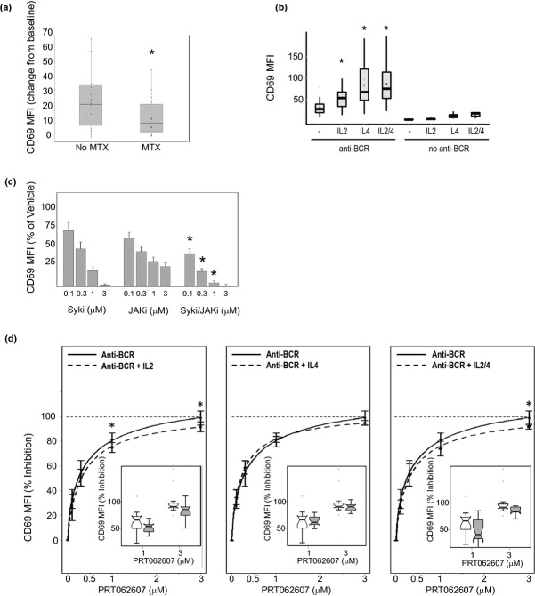
Background: Selective disruption of the spleen tyrosine kinase (Syk) represents a novel strategy to control B-cell functional responses by inhibition of B-cell antigen receptor (BCR) signaling. PRT062607 (P505-15) is a highly selective small molecule Syk inhibitor that potently suppresses B-cell function in human and rodent blood, and reduces inflammation in rodent models of rheumatoid arthritis (RA).
Aims: In this study, we sought to determine the potency of Syk inhibition by PRT062607 in whole blood from RA patients, and elucidate covariates that affect the potency of immune-regulation by this compound.
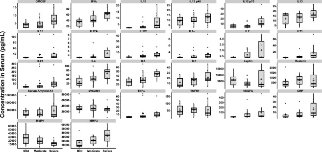
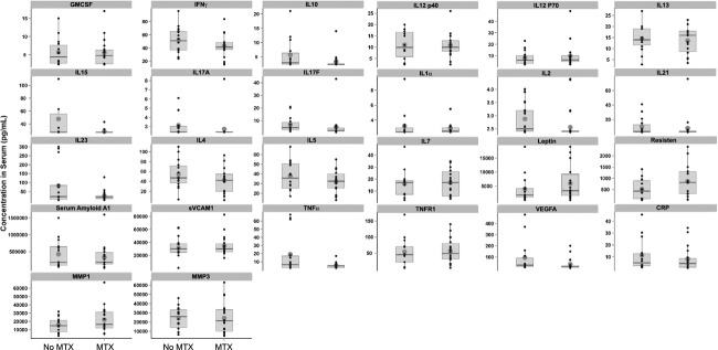
Materials and methods: Whole blood was collected from 30 patients diagnosed with RA as part of a single-center outpatient study. Disease severity, serum protein markers of inflammation, and co-medications were related to each other, and to PRT062607 activity in ex vivo Syk-mediated immune function assays.
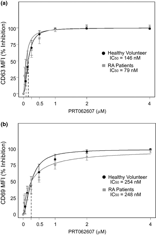
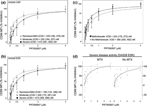
Results: We report here that PRT062607 exhibited greater potency in suppressing BCR mediated B-cell functional responses in whole blood from RA patients who received stable methotrexate (MTX) therapy. We demonstrate that the B-cell functional response to BCR ligation is influenced by cytokines and JAK/STAT signaling.
Discussion: MTX is a known cytokine modulating agent, and this mechanism may act in concert with PRT062607 to control B-cell function.
Conclusion: These data have important implications for the co-administration of Syk inhibitors and MTX for the treatment of RA.
Keywords: B cells; methotrexate; rheumatoid arthritis; spleen tyrosine kinase.
Figures
Similar articles
-
PRT062607 Achieves Complete Inhibition of the Spleen Tyrosine Kinase at Tolerated Exposures Following Oral Dosing in Healthy Volunteers.J Clin Pharmacol. 2017 Feb;57(2):194-210. doi: 10.1002/jcph.794. Epub 2016 Aug 17. J Clin Pharmacol. 2017. PMID: 27406873 Free PMC article.
-
Selective spleen tyrosine kinase inhibition delays autoimmune arthritis in mice.Mol Med Rep. 2015 Aug;12(2):2902-6. doi: 10.3892/mmr.2015.3759. Epub 2015 May 8. Mol Med Rep. 2015. PMID: 25955571
-
Activation of Syk in peripheral blood B cells in patients with rheumatoid arthritis: a potential target for abatacept therapy.Arthritis Rheumatol. 2015 Jan;67(1):63-73. doi: 10.1002/art.38895. Arthritis Rheumatol. 2015. PMID: 25303149
-
Inhibitors of switch kinase 'spleen tyrosine kinase' in inflammation and immune-mediated disorders: a review.Eur J Med Chem. 2013 Sep;67:434-46. doi: 10.1016/j.ejmech.2013.04.070. Epub 2013 Jul 12. Eur J Med Chem. 2013. PMID: 23917087 Review.
-
Targeting Syk in Autoimmune Rheumatic Diseases.Front Immunol. 2016 Mar 7;7:78. doi: 10.3389/fimmu.2016.00078. eCollection 2016. Front Immunol. 2016. PMID: 27014261 Free PMC article. Review.
Cited by
-
Methotrexate Combined with 4-Hydroperoxycyclophosphamide Downregulates Multidrug-Resistance P-Glycoprotein Expression Induced by Methotrexate in Rheumatoid Arthritis Fibroblast-Like Synoviocytes via the JAK2/STAT3 Pathway.J Immunol Res. 2018 Feb 18;2018:3619320. doi: 10.1155/2018/3619320. eCollection 2018. J Immunol Res. 2018. PMID: 29670920 Free PMC article.
-
Immunological evaluation of rheumatoid arthritis patients treated with itolizumab.MAbs. 2016;8(1):187-95. doi: 10.1080/19420862.2015.1105416. Epub 2015 Oct 15. MAbs. 2016. PMID: 26466969 Free PMC article. Clinical Trial.
-
PRT062607 Achieves Complete Inhibition of the Spleen Tyrosine Kinase at Tolerated Exposures Following Oral Dosing in Healthy Volunteers.J Clin Pharmacol. 2017 Feb;57(2):194-210. doi: 10.1002/jcph.794. Epub 2016 Aug 17. J Clin Pharmacol. 2017. PMID: 27406873 Free PMC article.
References
-
- Baggott JE, Morgan SL, Sams WM, Linden J. Urinary adenosine and aminoimidazolecarboxamide excretion in methotrexate-treated patients with psoriasis. Arch Dermatol. 1999;135:813–817. - PubMed
-
- Braun D, Caramalho I, Demengeot J. IFN-alpha/beta enhances BCR-dependent B cell responses. Int Immunol. 2002;14:411–419. - PubMed
LinkOut - more resources
Full Text Sources
Other Literature Sources
Miscellaneous
