. 2014 Feb;2(1):e00028.
doi: 10.1002/prp2.28. Epub 2014 Mar 3.

Database search of spontaneous reports and pharmacological investigations on the sulfonylureas and glinides-induced atrophy in skeletal muscle

Affiliations

Database search of spontaneous reports and pharmacological investigations on the sulfonylureas and glinides-induced atrophy in skeletal muscle

Antonietta Mele et al. Pharmacol Res Perspect. 2014 Feb.

Abstract

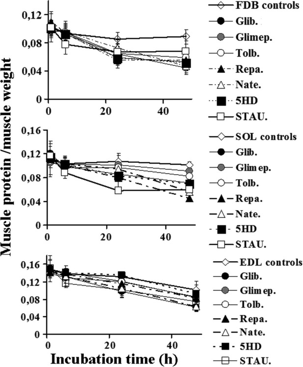
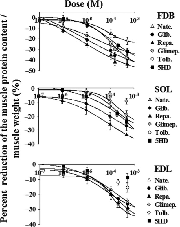
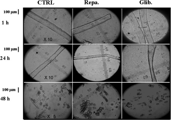
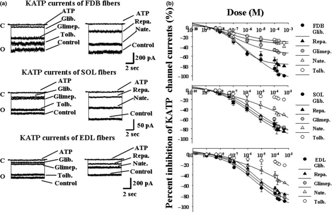
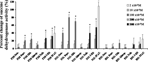
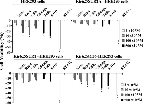
The ATP-sensitive K(+) (KATP) channel is an emerging pathway in the skeletal muscle atrophy which is a comorbidity condition in diabetes. The "in vitro" effects of the sulfonylureas and glinides were evaluated on the protein content/muscle weight, fibers viability, mitochondrial succinic dehydrogenases (SDH) activity, and channel currents in oxidative soleus (SOL), glycolitic/oxidative flexor digitorum brevis (FDB), and glycolitic extensor digitorum longus (EDL) muscle fibers of mice using biochemical and cell-counting Kit-8 assay, image analysis, and patch-clamp techniques. The sulfonylureas were: tolbutamide, glibenclamide, and glimepiride; the glinides were: repaglinide and nateglinide. Food and Drug Administration-Adverse Effects Reporting System (FDA-AERS) database searching of atrophy-related signals associated with the use of these drugs in humans has been performed. The drugs after 24 h of incubation time reduced the protein content/muscle weight and fibers viability more effectively in FDB and SOL than in the EDL. The order of efficacy of the drugs in reducing the protein content in FDB was: repaglinide (EC50 = 5.21 × 10(-6)) ≥ glibenclamide(EC50 = 8.84 × 10(-6)) > glimepiride(EC50 = 2.93 × 10(-5)) > tolbutamide(EC50 = 1.07 × 10(-4)) > nateglinide(EC50 = 1.61 × 10(-4)) and it was: repaglinide(7.15 × 10(-5)) ≥ glibenclamide(EC50 = 9.10 × 10(-5)) > nateglinide(EC50 = 1.80 × 10(-4)) ≥ tolbutamide(EC50 = 2.19 × 10(-4)) > glimepiride(EC50=-) in SOL. The drug-induced atrophy can be explained by the KATP channel block and by the enhancement of the mitochondrial SDH activity. In an 8-month period, muscle atrophy was found in 0.27% of the glibenclamide reports in humans and in 0.022% of the other not sulfonylureas and glinides drugs. No reports of atrophy were found for the other sulfonylureas and glinides in the FDA-AERS. Glibenclamide induces atrophy in animal experiments and in human patients. Glimepiride shows less potential for inducing atrophy.

Keywords: Atrophy; oral antidiabetic drugs; pharmacovigilance; skeletal muscle.


Figures

Figure 1
Figure 1
Effects of sulfonylureas, glinides, 5-hydroxydecanoate, and staurosporine on the ratio of muscle protein content/muscle weight of different muscle types of mice at different incubation time. The right hand flexor digitorum brevis (FDB), the extensor digitorum longus (EDL), and the soleus (SOL) muscles were incubated for 1, 6, 24, and 48 h, at 37°C, under 5% CO2/95% O2 atmosphere, with DMEM+ solution enriched with the glibenclamide (Glib.) (10−4 mol/L), glimepiride (Glimep.) (10−4 mol/L), tolbutamide (Tolb.) (5 × 10⁻⁴ mol/L), nateglinide (Nate.) (10−4 mol/L), repaglinide (Repa.) (10−4 mol/L), 5-hydroxydecanoate (5HD) (5 × 10⁻⁴ mol/L), and staurosporine (STAU.) (2 × 10⁻⁶ mol/L). The contralateral left hand muscles (controls) isolated from the same mice were incubated with a DMEM+ solution in the same experimental condition and used as controls. We found a significant reduction in the protein content/muscle weight after 24 h of incubation time of FDB muscle with all drugs under investigation with respect to the controls as determined by the two-way analysis of variance (P < 00.5). Repaglinide and glibenclamide were also effective in the SOL and EDL muscles after 24 h of incubation, while glimepiride did not produce a significant reduction in this parameter in these muscle types. The 5HD was effective as atrophic agent in FDB and SOL muscles but it was less effective in EDL. The staurosporine was capable to reduce the protein content in all muscles after 6 h of incubation time. Data are expressed as means±SEM. A minimum of five sample replicates were analyzed for each set of data points.
Figure 2
Figure 2
Concentration–response relationships of sulfonylureas, glinides, and 5-hydroxydecanoate concentrations versus the percent reduction in the protein content/muscle weight in all muscle types of mice. These experiments were performed in flexor digitorum brevis (FDB), soleus (SOL), and extensor digitorum longus (EDL) muscles after 24 h of incubation time at 37°C, under 5% CO2/95% O2 atmosphere, with DMEM+ solution enriched with increasing concentrations (10−7 to 5 × 10⁻⁴ mol/L) of glibenclamide (Glib.), glimepiride (Glimep.), tolbutamide (Tolb.), nateglinide (Nate.), repaglinide (Repa.), and 5-hydroxydecanoate (5HD). These drugs were more effective in FDB and SOL rather than in the EDL muscle. Repaglinide and glibenclamide were the most effective and potent drugs in reducing the protein content in all muscles as determined by the one-way analysis of variance (P < 0.05). The data were fitted using a four-parameter logistic Hill function. Data are expressed as means ± SEM. A minimum of five sample replicates were analyzed for each data point.
Figure 3
Figure 3
Images of enzymatically isolated fibers from flexor digitorum brevis (FDB) muscles after treatment with the glibenclamide (Glib.) and repaglinide (Repa.) at different incubation time. The fiber diameter and morphology were measured at a 10× magnification; mortality was evaluated at a 5× magnification using QuantiCell 900 integrated imaging system. Isolated fibers, before analysis, were equilibrated in DMEM (300 mOsmol L) for 15 min at 25°C. Fiber diameter and mortality following incubation for 1–48 h with DMEM solution used as control and DMEM solution + glibenclamide (10−5 mol/L) and repaglinide (10−5 mol/L). No effects were observed after 1 h incubation time in the presence of the drugs. The glibenclamide and repaglinide treatments reduced the fiber diameter after 24 h of incubation with respect to the controls. The fiber mortality increased with drug treatments with respect to the control after 48 h of incubation time.
Figure 4
Figure 4
Cytoprotective effects of diazoxide against the muscle atrophy induced by glibenclamide and repaglinide in mice. The right hand flexor digitorum brevis (FDB), the extensor digitorum longus (EDL), and the soleus (SOL) muscles were incubated for 24 h, at 37°C, under 5% CO2/95% O2 atmosphere, with DMEM+ solution enriched with the diazoxide (Diazo.) (250 × 10−6 mol/L), glibenclamide (Glib.) (10−4 mol/L), repaglinide (Repa.) (10−4 mol/L), and the combination of glibenclamide or repaglinide (10−4 mol/L) and diazoxide (250 × 10−6 mol/L). The contralateral left hand muscles (controls) isolated from the same mice were incubated with a DMEM+ solution in the same experimental condition and used as controls. Diazoxide treatment, per se, did not affect the protein content in all muscles. The coincubation of the muscles with diazoxide + glibenclamide or repaglinide prevented the atrophy induced by these drugs in all muscles. The diazoxide-repaglinide or diazoxide-glibenclamide data were significantly different in respect to the glibenclamide or repaglinide data in FDB muscle. No significant differences were observed between the diazoxide-repaglinide or diazoxide-glibenclamide data and controls. The data are expressed as means ± SEM of a minimum of five sample replicates. The data (*) were significantly different with respect to the controls and (▪) to the glibenclamide or repaglinide data (P < 0.05) as determined by student t-test.
Figure 5
Figure 5
Effects of the sulfonylurea and glinides on ATP-sensitive K+ channel (KATP) current in skeletal muscle fibers of mice. (A) Sample traces of KATP channel currents recorded at −60 mV (Vm) in excised macropatches from flexor digitorum brevis (FDB), soleus (SOL), and extensor digitorum longus (EDL) muscle fibers in the presence of 150 mmol/L KCl on both sides of the membrane patches, at 25°C. The drug solutions (10−4 mol/L) were applied on the internal side of the membrane patches. The glibenclamide (Glib.), glimepiride (Glimep.), tolbutamide (Tolb.), nateglinide (Nate.), and repaglinide (Repa.) reduced the KATP channel currents with different efficacy. C, closed channel level; O, open-channel level. (B) Concentration–response relationship of the percent reduction in the KATP channel current versus drug concentrations was performed on currents recorded, at −60 mV (Vm), in excised macropatches in the presence of 150 mmol/L KCl on both sides of the membrane patches, at 25°C, in the FDB, SOL, and EDL muscle fibers. Increasing concentrations (10−9 to 5 × 10⁻⁴ mol/L) of the drug solutions were applied to the internal sides of the membrane patch. The most potent and effective drugs in reducing the KATP channel current were repaglinide and glibenclamide in all muscle fibers. Nateglinide and tolbutamide were the less potent and effective drugs in all fibers. The data were fitted using a four-parameter logistic Hill function. Data are expressed as means ± SEM of a minimum of four macropatches.
Figure 6
Figure 6
Effects of the sulfonylureas, glinides, and 5-hydroxydecanoate on dehydrogenase activity of muscles in mice. The dehydrogenase activity was expressed as the percent changes of the enzyme activity with respect to that measured in the contralateral controls. The dehydrogenase activity was measured in the flexor digitorum brevis (FDB), extensor digitorum longus (EDL), and soleus (SOL) muscles incubated for 24 h with 5-hydroxydecanoato (5-HD) (200–500 × 10−6 mol/L), glibenclamide (Glib.) (10−5 to 10−4 mol/L), tolbutamide (Tolb.) (100–500 × 10−6 mol/L), and glimepiride (Glimep.) (10−5 to 10−4 mol/L), nateglinide (Nate.) (10−5 to 10−4 mol/L), repaglinide (Repa.) (10−5 to 10−4 mol/L). All drugs were capable to significantly enhance the dehydrogenase activity in the SOL muscle with respect to the controls but not glimepiride. Glibenclamide, repaglinide, tolbutamide, and 5HD caused a mild and significant increase in the dehydrogenase activity also in the FDB muscle. All drugs did not affect the dehydrogenase activity in the EDL muscle. The apoptotic agent staurosporine (STAU.) (2 × 10⁻⁶ mol/L) significantly enhanced this parameter in all muscle types. The data (*) were significantly different with respect to the controls (P < 0.05) as determined by student t-test.
Figure 7
Figure 7
Effects of the sulfonylureas, glinides, 5-hydroxydecanoate, and staurosporine on cell viability in HEK293 cells transfected with the KATP channel subunits. The drug effects on cell viability were investigated in not transfected HEK293 cells or in cells that were expressing the Kir6.2ΔC36, Kir6.2+SUR1, and Kir6.2+SUR2A channel subunits. The cells were incubated for 24 h in the presence of glibenclamide (Glib.), glimepiride (Glimep.), repaglinide (Repa.), nateglinide (Nate.) (10–100 × 10−6 mol/L), tolbutamide (Tolb.) (500 × 10−6 mol/L), 5-hydroxydecanoate (5-HD) (500 × 10−6 mol/L), and staurosporine (STAU.) (2 × 10⁻⁶ mol/L) solutions under 5% CO2–95% O2 atmosphere for the maintenance of aerobic conditions, at 37°C. The cell viability was evaluated in the presence of the drugs using the Cell-Counting Kit-8 and expressed as% changes of cell viability with respect to the controls. The glibenclamide and repaglinide exerted a mild cytotoxicity in cells transfected with SUR1-KIR6.2 leading to a reduction in the cell viability of about 18–20% in our experimental condition. In contrast, no effects of the drugs were observed in the cell line expressing the SUR2A-Kir6.2 subunits. The observed effects of the drugs in the not transfected cells and in Kir6.2ΔC36-HEK293 cells in the absence of SUR subunits can be related with off-target effects. The data (*) were significantly different with respect to the controls (P < 0.05) as determined by student t-test.

Similar articles

Cited by

References

    1. Arnoux JB, de Lonlay P, Ribeiro MJ, Hussain K, Blankenstein O, Mohnike K, et al. Congenital hyperinsulinism. Early Hum Dev. 2010;86:287–294. - PubMed
    1. Ashcroft FM. New uses for old drugs: neonatal diabetes and sulphonylureas. Cell Metab. 2010;11:179–181. - PubMed
    1. Babenko AP, Gonzalez G, Bryan J. Pharmaco-topology of sulfonylurea receptors. Separate domains of the regulatory subunits of K(ATP) channel isoforms are required for selective interaction with K(+) channel openers. J Biol Chem. 2000;275:717–720. - PubMed
    1. Bonaldo P, Sandri M. Cellular and molecular mechanisms of muscle atrophy. Dis Model Mech. 2013;6:25–39. - PMC - PubMed
    1. De la Cadena SG, Hernández-Fonseca K, Camacho-Arroyo I, Massieu L. Glucose deprivation induces reticulum stress by the PERK pathway and caspase-7- and calpain-mediated caspase-12 activation. Apoptosis. 2013 doi: 10.1007/s10495-013-0930-7. - DOI - PubMed

LinkOut - more resources