Review
. 2014 Nov 26:2:105.
doi: 10.3389/fchem.2014.00105. eCollection 2014.

Cancer immunotherapy: nanodelivery approaches for immune cell targeting and tracking

Affiliations
Review

Cancer immunotherapy: nanodelivery approaches for immune cell targeting and tracking

João Conniot et al. Front Chem. .

Abstract

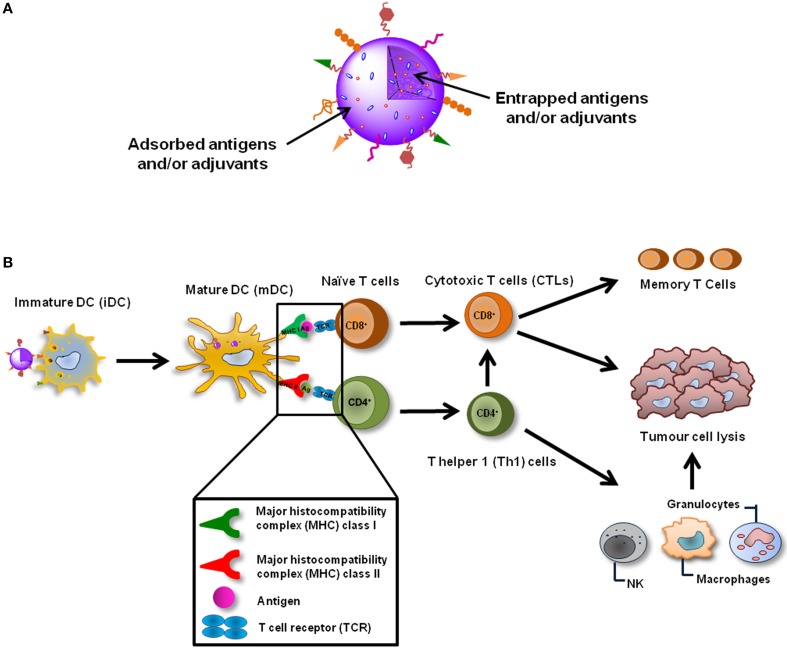
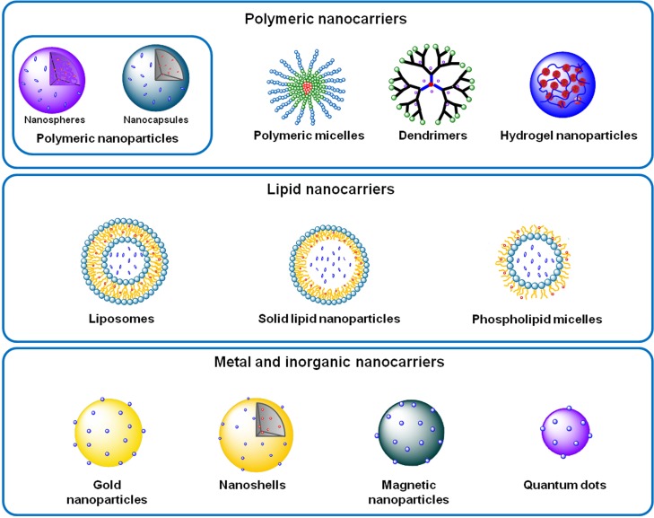
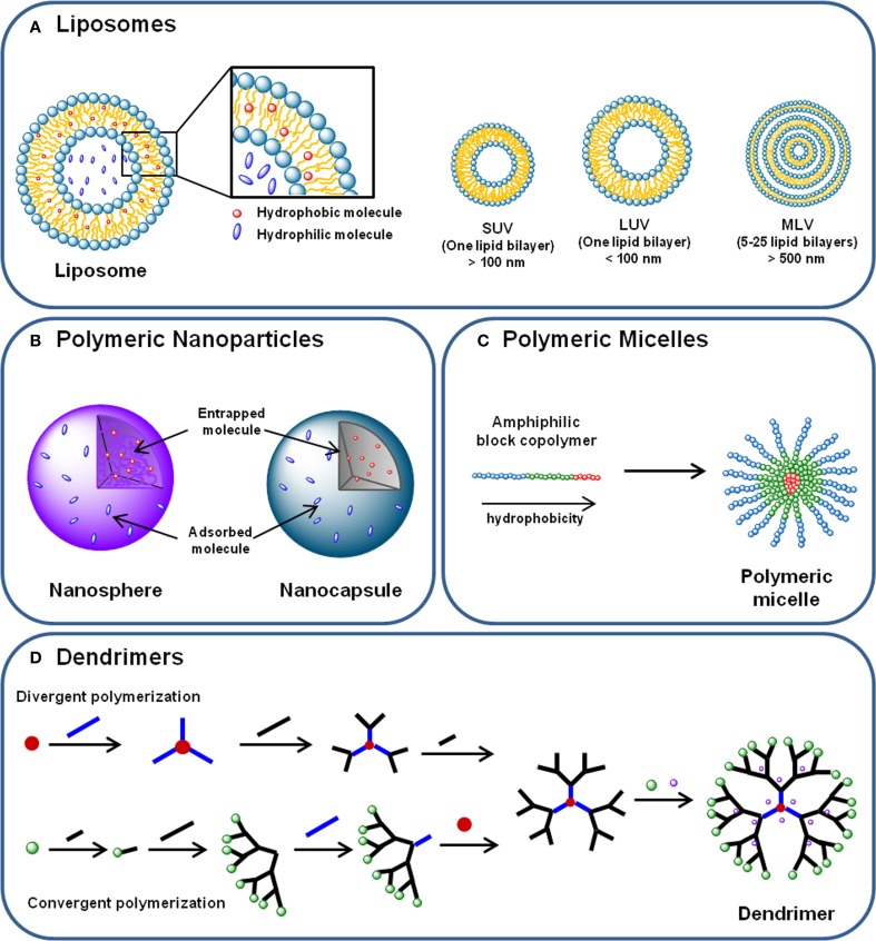
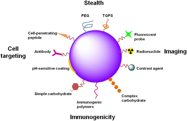
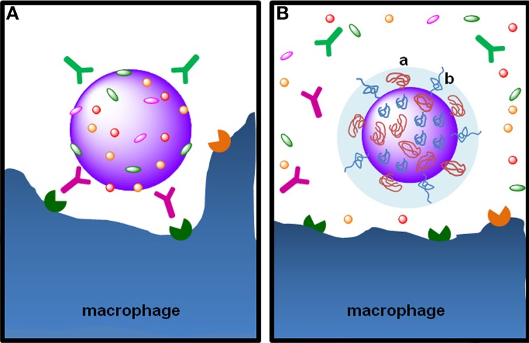
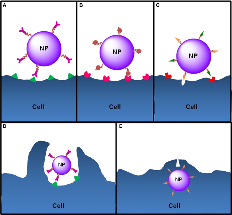
Cancer is one of the most common diseases afflicting people globally. New therapeutic approaches are needed due to the complexity of cancer as a disease. Many current treatments are very toxic and have modest efficacy at best. Increased understanding of tumor biology and immunology has allowed the development of specific immunotherapies with minimal toxicity. It is important to highlight the performance of monoclonal antibodies, immune adjuvants, vaccines and cell-based treatments. Although these approaches have shown varying degrees of clinical efficacy, they illustrate the potential to develop new strategies. Targeted immunotherapy is being explored to overcome the heterogeneity of malignant cells and the immune suppression induced by both the tumor and its microenvironment. Nanodelivery strategies seek to minimize systemic exposure to target therapy to malignant tissue and cells. Intracellular penetration has been examined through the use of functionalized particulates. These nano-particulate associated medicines are being developed for use in imaging, diagnostics and cancer targeting. Although nano-particulates are inherently complex medicines, the ability to confer, at least in principle, different types of functionality allows for the plausible consideration these nanodelivery strategies can be exploited for use as combination medicines. The development of targeted nanodelivery systems in which therapeutic and imaging agents are merged into a single platform is an attractive strategy. Currently, several nanoplatform-based formulations, such as polymeric nanoparticles, micelles, liposomes and dendrimers are in preclinical and clinical stages of development. Herein, nanodelivery strategies presently investigated for cancer immunotherapy, cancer targeting mechanisms and nanocarrier functionalization methods will be described. We also intend to discuss the emerging nano-based approaches suitable to be used as imaging techniques and as cancer treatment options.

Keywords: cancer; cell tracking; immunotherapy; nanosystems; targeted delivery.


Figures

Figure 1
Figure 1
Nanoparticulate cancer vaccines. (A) NPs are able to deliver several TAAs and adjuvants simultaneously, enabling a coordinated activation of DCs. NPs can also be functionalized in order to actively target DCs in vivo, increase their cellular internalization and immunogenicity or even target specific intracellular compartments. (B) NP-based cancer vaccines can be targeted to DCs in vivo and after their internalization induce the maturation of these cells. TAAs and adjuvants are simultaneously released inside the same DC which guaranties its coordinated activation. TAAs are presented trough MHC class I and class II molecules to CD8+ and CD4+ naïve T cells which recognize the processed antigens through TCRs. Activated CD8+ T cells differentiate into CTLs, which can destroy tumor cells, and memory T cells, that are important to avoid recidivism and metastasis. CD4+ T cells should differentiate in Th1 cells, which will potentiate the action of CTLs and will also activate cells of the innate immune system, such as NK cells, granulocytes and macrophages that play a role in the tumor destruction process as well.
Figure 2
Figure 2
Examples of polymeric, lipid, and metal and inorganic nanocarriers.
Figure 3
Figure 3
(A) Liposomes are phospholipid and cholesterol self-assembled bilayer membranes that enclose an aqueous core, where hydrophilic molecules can be incorporated. Hydrophobic compounds can also be incorporated in the lipid bilayer. Liposomes can be classified in (i) small unilamellar vesicles (SUVs); (ii) large unilamellar vesicles (LUVs) and (iii) multilamellar vesicles (MLVs), according to their size and lamellarity. (B) Polymeric nanoparticles are submicron spherical entities composed by a polymeric compact net than can either constitute a polymeric matrix—in the case of nanospheres—or a polymeric wall surrounding a vesicular core—nanocapsules. Nanoparticles can transport hydrophilic and hydrophobic molecules either entrapped in the polymeric matrix or core, or adsorbed to their surface. (C) Polymeric micelles are self-assembled spherical nanocarriers formed by amphiphilic block copolymers. In aqueous medium, the block copolymers arrange themselves in a disposition where the most hydrophobic parts of their chains form a hydrophobic core—where hydrophobic molecules can be incorporated –, and the most hydrophilic regions of the polymer chain are displayed outoward. (D) Dendrimers are hyperbranched nanocarriers formed by a central core, branching monomers and functionalized peripheral groups. Dendrimer synthesis can start from the core element (divergent polymerization) or from the peripheral branching units (convergent polymerization), resulting in a structure with a hydrophilic surface and a hydrophobic central core. Molecules can be transported by dendrimers either incorporated in the core and branches, either conjugated to the terminal groups.
Figure 4
Figure 4
Examples of NP functionalization. NPs can be functionalized differently in order to attain distinct goals. PEG or TGPS functionalization provide stealth properties to NPs, avoiding capture by phagocytic cells and increasing their circulation time. Functionalization of NPs with imaging agents, such as fluorescent probes, radionuclides or contrast agents (e.g., gold or magnetic NPs), provide applicability of NPs to diagnostic, theranostic or even in vivo real-time imaging. The immunogenicity of NPs can be increased for immunotherapy or prophylactic vaccination. Different molecules can be used for that propose, such as PAMPs (several carbohydrates, lipids or nucleic acids) or immunogenic polymers (e.g., chitosan, alginate, poloxamers). Specific tissue and cell targeting can be achieved through the functionalization of NPs with antibodies directed to specific or overexpressed antigens. Cell-penetrating peptides can improve NP internalization. pH-sensitive coatings allow drug release in specific tissues or intracellular compartments in a pH-dependent manner.
Figure 5
Figure 5
The stealth effect from NP functionalization with PEG. (A) Particulate foreign entities in body fluids are promptly covered with opsonins, such as the immunoglobulins IgG and IgA and the complement proteins C3b C4b, in a process called opsonization. Opsonins mark the particulate entity to phagocytosis through their recognition by Fc receptors on phagocytic cells, such as macrophages. (B) Functionalization of NPs with PEG by grafting, conjugation or adsorption—note the “mushroom-like” (a) or “brush-like” (b) configuration of PEG chains—provides steric stabilization and stealth properties, preventing the adsorption of opsonins at the surface of nanoparticles. PEG hydrophilicity attracts water molecules to particle surface avoiding the adsorption of opsonins at NP surface, rendering them “invisible” to phagocytic cells.
Figure 6
Figure 6
Ligand-cell interaction and NP internalization. NPs can be functionalized with different ligands to increase cell targeting and NP internalization. (A) Functionalization of NPs with antibodies allows the targeting of antigens exclusively expressed or overexpressed by target cells (e.g., anti-CD205 antibody to target CD205 on DCs or anti-HER2 antibody to target HER2 on breast cancer cells). (B) In order to target DCs, NPs can be functionalized with molecules that mimic PAMPs, normally carbohydrates, nucleic acids or lipids, which are recognized by PRRs expressed by DCs. For instance, mannose or fucose residues are recognized by the mannose receptors—a C-lectin receptor. Bacterial lipopolysaccharide or flagellin target TLR4 and TLR5 on DCs, respectively. (C) Cell-penetrating peptides are small amino acid sequences normally used by viruses or bacteria to facilitate cellular invasion by those pathogens and can be used to increase the internalization of NPs. Functionalized NPs see their internalization by target cells increased essentially by two mechanisms: induction of endocytosis upon ligand-receptor binding, which happens to NPs functionalized with ligands such as antibodies, PAMPs or some penetrating peptides that induce receptor-mediated endocytosis (e.g., integrins) or (D) through direct cell penetration across the plasma membrane (e.g., antimicrobial peptides or histidine-rich peptides) (E) or both (e.g., HIV TAT peptide).

Similar articles

Cited by

References

    1. Abraham S. A., Waterhouse D. N., Mayer L. D., Cullis P. R., Madden T. D., Bally M. B. (2005). The liposomal formulation of doxorubicin. Meth. Enzymol. 391, 71–97. 10.1016/S0076-6879(05)91004-5 - DOI - PubMed
    1. Agemy L., Sugahara K. N., Kotamraju V. R., Gujraty K., Girard O. M., Kono Y., et al. . (2010). Nanoparticle-induced vascular blockade in human prostate cancer. Blood 116, 2847–2856. 10.1182/blood-2010-03-274258 - DOI - PMC - PubMed
    1. Ahlers J. D., Belyakov I. M. (2010). Memories that last forever: strategies for optimizing vaccine T-cell memory. Blood 115, 1678–1689. 10.1182/blood-2009-06-227546 - DOI - PMC - PubMed
    1. Ahmad M., Rees R. C., Ali S. A. (2004). Escape from immunotherapy: possible mechanisms that influence tumor regression/progression. Cancer Immunol. Immunother. 53, 844–854. 10.1007/s00262-004-0540-x - DOI - PMC - PubMed
    1. Ahrens E. T., Bulte J. W. (2013). Tracking immune cells in vivo using magnetic resonance imaging. Nat. Rev. Immunol. 13, 755–763. 10.1038/nri3531 - DOI - PMC - PubMed