Review
. 2014 Nov 25:2:69.
doi: 10.3389/fcell.2014.00069. eCollection 2014.

The neural crest lineage as a driver of disease heterogeneity in Tuberous Sclerosis Complex and Lymphangioleiomyomatosis

Affiliations
Review

The neural crest lineage as a driver of disease heterogeneity in Tuberous Sclerosis Complex and Lymphangioleiomyomatosis

Sean P Delaney et al. Front Cell Dev Biol. .

Abstract

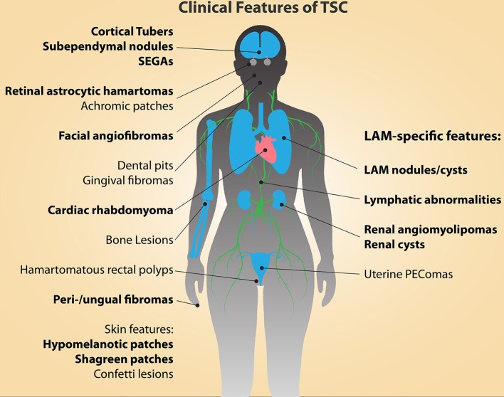
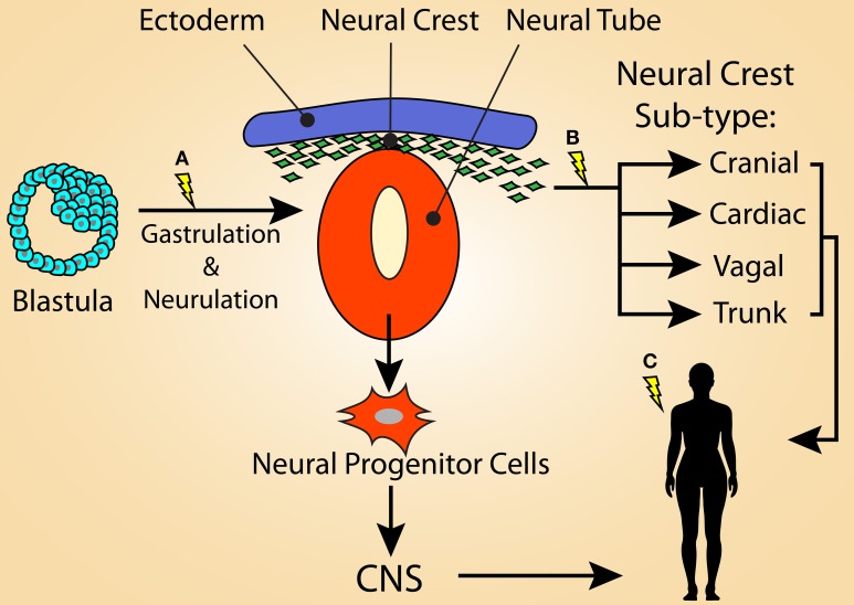
Lymphangioleiomyomatosis (LAM) is a rare neoplastic disease, best characterized by the formation of proliferative nodules that express smooth muscle and melanocytic antigens within the lung parenchyma, leading to progressive destruction of lung tissue and function. The pathological basis of LAM is associated with Tuberous Sclerosis Complex (TSC), a multi-system disorder marked by low-grade tumors in the brain, kidneys, heart, eyes, lung and skin, arising from inherited or spontaneous germ-line mutations in either of the TSC1 or TSC2 genes. LAM can develop either in a patient with TSC (TSC-LAM) or spontaneously (S-LAM), and it is clear that the majority of LAM lesions of both forms are characterized by an inactivating mutation in either TSC1 or TSC2, as in TSC. Despite this genetic commonality, there is considerable heterogeneity in the tumor spectrum of TSC and LAM patients, the basis for which is currently unknown. There is extensive clinical evidence to suggest that the cell of origin for LAM, as well as many of the TSC-associated tumors, is a neural crest cell, a highly migratory cell type with extensive multi-lineage potential. Here we explore the hypothesis that the types of tumors that develop and the tissues that are affected in TSC and LAM are dictated by the developmental timing of TSC gene mutations, which determines the identities of the affected cell types and the size of downstream populations that acquire a mutation. We further discuss the evidence to support a neural crest origin for LAM and TSC tumors, and propose approaches for generating humanized models of TSC and LAM that will allow cell of origin theories to be experimentally tested. Identifying the cell of origin and developing appropriate humanized models is necessary to truly understand LAM and TSC pathology and to establish effective and long-lasting therapeutic approaches for these patients.

Keywords: Lymphangioleiomyomatosis; Tuberous Sclerosis; cell of origin; disease modeling; neural crest.


Figures

Figure 1
Figure 1
The clinical manifestations of TSC and LAM are diverse and affect multiple organs and tissues. The major diagnostic features of TSC are indicated in bold type (Northrup et al., 2013). Cortical tubers and cardiac rhabdomyomas occur during fetal development. Facial angiofibromas, hypomelanic macules, and retinal astrocytic hamartomas can be detected in infancy, while the other features continue to present themselves throughout development into adulthood.
Figure 2
Figure 2
Model illustrating that the variation of tumor heterogeneity in TSC may be related to the stage of development in which loss of function mutations in TSC1 or TSC2 occurs. (A) For TSC patients with germline mutations, second-hit mutations may be acquired during early stages of embryogenesis, such as during gastrulation or neurulation. This would result in a large population of cells that carry the mutation and would lead to a severe TSC phenotype, possibly displaying the full spectrum of TSC manifestations. First-hit mutations at this time point would result in mosaicism. The affected cells could contribute not only to the neural crest lineage, but also the neural progenitor population of the neural tube, resulting in the CNS manifestations of TSC. (B) Second-hit mutations occurring in the emerging neural crest population likely results in many of the non-CNS symptoms of TSC, potentially including LAM. First-hit mutations at this time-point may result in S-LAM, as only the neural crest lineage would be vulnerable to second-hit mutations. It is possible that a sub-population of NCCs, such as the cardiac NC, is responsible for the more restricted lesions associated with S-LAM. (C) Alternatively, first- and/or second-hit mutations acquired within an adult NCC could also be responsible for driving LAM and some TSC-associated lesions (e.g., skin lesions).

Similar articles

Cited by

  • Notch transactivates Rheb to maintain the multipotency of TSC-null cells.
    Cho JH, Patel B, Bonala S, Manne S, Zhou Y, Vadrevu SK, Patel J, Peronaci M, Ghouse S, Henske EP, Roegiers F, Giannikou K, Kwiatkowski DJ, Mansouri H, Markiewski MM, White B, Karbowniczek M. Cho JH, et al. Nat Commun. 2017 Nov 29;8(1):1848. doi: 10.1038/s41467-017-01845-1. Nat Commun. 2017. PMID: 29184052 Free PMC article.
  • Renal neoplasms in tuberous sclerosis mice are neurocristopathies.
    Unachukwu U, Shiomi T, Goldklang M, Chada K, D'Armiento J. Unachukwu U, et al. iScience. 2021 Jun 4;24(7):102684. doi: 10.1016/j.isci.2021.102684. eCollection 2021 Jul 23. iScience. 2021. PMID: 34222844 Free PMC article.
  • The genomic landscape of tuberous sclerosis complex.
    Martin KR, Zhou W, Bowman MJ, Shih J, Au KS, Dittenhafer-Reed KE, Sisson KA, Koeman J, Weisenberger DJ, Cottingham SL, DeRoos ST, Devinsky O, Winn ME, Cherniack AD, Shen H, Northrup H, Krueger DA, MacKeigan JP. Martin KR, et al. Nat Commun. 2017 Jun 15;8:15816. doi: 10.1038/ncomms15816. Nat Commun. 2017. PMID: 28643795 Free PMC article.
  • Advances in physiological and clinical relevance of hiPSC-derived brain models for precision medicine pipelines.
    Imani Farahani N, Lin L, Nazir S, Naderi A, Rokos L, McIntosh AR, Julian LM. Imani Farahani N, et al. Front Cell Neurosci. 2025 Jan 6;18:1478572. doi: 10.3389/fncel.2024.1478572. eCollection 2024. Front Cell Neurosci. 2025. PMID: 39835290 Free PMC article. Review.
  • Lymphangioleiomyomatosis Biomarkers Linked to Lung Metastatic Potential and Cell Stemness.
    Ruiz de Garibay G, Herranz C, Llorente A, Boni J, Serra-Musach J, Mateo F, Aguilar H, Gómez-Baldó L, Petit A, Vidal A, Climent F, Hernández-Losa J, Cordero Á, González-Suárez E, Sánchez-Mut JV, Esteller M, Llatjós R, Varela M, López JI, García N, Extremera AI, Gumà A, Ortega R, Plà MJ, Fernández A, Pernas S, Falo C, Morilla I, Campos M, Gil M, Román A, Molina-Molina M, Ussetti P, Laporta R, Valenzuela C, Ancochea J, Xaubet A, Casanova Á, Pujana MA. Ruiz de Garibay G, et al. PLoS One. 2015 Jul 13;10(7):e0132546. doi: 10.1371/journal.pone.0132546. eCollection 2015. PLoS One. 2015. PMID: 26167915 Free PMC article.

References

    1. Arbiser J. L., Brat D., Hunter S., D'Armiento J., Henske E. P., Arbiser Z. K., et al. . (2002). Tuberous sclerosis-associated lesions of the kidney, brain, and skin are angiogenic neoplasms. J. Am. Acad. Dermatol. 46, 376–380. 10.1067/mjd.2002.120530 - DOI - PubMed
    1. Astrinidis A., Khare L., Carsillo T., Smolarek T., Au K. S., Northrup H., et al. . (2000). Mutational analysis of the tuberous sclerosis gene TSC2 in patients with pulmonary lymphangioleiomyomatosis. J. Med. Genet. 37, 55–57. 10.1136/jmg.37.1.55 - DOI - PMC - PubMed
    1. Au K. S., Williams A. T., Roach E. S., Batchelor L., Sparagana S. P., Delgado M. R., et al. . (2007). Genotype/phenotype correlation in 325 individuals referred for a diagnosis of tuberous sclerosis complex in the United States. Genet. Med. 9, 88–100. 10.1097/GIM.0b013e31803068c7 - DOI - PubMed
    1. Aybar M. J., Nieto M. A., Mayor R. (2003). Snail precedes slug in the genetic cascade required for the specification and migration of the Xenopus neural crest. Development 130, 483–494. 10.1242/dev.00238 - DOI - PubMed
    1. Barnes E. A., Kenerson H. L., Jiang X., Yeung R. S. (2010). Tuberin regulates E-cadherin localization: implications in epithelial-mesenchymal transition. Am. J. Pathol. 177, 1765–1778. 10.2353/ajpath.2010.090233 - DOI - PMC - PubMed

LinkOut - more resources