

doi: 10.1155/2014/928079.
Epub 2014 Nov 19.
Management of posterior reversible syndrome in preeclamptic women
Affiliations
- PMID: 25506009
- PMCID: PMC4254080
- DOI: 10.1155/2014/928079
Item in Clipboard
Management of posterior reversible syndrome in preeclamptic women
Case Rep Obstet Gynecol.
2014.
Abstract
Posterior reversible encephalopathy syndrome (PRES) is a neurological syndrome associated with a number of conditions including preeclampsia. It is characterized by seizures, alteration of consciousness, visual disturbances, and symmetric white matter abnormalities, typically in the posterior parietooccipital regions of the cerebral hemispheres, at computed tomography (CT) and magnetic resonance (MRI). We report three new cases of PRES in preeclamptic patients and describe the management of these patients. We present a brief review of other cases in the literature, with particular attention to the anesthetic management.
Figures
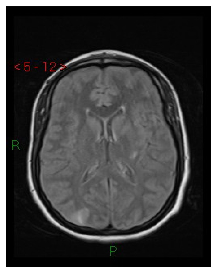
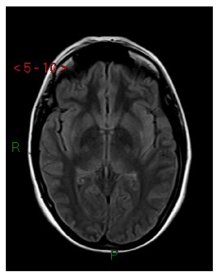
Case 1. Axial FLAIR MRI: cortical-subcortical hyperintense lesions in parietal-occipital regions and in the posterior lateral left putamen.
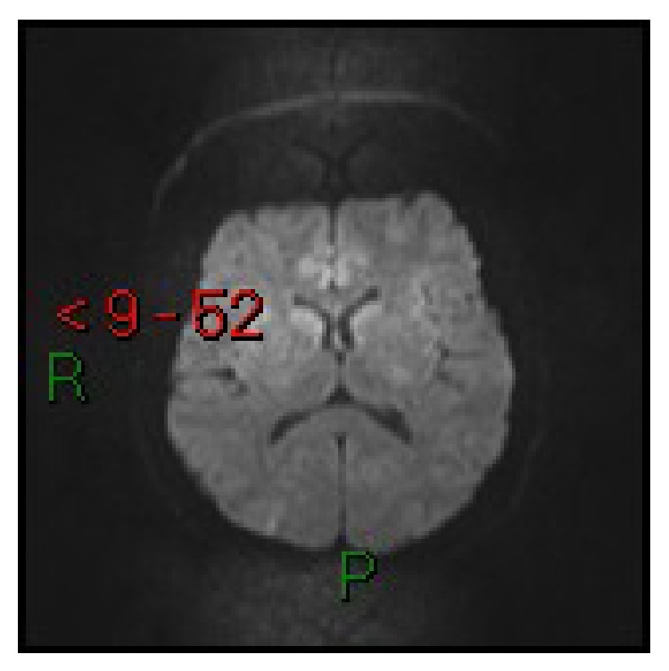
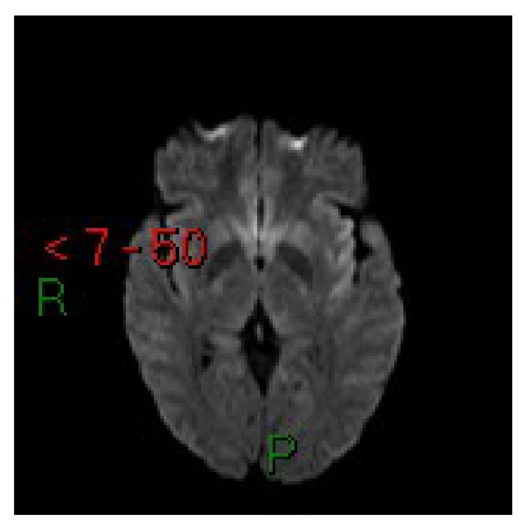
Case 1. Axial diffusion-weighted MRI: lesions do not present diffusion restriction.
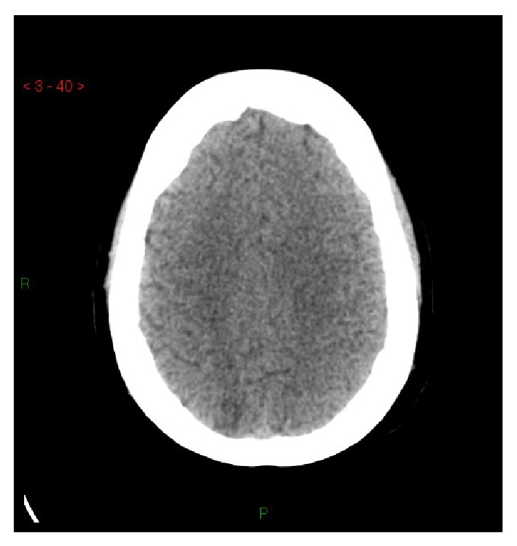
Case 2. Basal CT: hypodense cortical-subcortical lesions of the medial parietal region.
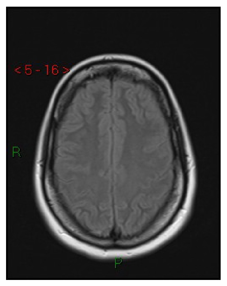
Case 2. Axial T2-FLAIR MRI: absence of lesions after 7 days.
Case 3. Axial T2-FLAIR MRI: cortical-subcortical hyperintense lesions at the insular cortex and bilaterally at the lateral parts of the putamen.
Case 3. Axial diffusion-weighted MRI: lesions showed a slight signal elevation.
References
LinkOut - more resources
Full Text Sources
Other Literature Sources
