

Immunoliposome co-delivery of bufalin and anti-CD40 antibody adjuvant induces synergetic therapeutic efficacy against melanoma
- PMID: 25506218
- PMCID: PMC4260685
- DOI: 10.2147/IJN.S73651
Immunoliposome co-delivery of bufalin and anti-CD40 antibody adjuvant induces synergetic therapeutic efficacy against melanoma
Abstract
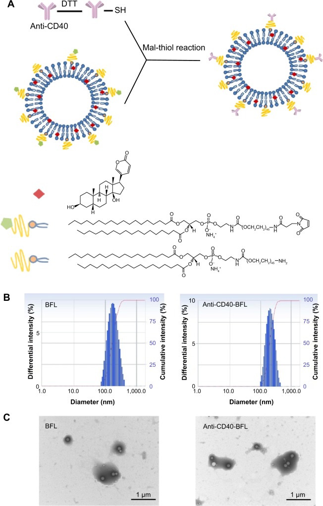
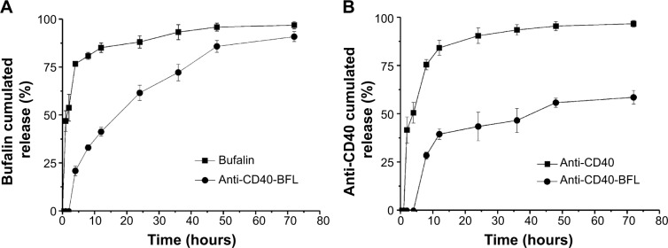
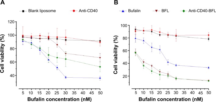
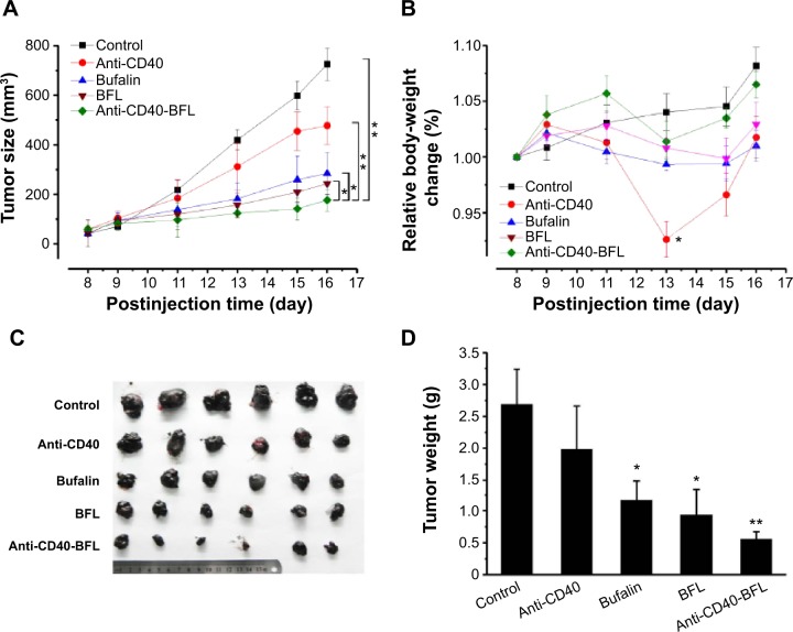
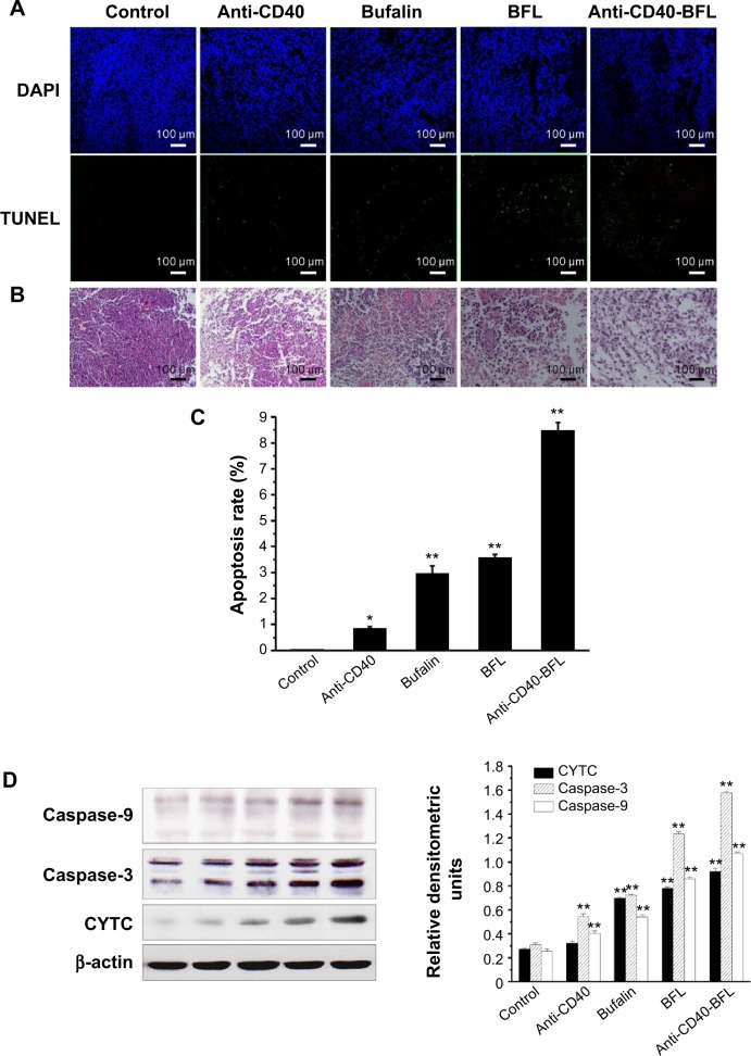
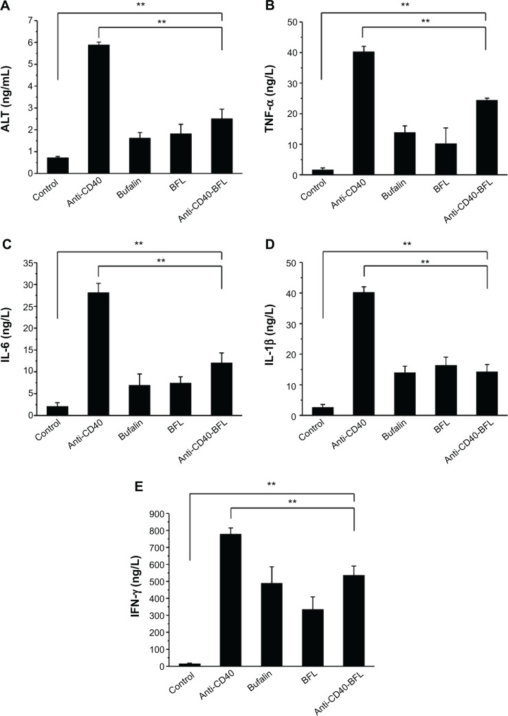
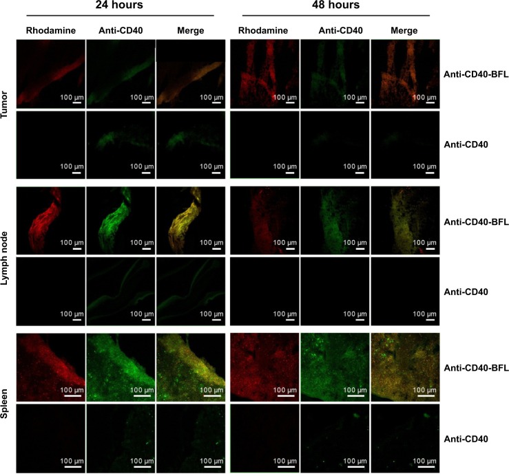
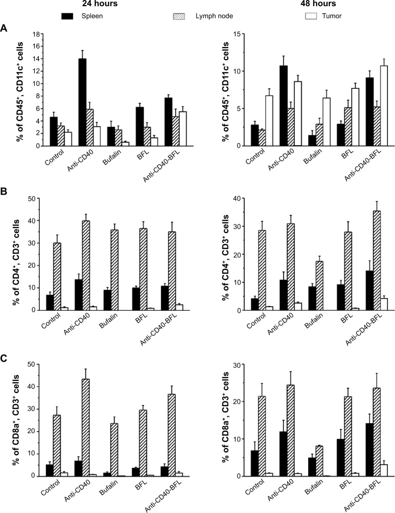
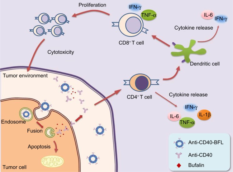
Liposomes constitute one of the most popular nanocarriers for improving the delivery and efficacy of agents in cancer patients. The purpose of this study was to design and evaluate immunoliposome co-delivery of bufalin and anti-CD40 to induce synergetic therapeutic efficacy while eliminating systemic side effects. Bufalin liposomes (BFL) conjugated with anti-CD40 antibody (anti-CD40-BFL) showed enhanced cytotoxicity compared with bufalin alone. In a mouse B16 melanoma model, intravenous injection of anti-CD40-BFL achieved smaller tumor volume than did treatment with BFL (average: 117 mm(3) versus 270 mm(3), respectively); the enhanced therapeutic efficacy through a caspase-dependent pathway induced apoptosis, which was confirmed using terminal deoxynucleotidyl transferase-mediated dUTP-Fluorescein nick end labeling and Western blot assay. Meanwhile, anti-CD40-BFL elicited unapparent body-weight changes and a significant reduction in serum levels of tumor necrosis factor-α, interleukin-1β, interleukin-6, interferon-γ, and hepatic enzyme alanine transaminase, suggesting minimized systemic side effects. This may be attributed to the mechanism by which liposomes are retained within the tumor site for an extended period of time, which is supported by the following biodistribution and flow cytometric analyses. Taken together, the results demonstrated a highly promising strategy for liposomal vehicle transport of anti-CD40 plus bufalin that can be used to enhance antitumor effects via synergetic systemic immunity while blocking systemic toxicity.
Keywords: anti-CD40; bufalin; chemoimmunotherapy; liposomes.
Figures
Similar articles
-
Locally delivered CD40 agonist antibody accumulates in secondary lymphoid organs and eradicates experimental disseminated bladder cancer.Cancer Immunol Res. 2014 Jan;2(1):80-90. doi: 10.1158/2326-6066.CIR-13-0067. Epub 2013 Oct 21. Cancer Immunol Res. 2014. PMID: 24778163
-
Induction of potent anti-tumor responses while eliminating systemic side effects via liposome-anchored combinatorial immunotherapy.Biomaterials. 2011 Aug;32(22):5134-47. doi: 10.1016/j.biomaterials.2011.03.067. Epub 2011 Apr 22. Biomaterials. 2011. PMID: 21514665 Free PMC article.
-
Anti-VEGF antibody enhances the antitumor effect of CD40.Int J Cancer. 2014 Oct 15;135(8):1983-8. doi: 10.1002/ijc.28833. Epub 2014 Mar 14. Int J Cancer. 2014. PMID: 24604357
-
Amphoteric liposomes enable systemic antigen-presenting cell-directed delivery of CD40 antisense and are therapeutically effective in experimental arthritis.Arthritis Rheum. 2009 Apr;60(4):994-1005. doi: 10.1002/art.24434. Arthritis Rheum. 2009. PMID: 19333921
-
Bufalin induces G0/G1 phase arrest through inhibiting the levels of cyclin D, cyclin E, CDK2 and CDK4, and triggers apoptosis via mitochondrial signaling pathway in T24 human bladder cancer cells.Mutat Res. 2012 Apr 1;732(1-2):26-33. doi: 10.1016/j.mrfmmm.2011.09.010. Epub 2012 Jan 20. Mutat Res. 2012. PMID: 22285700
Cited by
-
Tumor-Targeted Delivery of Bufalin-Loaded Modified Albumin-Polymer Hybrid for Enhanced Antitumor Therapy and Attenuated Hemolysis Toxicity and Cardiotoxicity.AAPS PharmSciTech. 2021 Apr 20;22(4):137. doi: 10.1208/s12249-021-02000-2. AAPS PharmSciTech. 2021. PMID: 33880681
-
Nanoparticle Design Strategies for Effective Cancer Immunotherapy.J Biomed (Syd). 2017;2(2):64-77. doi: 10.7150/jbm.18877. J Biomed (Syd). 2017. PMID: 28503405 Free PMC article.
-
Tumor-Targeting Polymer-Drug Conjugate for Liver Cancer Treatment In Vitro.Polymers (Basel). 2022 Oct 25;14(21):4515. doi: 10.3390/polym14214515. Polymers (Basel). 2022. PMID: 36365509 Free PMC article.
-
Delivery of sodium morrhuate to hemangioma endothelial cells using immunoliposomes conjugated with anti-VEGFR2/KDR antibody.Int J Nanomedicine. 2017 Sep 19;12:6963-6972. doi: 10.2147/IJN.S144056. eCollection 2017. Int J Nanomedicine. 2017. PMID: 29033564 Free PMC article.
-
Advances in Lipid-Based Nanoparticles for Cancer Chemoimmunotherapy.Pharmaceutics. 2021 Apr 9;13(4):520. doi: 10.3390/pharmaceutics13040520. Pharmaceutics. 2021. PMID: 33918635 Free PMC article. Review.
References
-
- Xie CM, Chan WY, Yu S, Zhao J, Cheng CH. Bufalin induces autophagy-mediated cell death in human colon cancer cells through reactive oxygen species generation and JNK activation. Free Radic Biol Med. 2011;51(7):1365–1375. - PubMed
-
- Hsu CM, Tsai Y, Wan L, Tsai FJ. Bufalin induces G2/M phase arrest and triggers autophagy via the TNF, JNK, BECN-1 and ATG8 pathway in human hepatoma cells. Int J Oncol. 2013;43(1):338–348. - PubMed
-
- Tian X, Luo Y, Yan YB, Sui CG, Meng FD, Liu YP. Effect of bufalin on cellular proliferation and apoptosis in human esophageal squamous carcinoma EC9706 cells. Zhongguo Yi Xue Ke Xue Yuan Xue Bao. 2012;34(6):556–562. Chinese. - PubMed
-
- Pan S, Wang Y, Feng L, et al. Study on proteomics of Hela cell apoptosis in bufalin-induced human cervical carcinoma. Zhongguo Zhong Yao Za Zhi. 2012;37(13):1998–2004. Chinese. - PubMed
Publication types
MeSH terms
Substances
LinkOut - more resources
Full Text Sources
Other Literature Sources
Medical
Research Materials
