. 2014:2:14011.
doi: 10.1038/boneres.2014.11.

The heterodimeric structure of heterogeneous nuclear ribonucleoprotein C1/C2 dictates 1,25-dihydroxyvitamin D-directed transcriptional events in osteoblasts

Affiliations

The heterodimeric structure of heterogeneous nuclear ribonucleoprotein C1/C2 dictates 1,25-dihydroxyvitamin D-directed transcriptional events in osteoblasts

Thomas S Lisse et al. Bone Res. 2014.

Abstract

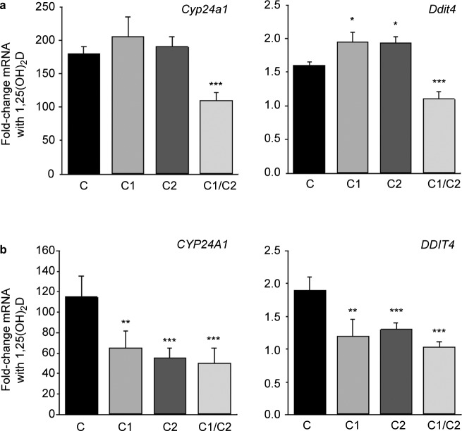
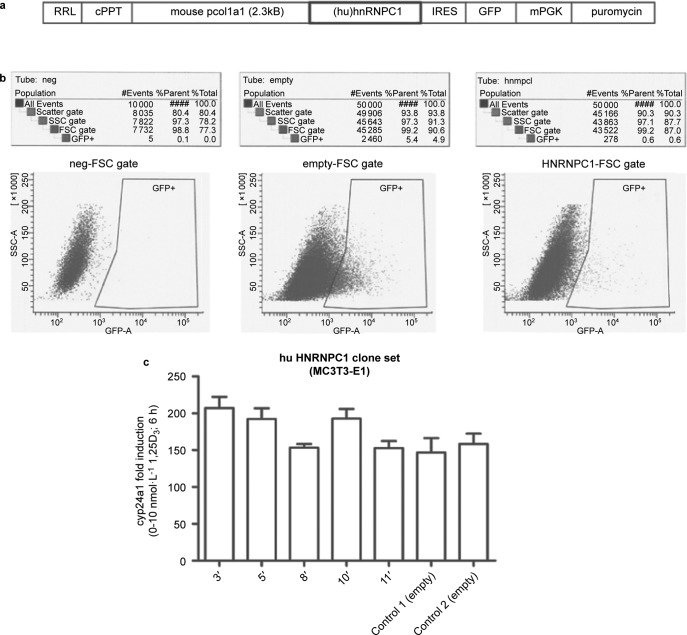
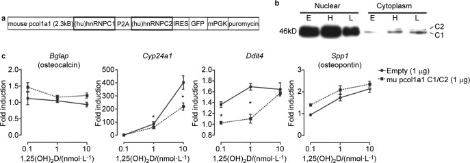
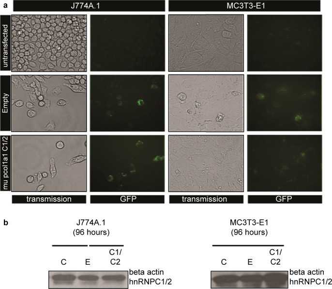
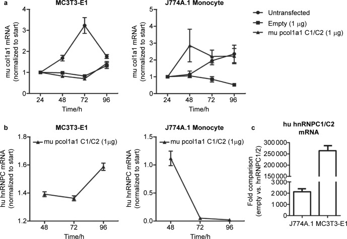
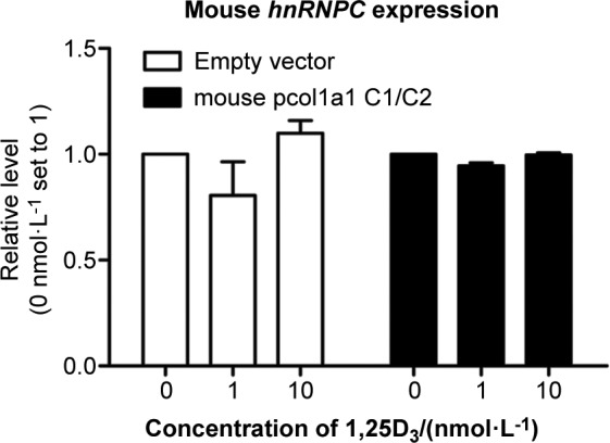
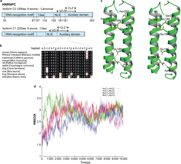
Heterogeneous nuclear ribonucleoprotein (hnRNP) C plays a key role in RNA processing. More recently hnRNP C has also been shown to function as a DNA binding protein exerting a dominant-negative effect on transcriptional responses to the vitamin D hormone,1,25-dihydroxyvitamin D (1,25(OH)2D), via interaction in cis with vitamin D response elements (VDREs). The physiologically active form of human hnRNPC is a tetramer of hnRNPC1 (huC1) and C2 (huC2) subunits known to be critical for specific RNA binding activity in vivo, yet the requirement for heterodimerization of huC1 and C2 in DNA binding and downstream action is not well understood. While over-expression of either huC1 or huC2 alone in mouse osteoblastic cells did not suppress 1,25(OH)2D-induced transcription, over-expression of huC1 and huC2 in combination using a bone-specific polycistronic vector successfully suppressed 1,25(OH)2D-mediated induction of osteoblast target gene expression. Over-expression of either huC1 or huC2 in human osteoblasts was sufficient to confer suppression of 1,25(OH)2D-mediated transcription, indicating the ability of transfected huC1 and huC2 to successfully engage as heterodimerization partners with endogenously expressed huC1 and huC2. The failure of the chimeric combination of mouse and human hnRNPCs to impair 1,25(OH)2D-driven gene expression in mouse cells was structurally predicted, owing to the absence of the last helix in the leucine zipper (LZ) heterodimerization domain of hnRNPC gene product in lower species, including the mouse. These results confirm that species-specific heterodimerization of hnRNPC1 and hnRNPC2 is a necessary prerequisite for DNA binding and down-regulation of 1,25(OH)2D-VDR-VDRE-directed gene transactivation in osteoblasts.


Figures

Figure 1
Figure 1
Effects of overexpressed human hnRNPC1 and C2 on VDR-mediated gene transcription in human and mouse osteoblast-like cells. Effect of overexpression of human hnRNPC1 (C1) and/or human hnRNPC2 (C2) on 1,25(OH)2D-induction of the vitamin D-target genes CYP24A1/Cyp24a1 and DDIT4/Ddit4 in (a) mouse MC3T3 osteoblastic cells and (b) human MG-63 osteoblastic cells. Cells were treated with or without 10 nmol⋅L−1 1,25(OH)2D for 6 h, 24 h post transfection with either empty vector (c), C1, C2 or C1 and C2 (C1/C2). Data show mean±s.d. (n=3 separate cultures) fold-change in mRNA expression for CYP24A1/Cyp24a1 and DDIT4/Ddit4 for each transfected cell type following treatment with 1,25(OH)2D relative to vehicle-treated control cells. ***P<0.001, statistically different from empty vector controls (C), **P<0.01, statistically different from C.
Figure 2
Figure 2
Stable expression of only human hnRNPC1 protein in mouse MC3T3-E1 osteoblasts does not confer resistance to 1,25(OH)2D. (a) Osteoblastic transduction was performed using the lentivirus RRL transfer vector harboring a stop codon-less human (hu) hnRNPC1 cDNA and a GFP cassette driven by the bone-specific mouse col1a1 2.3 kB promoter. The lentivector backbone also contains the cPPT from the HIV-1 integrase gene to increase the copy number of lentivirus integrating into the host genome to enhance viral titer. (b) FACS sorting of GFP-positive MC3T3-E1 osteoblasts transduced with the hu hnRNPC1 and empty lentivirus vectors for the isolation and generation of stable clones. (c) Fifteen hu hnRNPC1-overexpressing stable lines were created (data not shown), whereby five (clones 3″, 5″, 8″, 10″, 11″) lines with variable hnRNPC1 overexpression were further evaluated for 1,25(OH)2D (10 nmol⋅L−1, 6 h) mediated induction of endogenous mouse (mu) Cyp24a1 mRNA. cPPT, central polypurine tract.
Figure 3
Figure 3
Equimolar expression of human hnRNPC1 and C2 confers resistance to 1,25(OH)2D in mouse MC3T3-E1 osteoblasts. (a) Schematic representation of the mouse col1a1 2.3 kB promoter-driven piconoviral P2A sequence construct for the co-expression of human (hu) hnRNPC1 and C2 proteins with a GFP reporter. (b) Western blot analyses showing overexpression of hnRNPC1 and C2 proteins using the P2A construct enriched in the nuclear fraction of MC3T3-E1 cells at 96 h post transfection. Empty vector (E), and high (H, 2 μg per well) and low (L, 0.1 μg per well) levels of P2A expression vector cDNA were transfected into MC3T3 cells. (c) 1,25(OH)2D (0.1–10 nmol⋅L−1; 6 h) induction of mouse osteoblastic genes after transfection of empty vector or hnRNPC1/C2 P2A expression construct in MC3T3-E1 96 h post transfection.
Figure 4
Figure 4
Efficient transfection of the P2A hnRNPC1/C2 construct. (a) Transient transfection of P2A hnRNPC1/C2 construct with IRES-GFP reporter shows efficient labeling of both J774A.1 mouse monocytic and MC3T3-E1 osteoblastic cells after 96 h post transfection. The empty vector control also depicts efficient and comparable GFP labeling. (b) Western analysis of whole cell lysate for beta-actin and hnRNPC1/C2 expression 96 h post transfection.
Figure 5
Figure 5
Cell type-specific action and temporal regulation of the P2A hnRNPC1/C2 construct. (a) Endogenous mouse (mu) type I collagen expression was monitored over a 96-h period under the different transient transfection conditions. For both transfected constructs into MC3T3-E1 cells, col1a1 mRNA expression was delayed when compared to the untransfected controls. In J774A.1 mouse monocytes the expression of endogenous mu type I collagen was variable between the transfected P2A construct and untransfected control, and unregulated in the empty vector control. (b) Human (hu) hnRNPC mRNA levels were monitored in the mouse col1a1 promoter-driven P2A construct-transfected cells at 96 h post transfection. Hu hnRNPC mRNA was temporally regulated in a similar fashion as mu type I collagen in MC3T3-E1 osteoblasts. No temporal regulation of huhnRNPC mRNA was observed in J774A.1 mu monocytes transfected with the P2A construct, suggesting the mu col1a1 2.3 kB promoter was bone-specific. (c) After 96 h of transfection, MC3T3-E1 cells harboring the P2A hnRNPC1/C2 construct express huhnRNPC with mRNA levels approximately 125-fold higher relative to the matching empty vector control when compared to the J774A.1 mouse monocytic cell line.
Figure 6
Figure 6
Endogenous mouse hnRNPC not responsible for functional effects. Endogenous mouse (mu) hnRNPC expression was monitored in MC3T3-E1 cells after transfection and 6-h treatment with various concentrations of 1,25(OH)2D.
Figure 7
Figure 7
hnRNPC1/C2 tetramer stability based on species-specific amino-acid substitutions. (a) Schematic representation of human hnRNPC isoforms. Each protein contains a single RRM, a delineated NLS, a bZLM and an encompassing acidic auxiliary domain. hnRNPC2 contains an additional 13 aa and is expressed at one-third the level of hnRNPC1. An oligomerization domain (CLZ) is also present in C proteins. Numbers represent aa position. (b) Alignment of CLZ sequences derived from the Ensembl database (http://www.ensembl.org). Key asparagine (N) to serine (S) change within the mouse sequences occupying an interhelical contact surface heptad residue within the coiled coil core of hnRNP C. Similar residues are highlighted in black, while discrepant residues are highlighted in white. The heptad arrangement is in italics, with the key intermolecular residues forming antiparallel chain contacts underlined and bolded. Numbering based on the human C2 full-length isoform. (c) The hnRNPC protein CLZ tetramers of Hu and Mu C1 and C2 proteins. HuC13–HuC21 (left panel) and MuC13–HuC21 (right panel). The Asn200, Ser200 and Glu186 residues are shown in stick representation. In HuC13–HuC21 CLZ domains, pairs from the adjacent helices make four hydrogen bonds in the coiled coil tetramer. In contrast, in the MuC13–HuC21 tetramer, only one hydrogen bond is possible. The hydrogen bonds formed between Asn200 and Glu186 are shown in dashed lines. (d) Molecular dynamic simulations for CLZ tetramers. The root mean square fluctuations for the backbone atoms of the tetramer complexes for different combinations of human (Hu) and mouse (Mu) hnRNPC1 and C2. HuC13–HuC21, red; HuC13–MuC21, green; MuC13–HuC21, blue; and MuC13–MuC21, magenta. Data are based on experimental structure over 10-ns simulations. bZLM, basic leucine zipper-like motif; NLS, nuclear localization signal; RRM, RNA recognition motif.

References

    1. Carlberg C, Campbell MJ. Vitamin D receptor signaling mechanisms: integrated actions of a well-defined transcription factor. Steroids. 2013;78:127–136. - PMC - PubMed
    1. Pike JW, Meyer MB. The vitamin D receptor: new paradigms for the regulation of gene expression by 1, 25-dihydroxyvitamin D3. Rheum Dis Clin North Am. 2012;38:13–27. - PMC - PubMed
    1. Haussler MR, Whitfield GK, Kaneko I. Molecular mechanisms of vitamin d action. Calcif Tissue Int. 2013;92:77–98. - PubMed
    1. Haussler MR, Haussler CA, Whitfield GK. The nuclear vitamin D receptor controls the expression of genes encoding factors which feed the “Fountain of Youth” to mediate healthful aging. J Steroid Biochem Mol Biol. 2010;121:88–97. - PMC - PubMed
    1. Chen H, Hu B, Allegretto EA, Adams JS. The vitamin D response element-binding protein. A novel dominant-negative regulator of vitamin D-directed transactivation. J Biol Chem. 2000;275:35557–35564. - PubMed