

3D dynamic culture of rabbit articular chondrocytes encapsulated in alginate gel beads using spinner flasks for cartilage tissue regeneration
- PMID: 25506593
- PMCID: PMC4260432
- DOI: 10.1155/2014/539789
3D dynamic culture of rabbit articular chondrocytes encapsulated in alginate gel beads using spinner flasks for cartilage tissue regeneration
Abstract
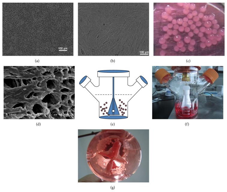
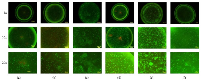
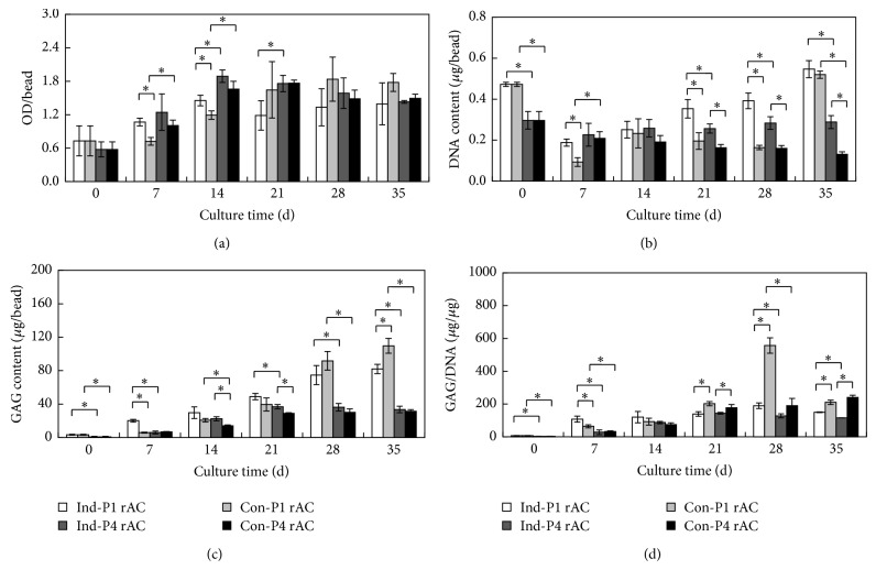
Cell-based therapy using chondrocytes for cartilage repair suffers from chondrocyte dedifferentiation. In the present study, the effects of an integrated three-dimensional and dynamic culture on rabbit articular chondrocytes were investigated. Cells (passages 1 and 4) were encapsulated in alginate gel beads and cultured in spinner flasks in chondrogenic and chondrocyte growth media. Subcutaneous implantation of the cell-laden beads was performed to evaluate the ectopic chondrogenesis. It was found that cells remained viable after 35 days in the three-dimensional dynamic culture. Passage 1 cells demonstrated a proliferative growth in both media. Passage 4 cells showed a gradual reduction in DNA content in growth medium, which was attenuated in chondrogenic medium. Deposition of glycosaminoglycans (GAG) was found in all cultures. While passage 1 cells generally produced higher amounts of GAG than passage 4 cells, GAG/DNA became similar on day 35 for both cells in growth media. Interestingly, GAG/DNA in growth medium was greater than that in chondrogenic medium for both cells. Based on GAG quantification and gene expression analysis, encapsulated passage 1 cells cultured in growth medium displayed the best ectopic chondrogenesis. Taken together, the three-dimensional and dynamic culture for chondrocytes holds great potential in cartilage regeneration.
Figures


Similar articles
-
Induced Redifferentiation of Human Chondrocytes from Articular Cartilage Lesion in Alginate Bead Culture After Monolayer Dedifferentiation: An Alternative Cell Source for Cell-Based Therapies?Tissue Eng Part A. 2018 Feb;24(3-4):275-286. doi: 10.1089/ten.TEA.2016.0505. Epub 2017 Jul 19. Tissue Eng Part A. 2018. PMID: 28610480
-
Autologous nasal chondrocytes delivered by injectable hydrogel for in vivo articular cartilage regeneration.Cell Tissue Bank. 2018 Mar;19(1):35-46. doi: 10.1007/s10561-017-9649-y. Epub 2017 Aug 16. Cell Tissue Bank. 2018. PMID: 28815373 Free PMC article.
-
Mesenchymal stem cells downregulate articular chondrocyte differentiation in noncontact coculture systems: implications in cartilage tissue regeneration.Stem Cells Dev. 2013 Jun 1;22(11):1657-69. doi: 10.1089/scd.2012.0605. Epub 2013 Feb 15. Stem Cells Dev. 2013. PMID: 23301843
-
Advances in autologous chondrocyte implantation and related techniques for cartilage repair.Dan Med J. 2013 Apr;60(4):B4600. Dan Med J. 2013. PMID: 23651721 Review.
-
Current in vitro culture systems for Pneumocystis.FEMS Immunol Med Microbiol. 1998 Sep;22(1-2):169-72. doi: 10.1111/j.1574-695X.1998.tb01202.x. FEMS Immunol Med Microbiol. 1998. PMID: 9792076 Review.
Cited by
-
Non-contact Coculture Reveals a Comprehensive Response of Chondrocytes Induced by Mesenchymal Stem Cells Through Trophic Secretion.Tissue Eng Regen Med. 2017 Sep 27;15(1):37-48. doi: 10.1007/s13770-017-0084-8. eCollection 2018 Feb. Tissue Eng Regen Med. 2017. PMID: 30603533 Free PMC article.
-
Multiple Cell Cultures for MRI Analysis.Int J Mol Sci. 2022 Sep 3;23(17):10109. doi: 10.3390/ijms231710109. Int J Mol Sci. 2022. PMID: 36077507 Free PMC article. Review.
-
Mesenchymal Stem Cells Reshape and Provoke Proliferation of Articular Chondrocytes by Paracrine Secretion.Sci Rep. 2016 Sep 6;6:32705. doi: 10.1038/srep32705. Sci Rep. 2016. PMID: 27596239 Free PMC article.
-
The Application of Bioreactors for Cartilage Tissue Engineering: Advances, Limitations, and Future Perspectives.Stem Cells Int. 2021 Jan 21;2021:6621806. doi: 10.1155/2021/6621806. eCollection 2021. Stem Cells Int. 2021. PMID: 33542736 Free PMC article. Review.
-
Calcium/Cobalt Alginate Beads as Functional Scaffolds for Cartilage Tissue Engineering.Stem Cells Int. 2016;2016:2030478. doi: 10.1155/2016/2030478. Epub 2016 Jan 6. Stem Cells Int. 2016. PMID: 27057167 Free PMC article.
References
-
- Redman S. N., Oldfield S. F., Archer C. W., Roughley P. J., Lee C. Current strategies for articular cartilage repair. European Cells and Materials. 2005;9:23–32. - PubMed
-
- Knutsen G., Drogset J. O., Engebretsen L., Grøntvedt T., Isaksen V., Ludvigsen T. C., Roberts S., Solheim E., Strand T., Johansen O. A randomized trial comparing autologous chondrocyte implantation with microfracture: findings at five years. Journal of Bone and Joint Surgery A. 2007;89(10):2105–2112. doi: 10.2106/JBJS.G.00003. - DOI - PubMed
Publication types
MeSH terms
Substances
LinkOut - more resources
Full Text Sources
Other Literature Sources
