Review
. 2015 Feb;93(2):97-110.
doi: 10.1139/cjpp-2014-0361. Epub 2014 Nov 25.

Interaction between endoplasmic/sarcoplasmic reticulum stress (ER/SR stress), mitochondrial signaling and Ca(2+) regulation in airway smooth muscle (ASM)

Affiliations
Review

Interaction between endoplasmic/sarcoplasmic reticulum stress (ER/SR stress), mitochondrial signaling and Ca(2+) regulation in airway smooth muscle (ASM)

Philippe Delmotte et al. Can J Physiol Pharmacol. 2015 Feb.

Abstract

Airway inflammation is a key aspect of diseases such as asthma. Several inflammatory cytokines (e.g., TNFα and IL-13) increase cytosolic Ca(2+) ([Ca(2+)]cyt) responses to agonist stimulation and Ca(2+) sensitivity of force generation, thereby enhancing airway smooth muscle (ASM) contractility (hyper-reactive state). Inflammation also induces ASM proliferation and remodeling (synthetic state). In normal ASM, the transient elevation of [Ca(2+)]cyt induced by agonists leads to a transient increase in mitochondrial Ca(2+) ([Ca(2+)]mito) that may be important in matching ATP production with ATP consumption. In human ASM (hASM) exposed to TNFα and IL-13, the transient increase in [Ca(2+)]mito is blunted despite enhanced [Ca(2+)]cyt responses. We also found that TNFα and IL-13 induce reactive oxidant species (ROS) formation and endoplasmic/sarcoplasmic reticulum (ER/SR) stress (unfolded protein response) in hASM. ER/SR stress in hASM is associated with disruption of mitochondrial coupling with the ER/SR membrane, which relates to reduced mitofusin 2 (Mfn2) expression. Thus, in hASM it appears that TNFα and IL-13 result in ROS formation leading to ER/SR stress, reduced Mfn2 expression, disruption of mitochondrion-ER/SR coupling, decreased mitochondrial Ca(2+) buffering, mitochondrial fragmentation, and increased cell proliferation.

Keywords: ER/SR stress; airway; asthma; asthme; inflammation; réponse aux protéines dépliées; stress du RE/RS; unfolded protein response; voies respiratoires.


Figures

Fig. 1
Fig. 1
Inflammation plays a central role in asthma pathophysiology inducing both increased cell proliferation (synthetic state) and an enhanced contractile response to agonist stimulation (hyper-reactive state) in airway smooth muscle (ASM). We hypothesize that increased ASM cell proliferation is associated with decreased mitofusin-2 (Mfn2) expression and mitochondrial fragmentation, while ASM hyper-reactivity results from an increased [Ca2+]cyt due to an uncoupling of mitochondria from the endoplasmic reticulum/sarcoplasmic reticulum membrane (decreased Mfn2 expression) and decreased mitochondrial Ca2+ buffering (reduced [Ca2+]mito response).
Fig. 2
Fig. 2
Exposure to tumor necrosis factor α (TNFα; 20 ng/mL for 24 h) increases the isometric force response of permeabilized (Triton X-100; 1%) porcine tracheal smooth muscle activated by 10 µmol·L−1 free Ca2+ (n = 4).
Fig. 3
Fig. 3
In a mouse lung slice, tumor necrosis factor α (TNFα) exposure (20 ng/mL for 12 h) increases constriction of small bronchioles induced by 500 nmol·L−1 acetylcholine (ACh) (n = 2 mice).
Fig. 4
Fig. 4
(A) Changes in isometric force and ATP hydrolysis rate (measured via extinction of NADH fluorescence) in Triton X-100 (0.1%)-permeabilized canine tracheal smooth muscle activated by various levels of free Ca2+ (1.0 nmol·L−1 to 10.0 µmol·L−1). Note the linear relationship between isometric force and ATP hydrolysis rate. (B) Time course of changes in isometric force, ATP hydrolysis rate, and tension cost of permeabilized canine tracheal smooth muscle activated by 10 µmol·L−1 free Ca2+. During normal isometric activation of airway smooth muscle (ASM), ATP hydrolysis rate is initially faster and then declines with time to a sustained level, even though isometric force is maintained. Tension cost (ATP consumption per unit force generation) of ASM activation also starts higher and then decreases with time. Phalloidin induced disruption of actin cytoskeletal remodeling results in force decline and blunted the time-dependent changes in ATP hydrolysis rate and tension cost. (Modified from Fig. 2 of Jones et al. 1999b and from Figs. 2 and 4 of Jones et al. 1999a).
Fig. 5
Fig. 5
In airway smooth muscle cells, the agonist-induced transient increase of [Ca2+]cyt is accompanied by activation of the MCU and a transient increase in [Ca2+]mito thereby increasing activities of intra-mitochondrial dehydrogenases of the TCA cycle and the amount of NADH available to the electron transport chain. Mitochondrial O2 consumption and ATP synthesis are also linked to cross-bridge cycling and ATP hydrolysis via changes in the ATP/ADP ratio and the ANT. A portion of the O2 consumed in the ETC is incompletely reduced resulting in ROS. MCU, mitochondrial Ca2+ uniporter; TCA, tricarboxylic acid; ANT, adenine nucleotide transporter; ETC, electron transport chain; ROS, reactive oxidant species; mNCX, mitochondrial Na–Ca exchanger; F1Fo, F-type ATPases; PTP, permeability transition pore.
Fig. 6
Fig. 6
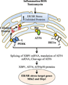
In hASM, inflammation, ROS, and (or) tunicamycin induce an accumulation of unfolded proteins and ER/SR stress (unfolded protein response). The molecular chaperone BiP (GRP78) is normally associated with PERK, ATF6, and IRE1α and maintains these proteins in an inactive state. As unfolded proteins accumulate in the ER/SR lumen, expression of BiP increases, and it dissociates from PERK, ATF6, and IRE1 leading to their activation and resulting in selective translation of the transcription factor ATF4; active transcription factor ATF6 (i.e., ATF6p50); and the active (spliced) XBP1 transcription factor. The IRE1, ATF6, and PERK pathways act to: (i) block protein translation, (ii) increase chaperone expression (e.g., calnexin), (iii) enhance SR-associated protein degradation pathways, and (iv) alter mitochondrial fusion (Mfn2) and fission (Drp1) protein expression. ROS, reactive oxidant species; hASM, human airway smooth muscle cells; ER/SR, endoplasmic reticulum/sarcoplasmic reticulum; BiP, binding immunoglobulin protein; PERK, protein kinase RNA-like endoplasmic reticulum kinase; ATF, active transcription factor; XBP1, X-box binding protein 1; IRE1, inositol-requiring enzyme 1; Mfn2, mitofusin-2; Drp1, dynamin related protein 1.
Fig. 7
Fig. 7
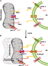
In hASM, agonist-induced elevations of [Ca2+]cyt are associated with corresponding elevations in [Ca2+]mito, reflecting mitochondrial Ca2+ buffering via activation of the MCU that requires close proximity of mitochondria to the ER/SR membrane. Activation of MCU is only achieved within localized microdomains (i.e., “Ca2+ hot spots”) in the vicinity of the IP3R or RyR. This mitochondrial coupling to the ER/SR membrane requires mitochondrial movement and tethering mediated by dimerization of Mfn2s (homodimer) or Mfn2 and Mfn1 (hetrodimer). Subsequent to agonist activation, [Ca2+]mito decreases via the mNCX. Refilling of the ER/SR is achieved by SERCA. hASM, human airway smooth muscle cells; MCU, mitochondrial Ca2+ uniporter; IP3R, inositol 1,4,5-triphosphate receptor; RyR, ryanodine receptor; ER/SR, endoplasmic reticulum/sarcoplasmic reticulum; mNCX, mitochondrial Na–Ca exchanger; SERCA, SR calcium transport ATPase.
Fig. 8
Fig. 8
Human ASM cells were loaded with 2.5 µmol·L−1 fluo3-AM (green) and 2.5 µmol·L−1 rhod-2-AM (red) to measure changes in [Ca2+]cyt and [Ca2+]mito, respectively (top: images at 3 different time points (A, B, and C) relative to ACh stimulation). Fluo3-AM and rhod-2-AM fluorescence increased in response to 1.0 µmol·L−1 ACh stimulation (bottom). Note that the transient [Ca2+]cyt response slightly preceded the [Ca2+]mito response, whereas the [Ca2+]mito response was more prolonged. ASM, airway smooth muscle cells; ACh, acetylcholine.
Fig. 9
Fig. 9
Human ASM cells were loaded with 500 nmol·L−1 MitoTraker Green AM to visualize the mitochondrial network. Control hASM cells displayed a filamentous/elongated mitochondrial network (fusion), which is promoted by Mfn1, Mfn2, and OPA1 proteins. In contrast, after 24 h exposure to TNFα (20 ng/mL for 12 h), mitochondria in hASM cells were more fragmented (fission). Mitochondria fragmentation (fission) is promoted by Drp1 and hFis1 proteins, and is associated with reduced mitochondrial Ca2+ uptake, ATP production, and increased ROS generation. ASM, airway smooth muscle; ROS, reactive oxidant species; Mfn, mitofusin; Drp1, dynamin related protein 1; OPA1, optic atrophy protein 1.
Fig. 10
Fig. 10
Inflammatory cytokines induce ROS generation in hASM that leads to ER/SR stress with downstream impact of reduced Mfn2 expression (increased Drp1), uncoupling of mitochondria to the ER/SR membrane, decreased [Ca2+]mito buffering, which contributes to increased [Ca2+]cyt and force responses to ACh stimulation (hyper-reactive state). A decrease in [Ca2+]mito buffering also leads to mitochondrial dysfunction and a further increase in ROS generation. Reduced Mfn2 expression also leads to mitochondrial fragmentation (fission) and increased cell proliferation and remodeling (synthetic state). ROS, reactive oxidant species; hASM, human airway smooth muscle cells; ER/SR, endoplasmic reticulum/sarcoplasmic reticulum; ACh, acetycholine; Mfn2, mitofusin-2; Drp1, dynamin related protein 1.

Similar articles

Cited by

References

    1. Adolph TE, Niederreiter L, Blumberg RS, Kaser A. Endoplasmic reticulum stress and inflammation. Dig. Dis. 2012;30(4):341–346. PMID: 22796794. - PMC - PubMed
    1. Ali F, Chin L, Pare PD, Seow CY. Mechanism of partial adaptation in airway smooth muscle after a step change in length. J. Appl. Physiol. (1985) 2007;103(2):569–577. PMID: 17495118. - PubMed
    1. Amrani Y. Modulation of airway smooth muscle contractile function by TNFα and IL-13 and airway hyperresponsiveness in asthma. In: Wang Y-X, editor. Calcium signaling in airway smooth muscle cells. Springer International Publishing; 2014. pp. 423–439.
    1. Amrani Y, Panettieri RA., Jr Modulation of calcium homeostasis as a mechanism for altering smooth muscle responsiveness in asthma. Curr. Opin. Allergy Clin. Immunol. 2002;2(1):39–45. PMID: 11964749. - PubMed
    1. Amrani Y, Chen H, Panettieri RA., Jr Activation of tumor necrosis factor receptor 1 in airway smooth muscle: a potential pathway that modulates bronchial hyper-responsiveness in asthma? Respir. Res. 2000;1(1):49–53. PMID: 11667965. - PMC - PubMed

Publication types

LinkOut - more resources