. 2015 Jan;43(1):661-73.
doi: 10.1093/nar/gku1314. Epub 2014 Dec 15.

Structural basis for the interaction of protein S1 with the Escherichia coli ribosome

Affiliations

Structural basis for the interaction of protein S1 with the Escherichia coli ribosome

Konstantin Byrgazov et al. Nucleic Acids Res. 2015 Jan.

Abstract

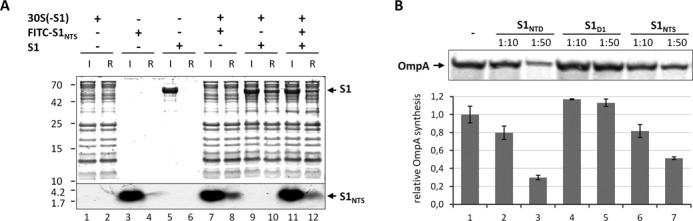
In Gram-negative bacteria, the multi-domain protein S1 is essential for translation initiation, as it recruits the mRNA and facilitates its localization in the decoding centre. In sharp contrast to its functional importance, S1 is still lacking from the high-resolution structures available for Escherichia coli and Thermus thermophilus ribosomes and thus the molecular mechanism governing the S1-ribosome interaction has still remained elusive. Here, we present the structure of the N-terminal S1 domain D1 when bound to the ribosome at atomic resolution by using a combination of NMR, X-ray crystallography and cryo-electron microscopy. Together with biochemical assays, the structure reveals that S1 is anchored to the ribosome primarily via a stabilizing π-stacking interaction within the short but conserved N-terminal segment that is flexibly connected to domain D1. This interaction is further stabilized by salt bridges involving the zinc binding pocket of protein S2. Overall, this work provides one hitherto enigmatic piece in the 'ribosome puzzle', namely the detailed molecular insight into the topology of the S1-ribosome interface. Moreover, our data suggest novel mechanisms that have the potential to modulate protein synthesis in response to environmental cues by changing the affinity of S1 for the ribosome.


Figures

Figure 1.
Figure 1.
The N-terminal segment is essential for protein S1 to interact with the ribosome in vivo. (A) Schematic of the domain organization of protein S1 and the C-terminally FLAG-tagged S1 variants used in the study. (B) The N-terminal domain D1 of protein S1 including the flexible N-terminal segment (NTS) and the C-terminal linker (CTL) is enlarged, and its variants used in the study are depicted below. (C) Equimolar amounts of S30 extracts (lanes 1 and 3) and 70S ribosomes (lanes 2 and 4) purified from E. coli strain JE28 synthesizing protein S119106 (lanes 1 and 2) or protein S1106 (lanes 3 and 4) were analysed for the presence of native S1 (panel a) and proteins S1106 and S119106 (panel b) by western blotting using antibodies directed against S1106 (18). Western blotting of protein S5 served as loading control (panel c). (D) Equal amounts of S30 extracts (extr.; lanes 1, 3, 5 and 7) and ribosomes (70S; lanes 2, 4, 6 and 8) purified from E. coli strain JE28 upon synthesis of FLAG-tagged proteins S1 (lanes 1 and 2), S119557 (lanes 3 and 4), S187557 (lanes 5 and 6) or S1NTФ106–557 (lanes 7 and 8) were analysed for the presence of the respective proteins by western blotting employing anti-FLAG antibodies (panel a). Protein S5 served as loading control (panel b).
Figure 2.
Figure 2.
Interaction between S1NTD and protein S2. (A) Overview showing the S2–S1NTD complex structure assembled from two protomers, with S1NTD in blue, S2 in yellow. Zn2+ is depicted as a green sphere. This colour code is used throughout the figures. (B) Stereo view showing the close up of the π-stacking interaction with the aromatic ring of Phe32 of protein S2 with Phe5 and Phe9 of S1NTH. (C) Stereo view showing the salt bridge interactions between the core domain S1D1 and the globular domain of S2 involving the Zn2+ binding pocket. The water molecules involved in the coordination of the Zn2+ ion are shown as red spheres.
Figure 3.
Figure 3.
Binding position of S1 on the E. coli 70S ribosome. (A) Cryo-EM structure of a translating E. coli 70S ribosome containing additional density for domain 1 (S1D1, blue) and domain 2 (S1D2, cyan) of ribosomal protein S1. Density for the large (grey) and small (pale yellow) ribosomal subunit, together with ribosomal protein S2 (bright yellow) is indicated. (B) Initial model for the position of S1NTD obtained by aligning S2 (yellow) of the chimeric S2–S1NTD with S2 (orange) from an E. coli 30S subunit (pdb accession code: 3ofo (40)) fitted to the cryo-EM map (grey mesh) as a rigid body. (C) Refined model for the complete S1NTD based on homology with eIF2α (pdb accession code: 1kl9 (43)) and fitted so as to maintain interactions between S1 and S2 as observed in the chimeric crystal structure, but also constrained by the electron density of the cryo-EM map (grey mesh). (D) The position of S1NTD (blue) relative to the E. coli 70S ribosome at 11 Å (EMD-1003 (45)) based on aligning S2 (yellow) of the chimeric S2–S1NTD with S2 (orange) from an E. coli 30S subunit (pdb accession code: 3ofo (40)) fitted to the cryo-EM map (grey mesh) as a rigid body.
Figure 4.
Figure 4.
The π-stacking interaction between S1NTH and S2 is pivotal for binding of S1 to the ribosome (A and B) and protein S2 (C and D). Schematic depiction of the co-purification experiments using either His-tagged ribosomes (33) (A) or FLAG-tagged protein S1NTD variants (C). (B) Equal amounts of S30 extract (Input; lanes 1, 3, 5, 7, 9, 11 and 13) and ribosomes (Elution; lanes 2, 4, 6, 8, 10, 12 and 14) purified from E. coli strain JE28 before (lanes 1 and 2) and after synthesis of proteins S1D1 (lanes 3 and 4), S1NTD (lanes 5 and 6), S1NTDF5A (lanes 7 and 8), S1NTDF9A (lanes 9 and 10), S1NTDK43E (lanes 11 and 12), S1NTDD39K (lanes 13 and 14) were tested for the presence of the respective S1 variants indicated to the right by western blot analysis using anti-S1106 antibodies (panels a–c). Protein S5 (panel d) served as loading control. (D) Under the same conditions exemplified in (B) S100 extracts were prepared and supplemented with purified HA-tagged protein S2 (input; lanes 1, 3, 5, 7, 9, 11 and 13). After incubation the FLAG-tagged protein S1 variants were immunoprecipitated by anti-FLAG antibodies (elution; lanes 2, 4, 6, 8, 10, 12 and 14; panel a) and the co-purification of protein S2 was determined by western blot analysis using anti-HA antibodies (panel b). The amounts of protein S1 variants were analysed employing anti-S1106 antibodies.
Figure 5.
Figure 5.
Free S1NTS binds to the ribosome and interferes with translation of the canonical ompA mRNA. (A) Purified S1-depleted 30S ribosomes (30S(-S1)) were incubated in the absence (lanes 1 and 2) or in the presence of FITC labelled S1NTS (lanes 7 and 8), native protein S1 (lanes 9 and 10) or both (lanes 11 and 12). Likewise, FITC labelled S1NTS (lanes 3 and 4) or native S1 (lanes 5 and 6) were incubated in the absence of ribosomes. Before (input; lanes 1, 3, 5, 7, 9 and 11) and after ultrafiltration using 100 kDa MWCO Amicon concentrators (Millipore) samples were taken and the presence of the respective proteins and the S1NTS peptide in the ribosome fraction (ribosome fraction; lanes 2, 4, 6, 8, 10 and 12) was determined by SDS-PAGE. (B) In vitro translation of ompA mRNA in the absence (lane 1) or in the presence of a 10- or 50-fold molar excess over ribosomes of S1NTD (lanes 2 and 3), S1D1 (lanes 4 and 5) or S1NTS (lanes 6 and 7), respectively. The assay was performed in triplicate and one representative autoradiograph is shown. Graph representing the quantification of three independent assays is given below. Error bars represent the standard deviation of the mean.
Figure 6.
Figure 6.
Schematic model showing the interaction of S1 with the 30S subunit. (A) In the free form the N-terminal segment of the multidomain protein S1 (spheres indicating the domains are colour-coded as in Figure 1A) is unstructured. (B) S1 can either interact with the globular domain of protein S2 (in yellow) on the 30S subunit (in light yellow) primarily via the N-terminal helix S1NTH, which adopts an α-helical conformation upon binding to S2 through a ‘folding upon binding’ mechanism (48). In this position, the protein can move in a ribosome-independent manner to scan for RNA molecules or (C) S1 can interact directly with the mRNA, facilitate unfolding of the mRNA, and its delivery to the ribosome (–7). (D) Binding of mRNA induces a rearrangement of the S1 domains D3–D5 (12) that might facilitate the correct positioning of the mRNA possibly supported by the salt bridges between S1D1 and S2, leading to the formation of the (E) translation initiation complex. It is still in question whether the presence of the Zn2+ ion (green sphere) affects the affinity or the topology of S1 on the ribosome, what could potentially influence the activity or selectivity of the ribosome for specific mRNAs. (F) Post-translational protein modifications within the region of the S1NTH or the S2 protein could likewise influence the affinity of S1 for the ribosome. Thereby, S1-depleted ribosomes that are selective for translation of lmRNAs could be present under specific conditions.

References

    1. Sørensen M.A., Fricke J., Pedersen S. Ribosomal protein S1 is required for translation of most, if not all, natural mRNAs in Escherichia coli in vivo. J. Mol. Biol. 1998;280:561–569. - PubMed
    1. Schuwirth B.S., Borovinskaya M.A., Hau C.W., Zhang W., Vila-Sanjurjo A., Holton J.M., Cate J.H. Structures of the bacterial ribosome at 3.5 A resolution. Science. 2005;310:827–834. - PubMed
    1. Sykes M.T., Williamson J.R. A complex assembly landscape for the 30S ribosomal subunit. Annu. Rev. Biophys. 2009;38:197–215. - PMC - PubMed
    1. Boni I.V., Isaeva D.M., Musychenko M.L., Tzareva N.V. Ribosome-messenger recognition: mRNA target sites for ribosomal protein S1. Nucleic Acids Res. 1991;19:155–162. - PMC - PubMed
    1. Qu X., Lancaster L., Noller H.F., Bustamante C., Tinoco I., Jr Ribosomal protein S1 unwinds double-stranded RNA in multiple steps. Proc. Natl. Acad. Sci. U.S.A. 2012;109:14458–14463. - PMC - PubMed

Publication types

MeSH terms