. 2015 Sep 24:1621:51-61.
doi: 10.1016/j.brainres.2014.12.010. Epub 2014 Dec 13.

Biochemical principles underlying the stable maintenance of LTP by the CaMKII/NMDAR complex

Affiliations

Biochemical principles underlying the stable maintenance of LTP by the CaMKII/NMDAR complex

John Lisman et al. Brain Res. .

Abstract

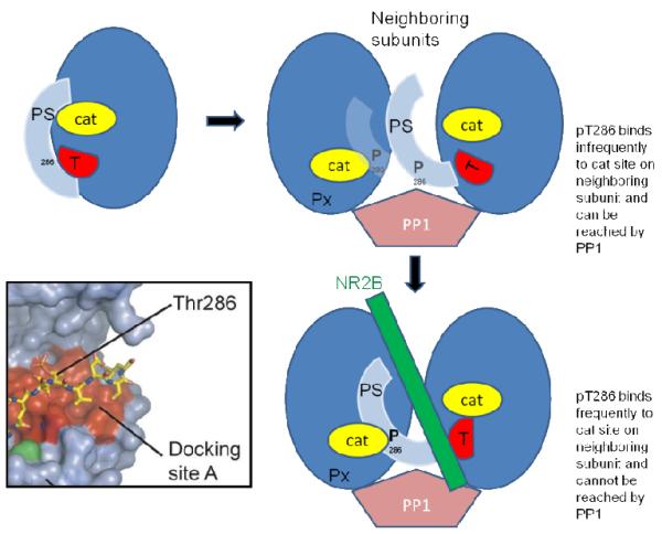
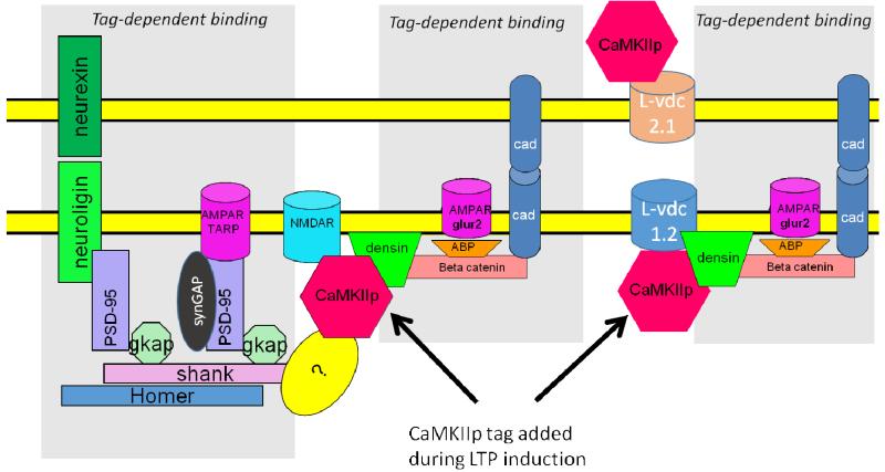
Memory involves the storage of information at synapses by an LTP-like process. This information storage is synapse specific and can endure for years despite the turnover of all synaptic proteins. There must, therefore, be special principles that underlie the stability of LTP. Recent experimental results suggest that LTP is maintained by the complex of CaMKII with the NMDAR. Here we consider the specifics of the CaMKII/NMDAR molecular switch, with the goal of understanding the biochemical principles that underlie stable information storage by synapses. Consideration of a variety of experimental results suggests that multiple principles are involved. One switch requirement is to prevent spontaneous transitions from the off to the on state. The highly cooperative nature of CaMKII autophosphorylation by Ca(2+) (Hill coefficient of 8) and the fact that formation of the CaMKII/NMDAR complex requires release of CaMKII from actin are mechanisms that stabilize the off state. The stability of the on state depends critically on intersubunit autophosphorylation, a process that restores any loss of pT286 due to phosphatase activity. Intersubunit autophosphorylation is also important in explaining why on state stability is not compromised by protein turnover. Recent evidence suggests that turnover occurs by subunit exchange. Thus, stability could be achieved if a newly inserted unphosphorylated subunit was autophosphorylated by a neighboring subunit. Based on other recent work, we posit a novel mechanism that enhances the stability of the on state by protection of pT286 from phosphatases. We posit that the binding of the NMNDAR to CaMKII forces pT286 into the catalytic site of a neighboring subunit, thereby protecting pT286 from phosphatases. A final principle concerns the role of structural changes. The binding of CaMKII to the NMDAR may act as a tag to organize the binding of further proteins that produce the synapse enlargement that underlies late LTP. We argue that these structural changes not only enhance transmission, but also enhance the stability of the CaMKII/NMDAR complex. Together, these principles provide a mechanistic framework for understanding how individual synapses produce stable information storage. This article is part of a Special Issue entitled SI: Brain and Memory.

Keywords: Kinase; LTP; Memory; Phosphatase.


Figures

Fig.1
Fig.1. Erasure of LTP by tatCN21, a peptide that interferes with the binding of CaMKII to NR2B
The postsynaptic response of CA1 neurons was measured by the slope of the field EPSP. At 20 min, LTP was induced by four tetani (100 Hz, 1 s) given to the input axons. This LTP is saturated, as evidenced by the lack of effect of an additional tetanus given at 40 min. tatCN21 was applied and then removed. It produced both a reversible effect and a persistent erasure of a large fraction of LTP. At right, a period of further recording showed that LTP could be reinduced in the same slices. In control experiments in which tatCN19 was not applied, LTP was not erased and no further LTP could be induced. From Fig.2 of (Sanhueza et al., 2011). Note that the application of tatCN21, in addition to persistently reversing LTP, has a reversible effect, probably due to the inhibition of presynaptic release (Waxham et al., 1993).
Fig.2
Fig.2. Contribution of autophosphorylation and subunit exchange to maintenance of the on state
(Left) Simplified model of CaMKII holoenzyme with only four shown subunits, each with a t286 site unphosphorylated. Such holoenzymes are in the cytoplasm, usually bound to actin. (Right) Upon activation during LTP induction, CaMKII translocates to the postsynaptic density, where it binds to the NMDAR. In this on state, each T286 site is phosphorylated. Two types of reactions must be counteracted to make the switch stable. (Right/Upper) Phosphatase may dephosphorylate a subunit; this is counteracted by autophosphorylation of that site by a neighboring active subunit. (Right/Lower) If protein turnover occurs by subunit exchange, a phosphorylated subunit may be replaced by an unphosphorylated subunit. This subunit is then phosphorylated by a neighboring subunit.
Fig.3
Fig.3. In vitro reconstitution of memory switch using purified CaMKII-alpha, protein phosphatase (PP1) and peptide corresponding to CaMKII binding site of NR2B (1289-1310)
Switch state is determined by the fraction of CaMKII phosphorylated on T286. Switch starts out off. At −10 min, Ca2+ is added and then lowered at t=0 to basal levels (0.56 micromolar). Without this Ca2+ elevation, the switch stays off (open circles). With Ca2+ elevation, the switch remains on after Ca2+ removal (closed black circles). This persistence requires autophosphorylation because if kinase is inhibited with K252a, the switch does not remain on (closed dark blue circles). Light blue circles show that the persistence is maintained if the solution change does not introduce a kinase inhibitor. From Fig.1 of (Urakubo et al., 2014).
Fig.4
Fig.4. In vitro experiments showing that dephosphorylation of CaMKII by PP1 is reduced by action of NR2B, but not by NR2A
Substrate is phospho-Thr286 of α-CaMKII. Phosphorylation sites on NR2A and NR2B have been mutated to alanines to prevent their phosphorylation (and associated complexity). Measurements were made with the kinase inhibited with staurosporine. Data normalized to level of CaMKII phosphorylation in absence of PP1. From Fig.4B of (Cheriyan et al., 2011).
Fig.5
Fig.5. Single-molecule assay reveals activation-dependent subunit exchange between CaMKII holoenzymes
TIRF was used to measure colocalization of labels within single holoenzymes that were originally labeled by a single fluorophore. The rate of increase in colocalization was faster at 37°C (red) compared to 25°C (blue) when Ca2+/CaM and ATP were added. At 37°C, the unactivated sample (i.e., with no addition of Ca2+/CaM and ATP) showed only a low level of exchange (green). From Fig.2B of (Stratton et al., 2014).
Fig.6
Fig.6. Reversal of phosphorylation reaction by resynthesis of ATP in catalytic site of CaMKII
CaMKII was initially autophosphorylated, leading to radioactive pT286. This was reduced by subsequent addition of ADP (left). When the same experiment was conducted under conditions in which the 286 site could not be phosphorylated (right), phosphorylation was low and ADP had no effect. From Fig.4A of (Kim et al., 2001).
Fig.7
Fig.7. Diagram showing how the binding of NR2B to the T site of CaMKII pushes pT286 into the catalytic site (cat) of a neighboring subunit, protecting pT286 from dephosphorylation by phosphatase (PP1)
PS is the pseudo-substrate region that is part of the regulatory region and that, in the off state, occupies and inhibits the catalytic site (cat). Inset shows crystal structure of pThr286 bound in the docking site A of the catalytic region of CaMKII (adapted from (Chao et al., 2010)). Px represents an undefined group of other phosphorylated sites that are not protected and which can be dephosphorylated by the PP1 in the PSD (Mullasseril et al., 2007).
Fig.8
Fig.8. Molecular model of trans-synaptic structural unit
Activated (phosphorylated) CaMKII binds to the NMDAR, forming the tag. Then other proteins, some newly synthesized, gradually (over the time course of an hour) bind to the synapse in a tag-dependent fashion. Three potential scenarios (not mutually exclusive) are shown in the three grey areas. The addition of adhesion molecules such as N-cadherin may serve to ensure that as the synapse grows, it has the same size presynaptically and postsynaptically. Although beta catenin is shown in the figure, both delta and beta forms bind to densin (Heikkila et al., 2007) and may thus be part of the structural unit. The figure shows a speculative binding interaction (?) that might explain how CaMKII binding to the NMDAR could serve as a seed for the observed addition of Shank and PSD-95.

References

    1. Abraham WC. How long will long-term potentiation last? Philos Trans R Soc Lond B Biol Sci. 2003;358:735–44. - PMC - PubMed
    1. Babiec WE, et al. Ionotropic NMDA receptor signaling is required for the induction of long-term depression in the mouse hippocampal CA1 region. J Neurosci. 2014;34:5285–90. - PMC - PubMed
    1. Barcomb K, Coultrap SJ, Bayer KU. Enzymatic activity of CaMKII is not required for its interaction with the glutamate receptor subunit GluN2B. Mol Pharmacol. 2013;84:834–43. - PMC - PubMed
    1. Barria A, Malinow R. Subunit-specific NMDA receptor trafficking to synapses. Neuron. 2002;35:345–53. - PubMed
    1. Barria A, Malinow R. NMDA receptor subunit composition controls synaptic plasticity by regulating binding to CaMKII. Neuron. 2005;48:289–301. - PubMed

Publication types

MeSH terms

Substances