. 2014 Dec 15:14:955.
doi: 10.1186/1471-2407-14-955.

Cancer-associated fibroblasts induce high mobility group box 1 and contribute to resistance to doxorubicin in breast cancer cells

Affiliations

Cancer-associated fibroblasts induce high mobility group box 1 and contribute to resistance to doxorubicin in breast cancer cells

Kamolporn Amornsupak et al. BMC Cancer. .

Abstract

Background: Cancer-associated fibroblasts and high mobility group box 1 (HMGB1) protein have been suggested to mediate cancer progression and chemotherapy resistance. The role of such fibroblasts in HMGB1 production in breast cancer is unclear. This study aimed to investigate the effects of cancer-associated fibroblasts on HMGB1 expression in breast cancer cells and its role in chemotherapeutic response.

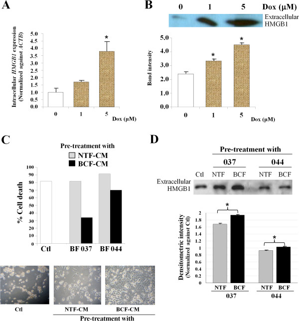
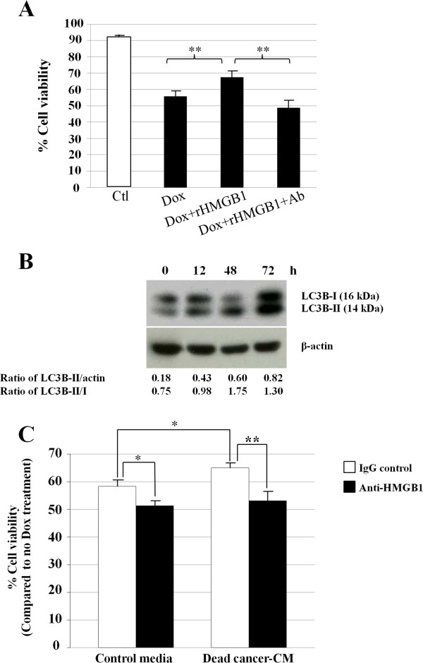
Methods: Breast cancer-associated fibroblasts (BCFs) and non-tumor-associated fibroblasts (NTFs) were isolated from human breast cancers or adjacent normal tissues and established as primary cultures in vitro. After confirmation of the activated status of these fibroblasts, conditioned-media (CM) were collected and applied to MDA-MB-231 human triple negative breast cancer cells. The levels of intracellular and extracellular HMGB1 were measured by real-time PCR and/or Western blot. The response of BCF-CM-pre-treated cancer cells to doxorubicin (Dox) was compared with those pre-treated with NTF-CM or control cultures. The effect of an HMGB1 neutralizing antibody on Dox resistance induced by extracellular HMGB1 from non-viable Dox-treated cancer cells or recombinant HMGB1 was also investigated.

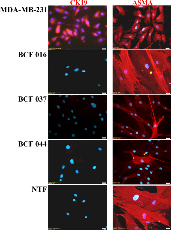
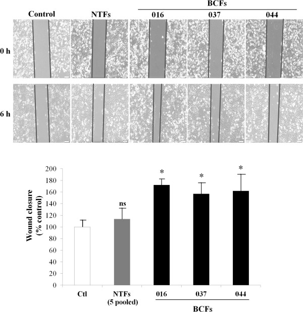
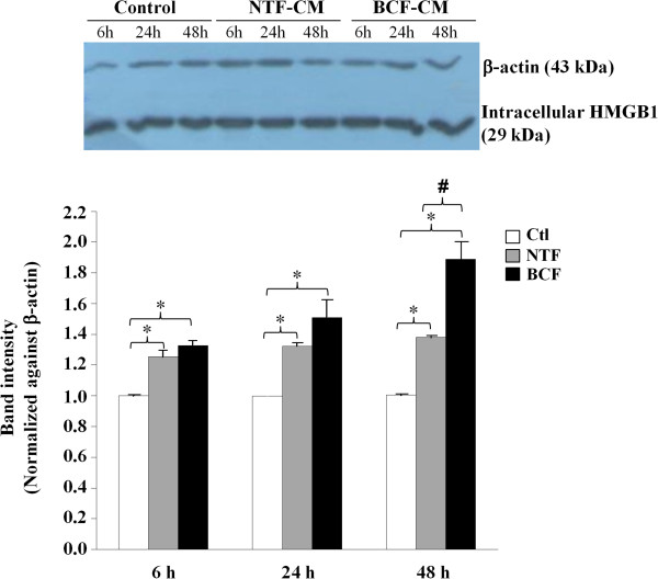
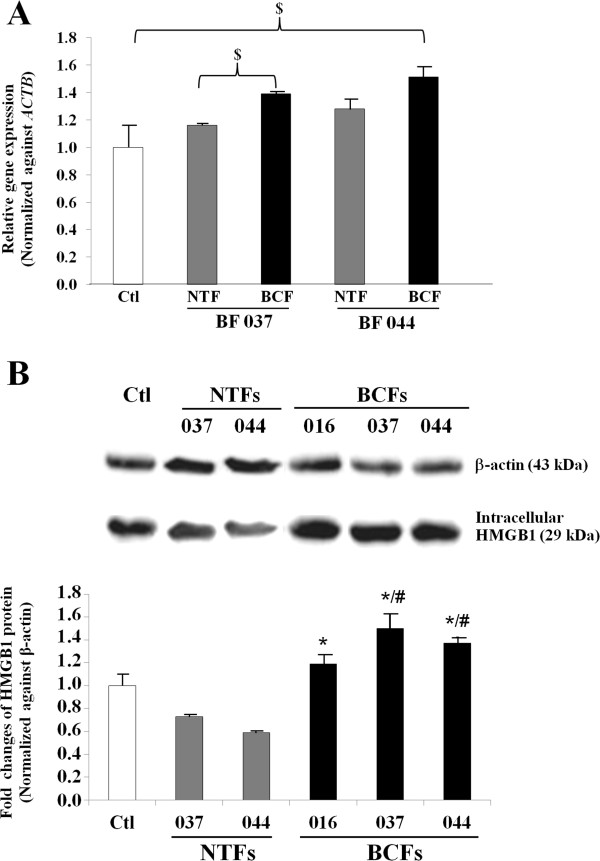
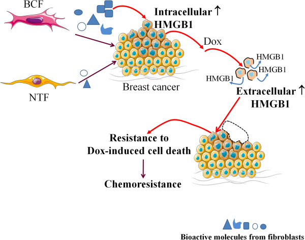
Results: Immunocytochemical analysis revealed that BCFs and NTFs were alpha-smooth muscle actin (ASMA) positive and cytokeratin 19 (CK19) negative cells: a phenotype consistent with that of activated fibroblasts. We confirmed that the CM from BCFs (but not NTFs), could significantly induce breast cancer cell migration. Intracellular HMGB1 expression was induced in BCF-CM-treated breast cancer cells and also in Dox-treated cells. Extracellular HMGB1 was strongly expressed in the CM after Dox-induced MDA-MB-231 cell death and was higher in cells pre-treated with BCF-CM than NTF-CM. Pre-treatment of breast cancer cells with BCF-CM induced a degree of resistance to Dox in accordance with the increased level of secreted HMGB1. Recombinant HMGB1 was shown to increase Dox resistance and this was associated with evidence of autophagy. Anti-HMGB1 neutralizing antibody significantly reduced the effect of extracellular HMGB1 released from dying cancer cells or of recombinant HMGB1 on Dox resistance.

Conclusions: These findings highlight the potential of stromal fibroblasts to contribute to chemoresistance in breast cancer cells in part through fibroblast-induced HMGB1 production.


Figures

Figure 1
Figure 1
Immunofluorescent staining of CK19 and ASMA in primary cultures of fibroblasts derived from breast cancers (BCF) and adjacent areas of normal breast from surgical specimens (NTF). BCF 016, BCF 037 and BCF 044 are derived from different patients, whereas NTF are matched normal tissue fibroblasts pooled from patients 037 and 044. Breast cancer cell line MDA-MB-231 was used as a positive control for CK19. Hoechst (blue) staining shows the nuclei. Original magnification of 400x. Bars represent 20 μm.
Figure 2
Figure 2
BCF-CMs enhance MDA-MD-231 cell migration. MDA-MB-231 breast cancer cells were exposed to 3 different BCF-CMs and pooled samples of 5 NTF-CMs and a scratch wound motility assay was performed over 6 h to measure the ability of CM to induce cancer cell migration. Bar graphs represent mean ± SD of two independent experiments. The migration of cells in control fresh media (Ctl) was set at 100%. * = p-value of less than 0.05 compared to the migration of cancer cells under control conditions. ns = not significance.
Figure 3
Figure 3
Western blot analysis of intracellular HMGB1 in MDA-MB-231 human breast cancer cells treated with fibroblast CMs (BCF 044 and NTF 044) for 6, 24, and 48 h. Cancer cells cultured in fresh medium were used as a negative control. The intensity of each HMGB1 band is shown after normalization against the β-actin internal loading control protein. Bar graphs represent mean ± SD of two independent experiments. * = p-value of less than 0.05 comparing HMGB1 levels in the CM-treated cells with controls at each time point; # = p-value of less than 0.05 comparing HMGB1 levels in BCF-CM-treated cells with NTF-CM treatment.
Figure 4
Figure 4
HMGB1 expression in MDA-MB-231 cells treated with fibroblast CM. Real time PCR for HMGB1 expression in MDA-MB-231 cells treated with NTF-CMs and BCF-CMs for 48 h using paired fibroblasts isolated from the same patient.The levels of HMGB1 transcript (A) and protein levels (B) are shown after normalization against the internal control β-actin. Controls (Ctl) are cells cultured in fresh medium with no CM treatment. Bars represent the mean ± SD of triplicate experiments. $ = p-value of less than 0.05. * = p-value of less than 0.05 compared to the average HMGB1 of the two NTFs-CM treatment conditions whereas # = p-value of less than 0.05 compared to HMGB1 of the matched NTF-CM treatment.
Figure 5
Figure 5
Dox-induced HMGB1 in MDA-MB-231 cells. (A) Intracellular HMGB1 expression was measured by real time PCR. Bars represent mean ± SD of HMGB1 expression level normalized against ACTB and relative to no drug treatment. Three independent experiments were performed. (B) Extracellular HMGB1 protein was detected by Western blot analysis in the culture media from cells treated with different concentrations of Dox for 48 h. (A and B) *p-value of less than 0.05 compared to the controls without Dox treatment. (C) BCF-CM induced resistance to Dox-mediated cell death in MDA-MB-231 breast cancer cells. Bars represent % cell death of each pre-treatment condition and the images show corresponding cell density. (D) Culture media from each condition in (C) were measured for extracellular HMGB1 by Western blot analysis. Equal amounts of proteins were loaded. Bars represent the mean band intensity (± SD) measured by densitometry. The band intensity of control cultures without CM pre-treatment (Ctl) was set as 1. *p-value of less than 0.05.
Figure 6
Figure 6
Effect of recombinant HMGB1 (rHMGB1) on the response of MDA-MB-231 to Dox. (A) MDA-MB-231 human breast cancer cells were treated with 5 μM Dox with or without addition of 100 ng/ml rHMGB1 and/or anti-HMGB1 neutralizing antibody for 24 h. The % cell viability is shown and the bars represent the mean ± SD of triplicate experimental wells. (B) Autophagy-related protein LC3B is induced in MDA-MB-231 breast cancer cells. Densitometric analysis of LC3B-II normalized against the protein loading control β-actin and the conversion of LC3B-I to LC3B-II is shown. (C) MDA-MB-231 cells treated with ‘dead cancer-CM’ showed increased viability compared with controls and this effect was attenuated by anti-HMGB1 neutralizing antibody. Bars represent mean ± SD of three independent experiments. * = p-value of less than 0.05 whereas ** is less than 0.01.
Figure 7
Figure 7
Schematic diagram illustrating the potential of secreted substances from breast cancer-associated fibroblasts (BCFs) to induce expression of intracellular HMGB1 in breast cancer cells. After exposure to a chemotherapeutic agent, in this case doxorubicin, (Dox), this increased intracellular HMGB1 can be released and may function in a paracrine manner to induce acquired chemoresistance of the nearby surviving cancer cells.

References

    1. Jemal A, Bray F, Center MM, Ferlay J, Ward E, Forman D. Global cancer statistics. CA Cancer J Clin. 2011;61(2):69–90. doi: 10.3322/caac.20107. - DOI - PubMed
    1. Attasara P, Buasom R. Hospital-based cancer registry 2010. Natl Cancer Inst Thai. 2010;24:1–52.
    1. Hernandez-Aya LF, Gonzalez-Angulo AM. Adjuvant systemic therapies in breast cancer. Surg Clin North Am. 2013;93(2):473–491. doi: 10.1016/j.suc.2012.12.002. - DOI - PubMed
    1. Gonzalez-Angulo AM, Morales-Vasquez F, Hortobagyi GN. Overview of resistance to systemic therapy in patients with breast cancer. Adv Exp Med Biol. 2007;608:1–22. doi: 10.1007/978-0-387-74039-3_1. - DOI - PubMed
    1. Sebens S, Schafer H. The tumor stroma as mediator of drug resistance–a potential target to improve cancer therapy? Curr Pharm Biotechnol. 2012;13(11):2259–2272. doi: 10.2174/138920112802501999. - DOI - PubMed
Pre-publication history
    1. The pre-publication history for this paper can be accessed here:http://www.biomedcentral.com/1471-2407/14/955/prepub

Publication types

MeSH terms

LinkOut - more resources