. 2015 Jan 12;212(1):15-22.
doi: 10.1084/jem.20130062. Epub 2014 Dec 15.

Neutrophils are required for both the sensitization and elicitation phase of contact hypersensitivity

Affiliations

Neutrophils are required for both the sensitization and elicitation phase of contact hypersensitivity

Felix C Weber et al. J Exp Med. .

Abstract

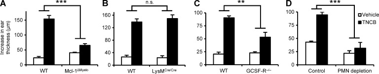
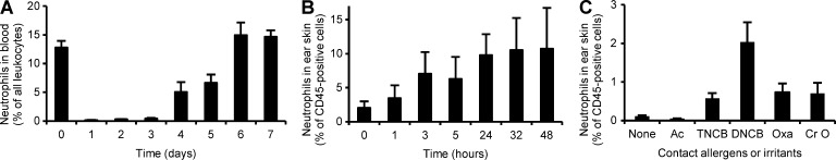
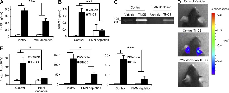
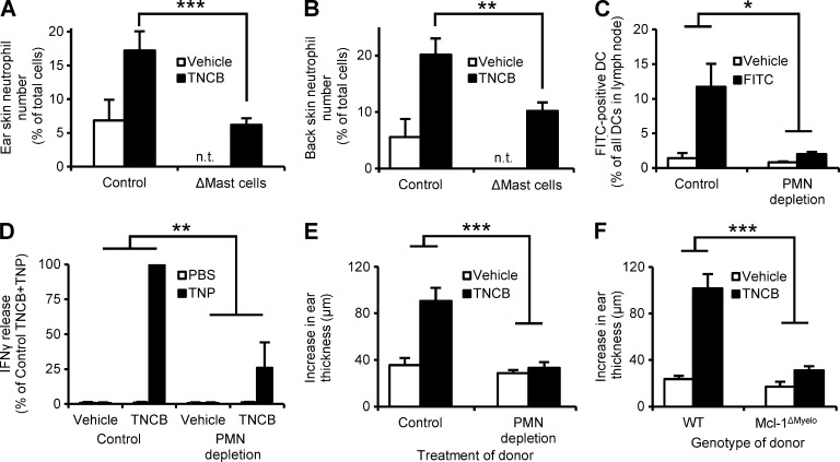
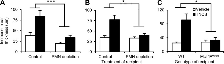
Allergic contact dermatitis and its animal model, contact hypersensitivity (CHS), are T cell-mediated inflammatory skin diseases induced by contact allergens. Though numerous cellular and molecular players are known, the mechanism of chemical-induced sensitization remains poorly understood. Here, we identify neutrophils as crucial players in the sensitization phase of CHS. Genetic deficiency of neutrophils caused by myeloid-specific deletion of Mcl-1 or antibody-mediated depletion of neutrophils before sensitization abrogated the CHS response. Neutrophil deficiency reduced contact allergen-induced cytokine production, gelatinase release, and reactive oxygen species production in naive mice. Mast cell deficiency inhibited neutrophil accumulation at the site of sensitization. In turn, neutrophils were required for contact allergen-induced release of further neutrophil-attracting chemokines, migration of DCs to the draining lymph nodes, and priming of allergen-specific T cells. Lymph node cells from mice sensitized in the absence of neutrophils failed to transfer sensitization to naive recipients. Furthermore, no CHS response could be induced when neutrophils were depleted before elicitation or when normally sensitized lymph node cells were transferred to neutrophil-deficient recipients, indicating an additional role for neutrophils in the elicitation phase. Collectively, our data identify neutrophils to be critically involved in both the sensitization and elicitation phase of CHS.


Figures

Figure 1.
Figure 1.
Neutrophils are essential for the CHS response. Mice were sensitized with TNCB or acetone and were challenged with TNCB 5 d after sensitization. The increase in ear thickness 24 h after challenge is depicted. (A and B) CHS response in WT, Mcl-1ΔMyelo, and LysMCre/Cre mice. (C) CHS response in bone marrow chimeras with WT or GCSF-R−/− hematopoietic compartment. (D) CHS response in WT mice treated with a neutrophil-depleting anti-Ly6G antibody (PMN depletion) or a control rat IgG 24 h before sensitization. The graphs show mean and SEM from 8–13 (A), 4–6 (B), 5–8 (C), or 5–24 (D) individual mice per group from 3 (A–C) or 5 (D) independent experiments. **, P < 0.01; ***, P < 0.002; n.s., statistically not significant.
Figure 2.
Figure 2.
Neutrophils in the CHS sensitization phase. (A) Analysis of the number of circulating neutrophils before or at the indicated times after treatment with the neutrophil-depleting anti-Ly6G antibody. (B and C) Infiltration of neutrophils was tested at the indicated times after sensitization with TNCB (B) or 24 h after sensitization with TNCB, DNCB, oxazolone (Oxa), or croton oil (Cr O) on the ears. Neutrophil infiltration was assessed by digestion of the ear skin followed by flow cytometry. The graphs show mean and SEM from 7–26 (A), 8–11 (B), or 9 (C) mice per group from 6 (A) or 4 (B and C) independent experiments.
Figure 3.
Figure 3.
Neutrophil depletion suppresses the contact allergen-induced inflammatory response. Naive mice were treated with a neutrophil-depleting anti-Ly6G antibody (PMN depletion) or control rat IgG, sensitized with TNCB, DNCB, oxazolone (Oxa), or vehicle control on both ears 24 h later, and the development of an inflammatory environment was tested after an additional 24 h. (A and B) Analysis of IL-1β and MIP-2 levels in the ear tissue by ELISA. (C) Analysis of gelatinase levels by in-gel zymography. (D and E) Analysis of ROS production by in vivo bioluminescence. C and D show representative images from 3 independent experiments. Bar graphs show mean and SEM from 4–5 (A and B) or 5–16 (E) individual mice from 3 (A and B) or 2–3 (E) independent experiments. *, P < 0.05; ***, P < 0.002.
Figure 4.
Figure 4.
Neutrophils are recruited by mast cells and trigger DC migration and T cell priming. (A and B) Mice with a DT-induced mast cell deficiency (ΔMast cells) or control animals were sensitized with TNCB on the ears (A) or the back skin (B) and the number of infiltrating neutrophils was determined 24 h later. (C) Mice were treated with the neutrophil-depleting anti-Ly6G antibody (PMN depletion) or control antibody, and FITC or vehicle was applied to the skin 24 h later, and the number of FITC-positive DCs in the draining lymph nodes was determined after an additional 24 h. (D) Neutrophil-depleted and control mice were sensitized with TNCB or acetone. Their draining lymph node cells were isolated 5 d after sensitization, restimulated by TNP-modification, and their IFN-γ production was measured by ELISA. (E and F) Neutrophil-depleted (E), Mcl-1ΔMyelo (F), or the appropriate control mice were sensitized with TNCB or acetone, their draining lymph node cells were isolated 5 d after sensitization, and transferred to naive mice, which were then challenged with TNCB and the increase in their ear thickness was measured. Graphs show mean and SEM from 4–15 (A-B), 6–8 (C), 8–9 (E), and 7–9 (F) individual mice or 3 independent samples (D) per group from 2 (A–C) or 3 (D–F) independent experiments. In (D), data are expressed in percentage of the IFN-γ release in nondepleted TNCB+TNP samples, which corresponded to 1.5 ± 0.6 ng IFN-γ per 106 cells. n.t., not tested. *, P < 0.05; **, P < 0.01; ***, P < 0.002.
Figure 5.
Figure 5.
Neutrophils are required for the elicitation phase of CHS. (A) Mice were sensitized by TNCB or acetone, treated with the neutrophil-depleting anti-Ly6G antibody (PMN depletion) 4 d later, and challenged with TNCB after an additional 24 h. The increase of ear thickness during an additional 24 h was measured. (B and C) Nondepleted WT naive mice were sensitized by TNCB or acetone, and their lymph node cells were isolated 5 d later and injected into neutrophil-depleted (B), Mcl-1ΔMyelo (C), or appropriate control recipients which were then challenged with TNCB and the increase of their ear thickness after 24 h was measured. Graphs show mean and SEM from 3–12 (A) 11–12 (B), or 4–9 (C) individual mice per group from 3 (A and C) or 4 (B) experiments. *, P < 0.05; ***, P < 0.002.

References

    1. Charmoy M., Milon G., and Tacchini-Cottier F.. 2011. Role of Neutrophils in the Early Shaping of the Leishmania major Specific Immune Response in Experimental Murine Cutaneous Leishmaniasis. Neutrophils in Infectious Diseases. Tacchini-Cottier F., and van Zandbergen G., Bentham Science Publishers Ltd. pp. 49–58.
    1. Chtanova T., Schaeffer M., Han S.J., van Dooren G.G., Nollmann M., Herzmark P., Chan S.W., Satija H., Camfield K., Aaron H., et al. . 2008. Dynamics of neutrophil migration in lymph nodes during infection. Immunity. 29:487–496 10.1016/j.immuni.2008.07.012 - DOI - PMC - PubMed
    1. Clausen B.E., Burkhardt C., Reith W., Renkawitz R., and Förster I.. 1999. Conditional gene targeting in macrophages and granulocytes using LysMcre mice. Transgenic Res. 8:265–277 10.1023/A:1008942828960 - DOI - PubMed
    1. Cumberbatch M., Dearman R.J., Groves R.W., Antonopoulos C., and Kimber I.. 2002. Differential regulation of epidermal langerhans cell migration by interleukins (IL)-1alpha and IL-1beta during irritant- and allergen-induced cutaneous immune responses. Toxicol. Appl. Pharmacol. 182:126–135 10.1006/taap.2002.9442 - DOI - PubMed
    1. Dudeck A., Dudeck J., Scholten J., Petzold A., Surianarayanan S., Köhler A., Peschke K., Vöhringer D., Waskow C., Krieg T., et al. . 2011. Mast cells are key promoters of contact allergy that mediate the adjuvant effects of haptens. Immunity. 34:973–984 10.1016/j.immuni.2011.03.028 - DOI - PubMed

Publication types

MeSH terms