. 2014 Dec 30;111(52):18572-7.
doi: 10.1073/pnas.1417816112. Epub 2014 Dec 15.

Identification of S-nitroso-CoA reductases that regulate protein S-nitrosylation

Affiliations

Identification of S-nitroso-CoA reductases that regulate protein S-nitrosylation

Puneet Anand et al. Proc Natl Acad Sci U S A. .

Abstract

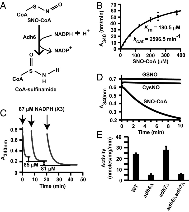
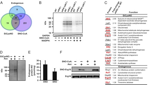
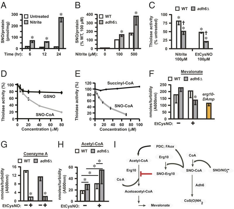
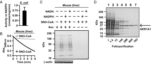
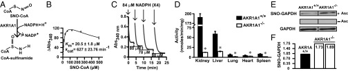
Coenzyme A (CoA) mediates thiol-based acyl-group transfer (acetylation and palmitoylation). However, a role for CoA in the thiol-based transfer of NO groups (S-nitrosylation) has not been considered. Here we describe protein S-nitrosylation in yeast (heretofore unknown) that is mediated by S-nitroso-CoA (SNO-CoA). We identify a specific SNO-CoA reductase encoded by the alcohol dehydrogenase 6 (ADH6) gene and show that deletion of ADH6 increases cellular S-nitrosylation and alters CoA metabolism. Further, we report that Adh6, acting as a selective SNO-CoA reductase, protects acetoacetyl-CoA thiolase from inhibitory S-nitrosylation and thereby affects sterol biosynthesis. Thus, Adh6-regulated, SNO-CoA-mediated protein S-nitrosylation provides a regulatory mechanism paralleling protein acetylation. We also find that SNO-CoA reductases are present from bacteria to mammals, and we identify aldo-keto reductase 1A1 as the mammalian functional analog of Adh6. Our studies reveal a novel functional class of enzymes that regulate protein S-nitrosylation from yeast to mammals and suggest that SNO-CoA-mediated S-nitrosylation may subserve metabolic regulation.

Keywords: AKR1A1; Adh6; S-nitrosylation; denitrosylase; denitrosylation.


Conflict of interest statement

The authors declare no conflict of interest.

Figures

Fig. 1.
Fig. 1.
Identification of Adh6 as the NADPH-dependent SNO-CoA reductase in yeast. (A) SNO-CoA–dependent NADPH consumption in yeast (S. cerevisiae). Extracts (800 µg/mL) were incubated with 100 μM SNO-CoA and 100 μM NADPH, and NADPH consumption (absorbance at 340 nm) was monitored continuously. (B) SNO-CoA–metabolizing activity in yeast extracts requires NADPH and not NADH. Extracts were incubated with 200 μM SNO-CoA and 100 μM NADPH or NADH and monitored continuously for 1 min. Data are presented as mean ± SD; n = 3. (C) SNO-CoA–mediated protein S-nitrosylation. Representative Coomassie-stained SDS/PAGE gel displaying SNO-proteins isolated by SNO-RAC following incubation of yeast lysate for 10 min with SNO-CoA (60 μM) alone or in combination with NADPH or NADH (100 μM). Ascorbate (Asc) was omitted from the SNO-RAC assay as a specificity control. Results are representative of three independent experiments. (D) Isolation and identification of SNO-CoA reductase. Representative Coomassie-stained SDS/PAGE gel corresponding to the five-step chromatographic purification scheme detailed in Table S1, which yielded from a crude extract (lane 1) 2,826-fold enrichment of NADPH-dependent SNO-CoA reductase activity identified by MS as Adh6 (lane 6).
Fig. 2.
Fig. 2.
Characterization of the yeast SNO-CoA reductase, Adh6. (A) CoA-sulfinamide was identified by MS as the major stable product of SNO-CoA reduction by purified Adh6 (see Fig. S2 A, B, and D for product analysis). (B) Kinetic analysis of SNO-CoA reductase activity of purified Adh6. (C) Stoichiometry of NADPH:SNO-CoA in Adh6-catalyzed SNO-CoA reduction. Sequential additions of 87 μM NADPH to an excess of SNO-CoA led to consumption of 79–85 μM (mean 82 ± 3 μM; n = 6 additions) of SNO-CoA, demonstrating a stoichiometry of 1:1. Results shown are representative of two independent experiments. (D) Specificity of Adh6 for SNO-CoA. Purified Adh6 (Table S1 and Fig. 1D) (20 nM) was incubated with NADPH (100 µM) and SNO-CoA, GSNO, or CysNO (100 µM), and NADPH consumption was measured over time. (E) Adh6 is the principal source of NADPH-dependent SNO-CoA reductase activity. Activity was assayed in lysates from WT yeast and adh6Δ, adh7Δ, and adh6Δadh7Δ yeast. Data are presented as mean ± SD; n = 3.
Fig. 3.
Fig. 3.
Adh6 regulates protein S-nitrosylation mediated by SNO-CoA. (A) A Venn diagram illustrates the relationships between the sets of SNO proteins identified in intact WT yeast under basal growth conditions (endogenous SNO proteins) or following treatment with EtCysNO, a cell-permeable S-nitrosylating agent, and in lysates treated with SNO-CoA. (B) Adh6-regulated protein S-nitrosylation. Representative Coomassie-stained SDS/PAGE gel illustrating SNO-proteins isolated by SNO-RAC following treatment of WT yeast lysates with SNO-CoA (in the presence or absence of NADPH) and the regulation by Adh6 of protein S-nitrosylation. Results are representative of three independent experiments. (C) SNO-CoA–mediated protein S-nitrosylation in situ. (Upper) SNO-proteins exhibiting enhanced S-nitrosylation in adh6Δ vs. WT yeast following treatment with EtCysNO. (Lower) Endogenous SNO proteins showing enhanced S-nitrosylation in adh6Δ vs. WT yeast under basal growth conditions. Shared targets identified following treatment of lysates with SNO-CoA are indicated in red, and metabolic enzymes are underlined. n = 3; P < 0.05 by Student t test; relative standard deviation < 35%. (D) Isolation of endogenous and in-situ-formed SNO-proteins. Representative Coomassie-stained SDS/PAGE gel illustrating endogenous SNO-proteins isolated by SNO-RAC from WT yeast (untreated) as well as SNO-proteins formed in situ (EtCysNO). Shared targets are shown in A. (Note that the demonstration of endogenous SNO-proteins in D but not B reflects different amounts of total protein used in the SNO-RAC assay (4 vs. 1 mg, respectively). Results are representative of three independent experiments. (E) Endogenous protein-bound NO. Total protein-bound NO (XNO; which includes FeNO and SNO) and SNO were quantified in WT yeast extracts by mercury-coupled photolysis-chemiluminescence. Data are presented as mean ± SEM (n = 7). (F) Endogenous S-nitrosylation of thiolase and its enhanced S-nitrosylation by SNO-CoA. Results of a representative analysis by SNO-RAC of endogenous and SNO-CoA–induced S-nitrosylation of Erg10. Data are representative of two independent experiments.
Fig. 4.
Fig. 4.
S-nitrosylation by SNO-CoA inhibits acetoacetyl-CoA thiolase (Erg10)-dependent metabolism. (A) Endogenous SNO-protein formation in yeast grown under hypoxia in the presence of nitrite. WT yeast were grown for the indicated times in the presence or absence of 100 µM nitrite. SNO levels in lysates were quantified by mercury-coupled photolysis chemiluminescence. Data are presented as mean ± SEM (n = 4). *P < 0.05 (by Student t test). (B) Adh6 regulates endogenous SNO-protein synthesis in yeast. Cells were grown under hypoxia for 24 h in the presence of 0, 100, or 500 µM nitrite, and SNO-protein levels in lysates were quantified as in A. Data are presented as mean ± SEM (n = 5). *P < 0.05 (by Student t test). (C) Erg10 activity is inhibited by Adh6-regulated, SNO-CoA–mediated S-nitrosylation. Erg10 activity was assayed in extracts of WT or adh6Δ yeast grown under hypoxia with or without 100 µM nitrite (as in A and B) or treated at normoxia with EtCysNO (100 µM). Data are mean ± SD (n = 3). *P < 0.05 vs. untreated; P < 0.05 adh6Δ vs. WT by ANOVA. (D and E) Selective inhibition of Erg10 by SNO-CoA vs. GSNO (D) or succinyl-CoA (E). Erg10 activity was assayed in extracts of WT yeast in the presence or absence of SNO-CoA (D and E), GSNO (D), or succinyl-CoA (E). (FH) Effects of enhanced S-nitrosylation on the metabolic profile of the mevalonate pathway. Midlog phase yeast were untreated or treated with 100 µM (F) or 500 µM (G and H) EtCysNO for 2, and metabolites (mevalonate, CoA, and acetyl-CoA) were measured as described in SI Materials and Methods. Data are mean ± SD (n = 5 or 6). *P < 0.05 (by Student t test in F and G and by ANOVA in H, normalized with respect to culture turbidity). Note in F that erg10DAmP yeast exhibits ∼50% of WT thiolase activity (Fig. S7D). (I) Schematic summary of the potential role of SNO-CoA–mediated protein S-nitrosylation in regulation of Erg10-dependent metabolism. Erg10 converts acetyl-CoA to free CoA and acetoacetyl-CoA, a precursor via 3-hydroxy-3-methyl-glutaryl-CoA (HMG-CoA) in mevalonate biosynthesis. S-nitrosylation of Erg10 by SNO-CoA, regulated by Adh6 acting as a SNO-CoA reductase, inhibits thiolase activity, resulting in decreased levels of mevalonate and may contribute to diminished levels of CoA and increased levels of acetyl-CoA. Altered levels of CoA and acetyl-CoA may reflect actions of Adh6-regulated SNO-CoA at additional loci (FAox, fatty acid β-oxidation; PDC, pyruvate dehydrogenase multienzyme complex). SNO-CoA reductase thus acts as a cognate denitrosylase for substrates of SNO-CoA by direct analogy to GSNOR, which acts as a denitrosylase for protein substrates of GSNO.
Fig. 5.
Fig. 5.
Identification of aldo-keto reductase 1A1 (AKR1A1) as the NADPH-dependent SNO-CoA reductase in mammals. (A and B) Conservation of SNO-CoA reductase activity from bacteria to mammals. NADPH-dependent SNO-CoA metabolizing activity in extracts from Escherichia coli (A) and mouse liver (B) is shown. Conditions in A were similar to Fig. 1B. In B, 160 µg/mL liver extract was incubated with 100 μM SNO-CoA and 100 μM NADPH. Note that high levels of basal NADH consumption (diaphorase activity) in E. coli under aerobic conditions prevented assessment of SNO-CoA dependence. (C) SNO-CoA–mediated protein S-nitrosylation. Representative Coomassie-stained SDS/PAGE gels displaying SNO proteins isolated by SNO-RAC following incubation of mouse liver extract (1 mg/mL in 1 mL of reaction volume) for 10 min with SNO-CoA (60 μM) alone or in combination with NADPH or NADH (100 μM). Ascorbate (Asc) was omitted from the SNO-RAC assay as a specificity control. (D) SNO-CoA reductase isolation. Representative SDS/PAGE gel corresponding to the six-step chromatographic purification scheme detailed in Table S1, which yielded from a crude extract (lane 1) 763-fold enrichment of SNO-CoA reductase activity identified by MS as AKR1A1 (lane 7).
Fig. 6.
Fig. 6.
Characterization of the mammalian SNO-CoA reductase AKR1A1. (A) CoA-sulfinamide was identified by MS as the major stable product of SNO-CoA reduction by purified AKR1A1 (see Fig. S2 A, C, and D for product analysis). (B) Kinetic analysis of SNO-CoA reductase activity of purified AKR1A1. (C) Stoichiometry of NADPH:SNO-CoA in AKR1A1-catalyzed SNO-CoA reduction. Sequential additions of 84 μM NADPH to an excess of SNO-CoA led to consumption of 75–82 μM (mean 79 ± 3 μM; n = 8 additions) SNO-CoA, demonstrating a stoichiometry of 1:1. Results shown are representative of two independent experiments. (D) SNO-CoA reductase activity in AKR1A1 knockout animals. NADPH-dependent SNO-CoA reductase activity across various tissues from WT or AKR1A1−/− mice is shown. Extracts were incubated with 100 µM NADPH and 0 or 200 µM SNO-CoA. Values are from three WT (filled) and three AKR1A1−/− (open) mice. *P < 0.05 (Student t test). (E and F) Regulation of endogenous protein S-nitrosylation by AKR1A1. Analysis of S-nitrosylated GAPDH (SNO-GAPDH) in kidney extracts of AKR1A1+/+ and AKR1A1−/− mice is shown. Changes in SNO-GAPDH levels were determined by SNO-RAC coupled to Western blotting (E; n = 2) or to iTRAQ-based MS (F; n = 2).

References

    1. Seth D, Hausladen A, Wang YJ, Stamler JS. Endogenous protein S-nitrosylation in E. coli: Regulation by OxyR. Science. 2012;336(6080):470–473. - PMC - PubMed
    1. Anand P, Stamler JS. Enzymatic mechanisms regulating protein S-nitrosylation: Implications in health and disease. J Mol Med (Berl) 2012;90(3):233–244. - PMC - PubMed
    1. Benhar M, Forrester MT, Stamler JS. Protein denitrosylation: Enzymatic mechanisms and cellular functions. Nat Rev Mol Cell Biol. 2009;10(10):721–732. - PubMed
    1. Liu L, et al. A metabolic enzyme for S-nitrosothiol conserved from bacteria to humans. Nature. 2001;410(6827):490–494. - PubMed
    1. Liu L, et al. Essential roles of S-nitrosothiols in vascular homeostasis and endotoxic shock. Cell. 2004;116(4):617–628. - PubMed

MeSH terms