. 2014 Dec 23;111(51):E5564-73.
doi: 10.1073/pnas.1419260111. Epub 2014 Dec 15.

Complementary genomic approaches highlight the PI3K/mTOR pathway as a common vulnerability in osteosarcoma

Affiliations

Complementary genomic approaches highlight the PI3K/mTOR pathway as a common vulnerability in osteosarcoma

Jennifer A Perry et al. Proc Natl Acad Sci U S A. .

Abstract

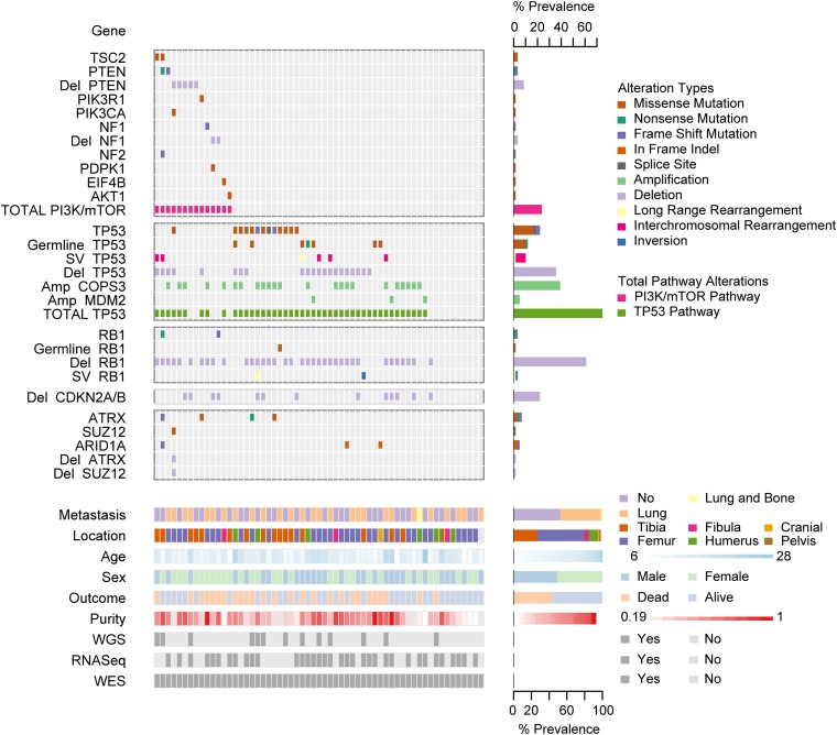
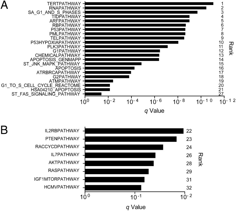
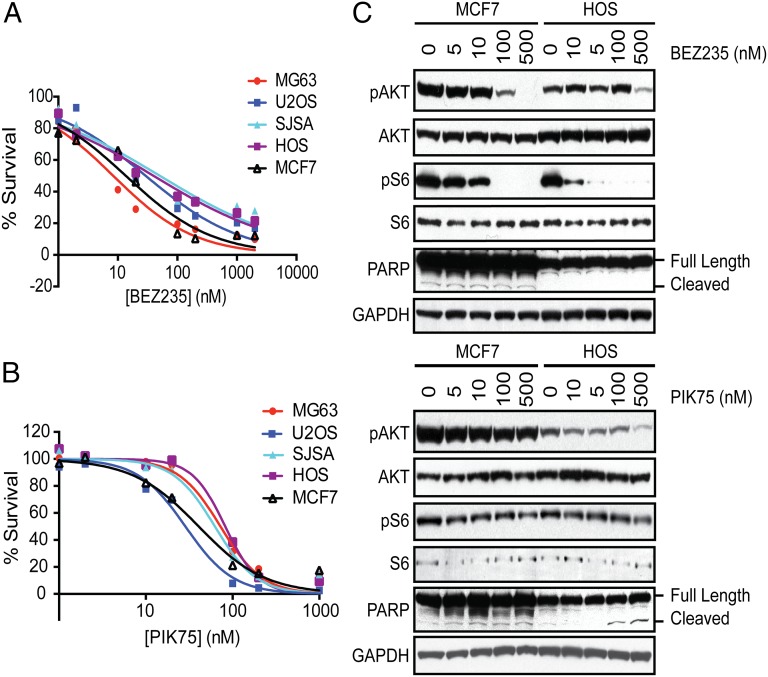
Osteosarcoma is the most common primary bone tumor, yet there have been no substantial advances in treatment or survival in three decades. We examined 59 tumor/normal pairs by whole-exome, whole-genome, and RNA-sequencing. Only the TP53 gene was mutated at significant frequency across all samples. The mean nonsilent somatic mutation rate was 1.2 mutations per megabase, and there was a median of 230 somatic rearrangements per tumor. Complex chains of rearrangements and localized hypermutation were detected in almost all cases. Given the intertumor heterogeneity, the extent of genomic instability, and the difficulty in acquiring a large sample size in a rare tumor, we used several methods to identify genomic events contributing to osteosarcoma survival. Pathway analysis, a heuristic analytic algorithm, a comparative oncology approach, and an shRNA screen converged on the phosphatidylinositol 3-kinase/mammalian target of rapamycin (PI3K/mTOR) pathway as a central vulnerability for therapeutic exploitation in osteosarcoma. Osteosarcoma cell lines are responsive to pharmacologic and genetic inhibition of the PI3K/mTOR pathway both in vitro and in vivo.

Keywords: PI3K; TP53; genomics; mTOR; osteosarcoma.


Conflict of interest statement

Conflict of interest statement: C.W.M.R. receives research support and consulting fees from the Novartis Institutes for Biomedical Research (NIBR) via the Dana–Farber Cancer Institute/NIBR drug discovery program.

Figures

Fig. 1.
Fig. 1.
Summary of sequencing results highlighting alterations in the PI3K/mTOR pathway, TP53, RB1, and TP53 and RB1 interacting genes, demographic and clinical variables, and sequencing characteristics. Each column represents a patient sample. The bottom section of the graph indicates the sequencing characteristics, demographic, and clinical data for each patient. The top section of the graph indicates the types of alteration for each gene or pathway per sample. Copy number alterations for PI3K/mTOR pathway genes, ATRX, SUZ12, and ARID1A were determined with the heuristic algorithm PHIAL. Copy number alterations for TP53, RB1, and TP53 and RB1 interacting genes were determined with GISTIC2. Amp, amplification; Del, deletion; SV, structural variation; long-range rearrangement, intrachromosomal rearrangements >1 Mb.
Fig. 2.
Fig. 2.
Associations between chromosomal rearrangements and clustered single-nucleotide mutations are evident in osteosarcoma. Representative Circos plots are labeled with sample ID. Chromosomes are depicted on the outer most track. C > T (green) and C > G (yellow) mutations are plotted according to the distance to the nearest mutation from 1 bp (inner circle) to 5 kb (toward outside of circle). Blue-red heatmap track indicates whole-genome sequence-based copy number ratio estimates. Arcs represent rearrangements and are depicted in the same color if they are statistically unlikely to have occurred independently of one another, as assessed by ChainFinder.
Fig. 3.
Fig. 3.
TP53 and PI3K/mTOR pathways are significantly mutated in osteosarcoma. GSEA combined with mutational analysis (MutSig2.0) identified 32 significantly mutated pathways (MSigDB Canonical Pathway gene sets) across 59 human osteosarcoma WES samples (q < 0.1). (A) Twenty-two pathways included the TP53 gene and (B) eight pathways implicated the PI3K/mTOR pathway by inclusion of PIK3CA and/or MTOR, while excluding TP53.
Fig. 4.
Fig. 4.
PI3K/mTOR identified as essential by in vitro and in vivo functional genomics screening. (A) Schematic of experimental design of genome-wide shRNA screen in mouse osteosarcoma (mOS) cell line. (B) Venn diagram outlining the overlap of genes identified by two methods of analysis: weighted second best and Kolmogorov–Smirnov statistic in GENE-E/RIGER. (C) The relative viability of mOS cell lines, determined by Alamar Blue assay, expressing four individual shRNAs each targeting Mtor, Pik3ca, or control shRNAs (GFP, LacZ, Luciferase, and RFP) compared with uninfected cells that were puromycin selected for 8 d (empty). Horizontal bars represent mean and SEM, n = 3. (D) Mean log-twofold change in shRNA abundance of control (GFP, LacZ, Luciferase, and RFP) or experimental shRNAs from five tumor samples compared with preinjection samples. *P < 0.01. (E) The relative viability of mOS cell lines expressing shRNAs targeting either Pik3ca, Pik3cb, or control (luciferase) at 9 d postinfection and puromycin selection determined by WST-1assay. Error bars are SEM, n = 3.
Fig. 5.
Fig. 5.
Small-molecule inhibitors of PI3K/mTOR inhibit osteosarcoma proliferation. Human OS cell lines (MG63, U2OS, SJSA, and HOS) and MCF7 cells (breast cancer cell line with an activating mutation in PIK3CA) were treated with (A) BEZ235 or (B) PIK75 or DMSO (control) for 24 h and viability was assessed with CellTiter-Glo. Experiments were conducted with eight replicates and displayed results are representative of three independent experiments. (C) Cells were treated with BEZ235 and PIK75 for 4 h and the effects on the PI3K/mTOR pathway were assessed. Cell extracts were analyzed by Western blotting with antibodies against phosphorylated AKT (Ser473), AKT, phosphorylated S6 (Ser235/236), S6, PARP, and GAPDH (loading control).

Comment in

  • Targeting osteosarcoma.
    Reed DE, Shokat KM. Reed DE, et al. Proc Natl Acad Sci U S A. 2014 Dec 23;111(51):18100-1. doi: 10.1073/pnas.1420596111. Epub 2014 Dec 15. Proc Natl Acad Sci U S A. 2014. PMID: 25512494 Free PMC article. No abstract available.

References

    1. Siegel R, Naishadham D, Jemal A. Cancer statistics, 2013. CA Cancer J Clin. 2013;63(1):11–30. - PubMed
    1. Mirabello L, Troisi RJ, Savage SA. Osteosarcoma incidence and survival rates from 1973 to 2004: Data from the Surveillance, Epidemiology, and End Results Program. Cancer. 2009;115(7):1531–1543. - PMC - PubMed
    1. Hansen MF, et al. Osteosarcoma and retinoblastoma: A shared chromosomal mechanism revealing recessive predisposition. Proc Natl Acad Sci USA. 1985;82(18):6216–6220. - PMC - PubMed
    1. McIntyre JF, et al. Germline mutations of the p53 tumor suppressor gene in children with osteosarcoma. J Clin Oncol. 1994;12(5):925–930. - PubMed
    1. Kansara M, Thomas DM. Molecular pathogenesis of osteosarcoma. DNA Cell Biol. 2007;26(1):1–18. - PubMed

Publication types