. 2014 Dec 30;111(52):18703-8.
doi: 10.1073/pnas.1422091112. Epub 2014 Dec 15.

pH modulates the activity and synergism of the airway surface liquid antimicrobials β-defensin-3 and LL-37

Affiliations

pH modulates the activity and synergism of the airway surface liquid antimicrobials β-defensin-3 and LL-37

Mahmoud H Abou Alaiwa et al. Proc Natl Acad Sci U S A. .

Abstract

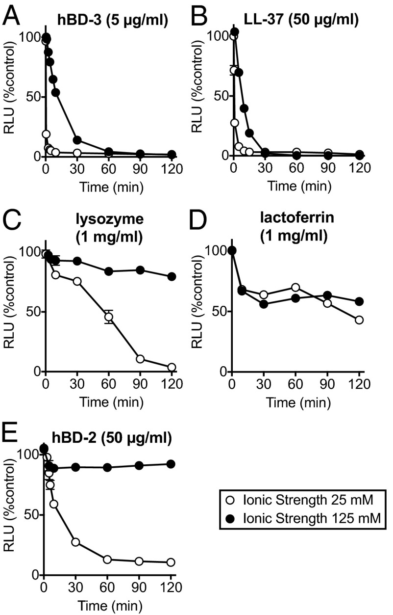
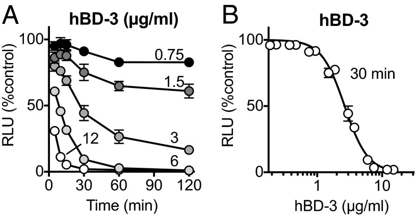
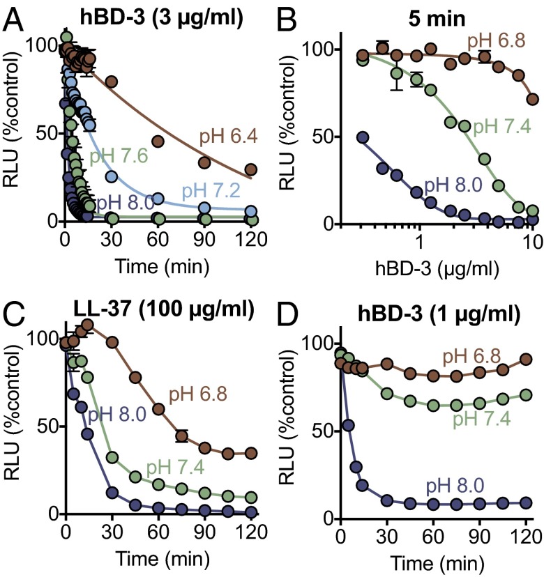
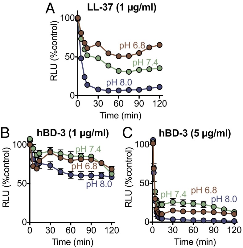
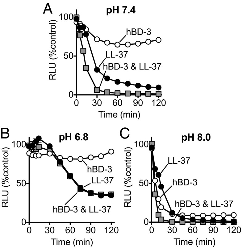
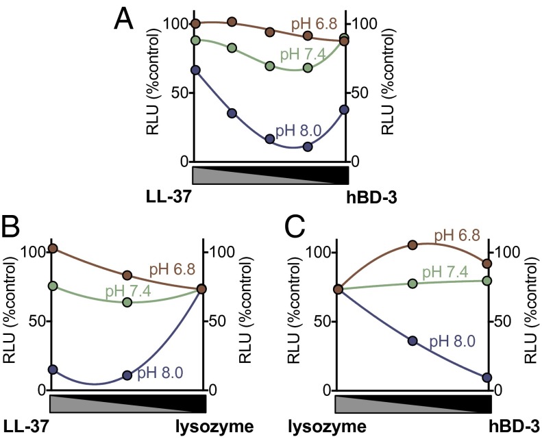
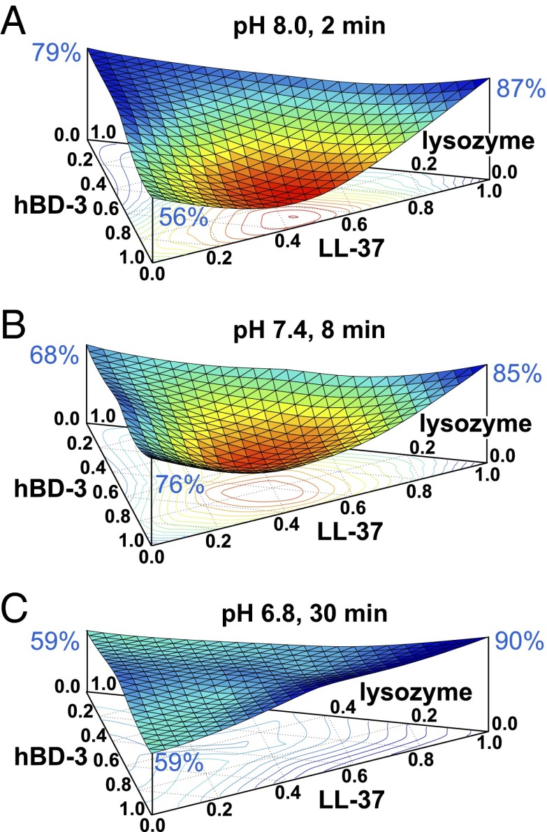
The pulmonary airways are continuously exposed to bacteria. As a first line of defense against infection, the airway surface liquid (ASL) contains a complex mixture of antimicrobial factors that kill inhaled and aspirated bacteria. The composition of ASL is critical for antimicrobial effectiveness. For example, in cystic fibrosis an abnormally acidic ASL inhibits antimicrobial activity. Here, we tested the effect of pH on the activity of an ASL defensin, human β-defensin-3 (hBD-3), and the cathelicidin-related peptide, LL-37. We found that reducing pH from 8.0 to 6.8 reduced the ability of both peptides to kill Staphylococcus aureus. An acidic pH also attenuated LL-37 killing of Pseudomonas aeruginosa. In addition, we discovered synergism between hBD-3 and LL-37 in killing S. aureus. LL-37 and lysozyme were also synergistic. Importantly, an acidic pH reduced the synergistic effects of combinations of ASL antibacterials. These results indicate that an acidic pH reduces the activity of individual ASL antimicrobials, impairs synergism between them, and thus may disrupt an important airway host defense mechanism.

Keywords: Pseudomonas aeruginosa; Staphylococcus aureus; cathelicidin; cystic fibrosis; host defense.


Conflict of interest statement

The authors declare no conflict of interest.

Figures

Fig. 1.
Fig. 1.
Antimicrobial activity of ASL factors at low and high ionic strength. Data are relative luminescence (in RLU) of S. aureus (Xen-29) as a percentage of control (no added antimicrobial and same buffer conditions) at an ionic strength of 25 mM (1% TSB, 10 mM potassium phosphate buffer: open circles) and at an ionic strength of 125 mM (1% TSB, 10 mM potassium phosphate buffer and 100 mM NaCl: closed circles) both at pH 7.4. (A) hBD-3 (5 μg/mL). (B) LL-37 (50 μg/mL). (C) Lysozyme (1 mg/mL). (D) Lactoferrin (1 mg/mL). (E) hBD-2 (50 μg/mL). Data are mean ± SEM; some error bars are hidden by symbols. Results are from a single experiment in triplicate. Each experiment was repeated at least three times with similar results.
Fig. 2.
Fig. 2.
Antimicrobial activity of hBD-3 is both time- and dose-dependent. (A) S. aureus luminescence as percentage of control measured over time at indicated concentrations of hBD-3 in μg/mL. (B) S. aureus luminescence with increasing concentrations of hBD-3 measured 30 min after addition. Data are mean ± SEM; some error bars are hidden by symbols. Results are from a single experiment in triplicate. Each experiment was repeated at least three times with similar results.
Fig. 3.
Fig. 3.
Effect of pH on antimicrobial activity of hBD-3 and LL-37 against S. aureus. (A) Effect of 3 μg/mL hBD-3 on S. aureus (Xen-29) luminescence measured over time at indicated pH. (B) Effect of hBD-3 concentration on S. aureus luminescence at three different pH values; measurements were made 5 min after addition. At pH 7.4, the IC50 for hBD-3 was 2.7 μg/mL, 95% CI (2.4–2.9). (C) Effect of 100 μg/mL LL-37 on S. aureus luminescence. (D) Effect of 1 μg/mL hBD-3 on S. aureus luminescence. Data are relative luminescence (in RLU) as a percentage of control S. aureus (Xen-29) that had no added antimicrobial but had same pH buffer conditions. Data are mean ± SEM; some error bars are hidden by symbols. Results are from a single experiment in triplicate. Each experiment was repeated at least three times with similar results.
Fig. 4.
Fig. 4.
Effect of pH on antimicrobial activity of hBD-3 and LL-37 against P. aeruginosa. Data are P. aeruginosa luminescence (in RLU). (A) LL-37 (1 μg/mL). (B) hBD-3 (1 μg/mL). (C) hBD-3 (5 μg/mL). Data are mean ± SEM; some error bars are hidden by symbols. Results are from a single experiment in triplicate. Each experiment was repeated at least three times with similar results.
Fig. 5.
Fig. 5.
Effect of pH on antimicrobial activity of hBD-3 and LL-37 in combination. Data are S. aureus (Xen-29) luminescence. Combination of 50 μg/mL of LL-37 and 0.5 μg/mL of hBD-3 at (A) pH 7.4, (B) pH 6.8, and (C) pH 8.0. Data are mean ± SEM; some error bars are hidden by symbols. Results are from a single experiment in triplicate. Each experiment was repeated at least three times with similar results. Graphs corresponding to the effect of individual factors in Fig. 4 A and B are replotted in A, B, and C for comparison.
Fig. 6.
Fig. 6.
Effect of pH on antimicrobial activity of combination of lysozyme, HBD-3, and LL-37. S. aureus (Xen-29) luminescence (in RLU) with varying ratios of two antimicrobial peptides. The x axis shows a ratio of antimicrobial peptides; from left to right there is an increasing ratio of one antimicrobial and a decreasing ratio of the other. (A) LL-37 and hBD-3 in combination (8 min incubation). (B) LL-37 and lysozyme in combination (30-min incubation). (C) Lysozyme and hBD-3 in combination (30-min incubation). Maximal combination (1.0) was 1 μg/mL for hBD-3, 100 μg/mL for LL-37, and 1 mg/mL for lysozyme. Data are mean ± SEM; some error bars are hidden by symbols. Results are from a single experiment in triplicate. Each experiment was repeated at least three times with similar results.
Fig. 7.
Fig. 7.
Ternary surface plot of the effect of pH antimicrobial activity on the combination of lysozyme, hBD-3, and LL-37. Data are S. aureus (Xen-29) luminescence (vertical axis, in RLU). The surface plot color and curvature correspond to changes in relative luminescence. Red and concave indicate less luminescence and low bacteria viability; blue and flat indicate more luminescence and high bacteria viability. Relative luminescence with each antimicrobial factor alone is indicated at top of each vertical axis in blue font. (Horizontal triangular axis indicates ratio of antimicrobial factors in triple combination). Maximal concentrations (1.0 on the axes) were 1 μg/mL for hBD-3, 100 μg/mL for LL-37, and 1 mg/mL for lysozyme. Data are shown at different times for each pH. (A) pH 8.0 after 2-min incubation. (B) pH 7.4 after 8-min incubation. (C) pH 6.8 after 30-min incubation. Results are an average of three experiments, each done in triplicate. Similar results were obtained with higher concentrations of antimicrobials (Fig. S1).

References

    1. Gallo RL, Hooper LV. Epithelial antimicrobial defence of the skin and intestine. Nat Rev Immunol. 2012;12(7):503–516. - PMC - PubMed
    1. Hancock RE, Diamond G. The role of cationic antimicrobial peptides in innate host defences. Trends Microbiol. 2000;8(9):402–410. - PubMed
    1. Boman HG. Peptide antibiotics and their role in innate immunity. Annu Rev Immunol. 1995;13:61–92. - PubMed
    1. Brogden KA. Antimicrobial peptides: Pore formers or metabolic inhibitors in bacteria? Nat Rev Microbiol. 2005;3(3):238–250. - PubMed
    1. Waterer GW. Airway defense mechanisms. Clin Chest Med. 2012;33(2):199–209. - PubMed

Publication types

MeSH terms