. 2014 Dec 30;111(52):18607-12.
doi: 10.1073/pnas.1416205112. Epub 2014 Dec 15.

Detergent-free isolation, characterization, and functional reconstitution of a tetrameric K+ channel: the power of native nanodiscs

Affiliations

Detergent-free isolation, characterization, and functional reconstitution of a tetrameric K+ channel: the power of native nanodiscs

Jonas M Dörr et al. Proc Natl Acad Sci U S A. .

Abstract

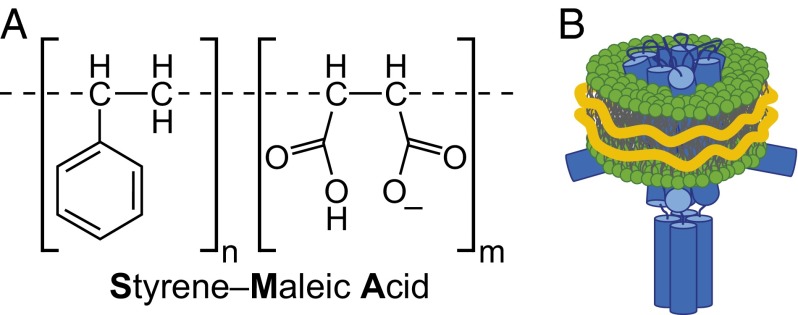
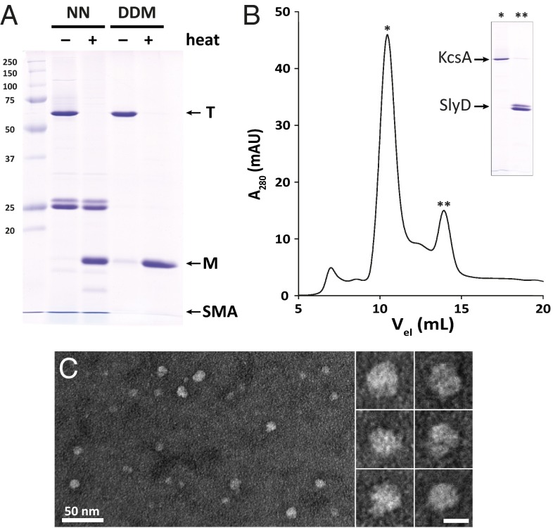
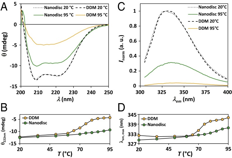
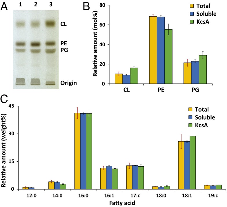
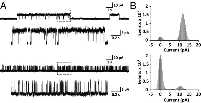
A major obstacle in the study of membrane proteins is their solubilization in a stable and active conformation when using detergents. Here, we explored a detergent-free approach to isolating the tetrameric potassium channel KcsA directly from the membrane of Escherichia coli, using a styrene-maleic acid copolymer. This polymer self-inserts into membranes and is capable of extracting membrane patches in the form of nanosize discoidal proteolipid particles or "native nanodiscs." Using circular dichroism and tryptophan fluorescence spectroscopy, we show that the conformation of KcsA in native nanodiscs is very similar to that in detergent micelles, but that the thermal stability of the protein is higher in the nanodiscs. Furthermore, as a promising new application, we show that quantitative analysis of the co-isolated lipids in purified KcsA-containing nanodiscs allows determination of preferential lipid-protein interactions. Thin-layer chromatography experiments revealed an enrichment of the anionic lipids cardiolipin and phosphatidylglycerol, indicating their close proximity to the channel in biological membranes and supporting their functional relevance. Finally, we demonstrate that KcsA can be reconstituted into planar lipid bilayers directly from native nanodiscs, which enables functional characterization of the channel by electrophysiology without first depriving the protein of its native environment. Together, these findings highlight the potential of the use of native nanodiscs as a tool in the study of ion channels, and of membrane proteins in general.

Keywords: ion channels; lipid–protein interactions; membrane–protein solubilization; nanodisc; styrene-maleic acid copolymer.


Conflict of interest statement

The authors declare no conflict of interest.

Figures

Fig. 1.
Fig. 1.
(A) Chemical structure of SMA polymers at neutral pH. For this study, a polymer with an average SMA ratio of n:m = 2:1 was used. (B) Schematic representation of a native nanodisc containing a KcsA tetramer (blue) and native lipids (green). The outer hydrophobic surface of the lipids is shielded by SMA (orange).
Fig. 2.
Fig. 2.
Purification of KcsA in native nanodiscs. (A) SDS/PAGE analysis of purified KcsA in native nanodiscs (NN) and DDM micelles at room temperature (−) and after incubation at 95 °C (+). Complete transitions from tetrameric (T) to monomeric (M) state are visible. (B) Size exclusion chromatogram showing effective separation of KcsA (*) from the soluble contaminant SlyD (**). (C) Negative stain transmission electron micrograph of native nanodiscs with KcsA after size exclusion chromatography. Round particles of an average size of 10 ± 2 nm are visible. (Scale bar in the enlarged images, 10 nm.)
Fig. 3.
Fig. 3.
Comparison of the thermal stability of KcsA in different environments. (A) Circular dichroism spectra of KcsA in native nanodiscs and DDM micelles at 20 °C and 95 °C. Data are offset corrected averages of 8–10 scans. (B) Corresponding thermal unfolding traces monitored by the ellipticity at 222 nm. Data are averages of two experiments with errors too small to be depicted. Solid lines are depicted to guide the eye. (C) Normalized fluorescence intensity of the intrinsic tryptophans of KcsA at 20 °C and 95 °C. Data are averages of three scans normalized to the intensity at 330 nm of the respective spectra at 20 °C. (D) Corresponding thermal unfolding traces monitored by the wavelength of maximum fluorescence emission. Solid lines are depicted to guide the eye.
Fig. 4.
Fig. 4.
Analysis of coisolated lipids in native nanodiscs. (A) TLC on extracted lipids from lysed whole bacterial cells after addition of SMA (1), complete soluble fraction separated by ultracentrifugation (2), and native nanodiscs with KcsA purified by Ni-affinity chromatography (3). (B) Molar composition of the extracted lipid species according to headgroup of total lysate (orange), soluble fraction (blue), and KcsA nanodiscs (green). Data are shown as average mol percentages with SDs (red error bars) of three independent nanodisc isolations. Data for the soluble fraction and KcsA nanodiscs were normalized to the corresponding total lysate. Black error bars denote SDs of the change compared with total lysate. (C) Gas chromatography analysis of the fatty acid composition of all isolated lipids. Depicted data are averages of two independent experiments. Errors are given as in B.
Fig. 5.
Fig. 5.
Functional characterization of KcsA reconstituted from native nanodiscs into a planar lipid bilayer of E. coli polar lipid extract. (A) Typical single-channel current traces on addition of KcsA nanodiscs to a setup with a symmetric 150-mM KCl solution at +100 mV. The top part represents a channel in the high open probability mode that is characterized by long opening dwell times, as shown in the enlarged section indicated by a box of dashed lines. Channels in the low open probability mode exhibit a distinctly different pattern, with short opening times resulting in sequences of bursts (Bottom). (B) All-point histograms for open/closed distribution of channels with high (Top) and low (Bottom) open probability calculated from the enlarged 2-s single-channel current traces from A.

References

    1. von Heijne G. The membrane protein universe: What’s out there and why bother? J Intern Med. 2007;261(6):543–557. - PubMed
    1. Garavito RM, Ferguson-Miller S. Detergents as tools in membrane biochemistry. J Biol Chem. 2001;276(35):32403–32406. - PubMed
    1. McQuade DT, et al. Rigid amphiphiles for membrane protein manipulation. Angew Chem Int Ed Engl. 2000;39(4):758–761. - PubMed
    1. Chae PS, et al. Maltose-neopentyl glycol (MNG) amphiphiles for solubilization, stabilization and crystallization of membrane proteins. Nat Methods. 2010;7(12):1003–1008. - PMC - PubMed
    1. Bowie JU. Stabilizing membrane proteins. Curr Opin Struct Biol. 2001;11(4):397–402. - PubMed

Publication types

MeSH terms