. 2015 Feb;35(4):728-36.
doi: 10.1128/MCB.01394-14. Epub 2014 Dec 15.

Novel long noncoding RNAs (lncRNAs) in myogenesis: a miR-31 overlapping lncRNA transcript controls myoblast differentiation

Affiliations

Novel long noncoding RNAs (lncRNAs) in myogenesis: a miR-31 overlapping lncRNA transcript controls myoblast differentiation

Monica Ballarino et al. Mol Cell Biol. 2015 Feb.

Erratum in

Abstract

Transcriptome analysis allowed the identification of new long noncoding RNAs differentially expressed during murine myoblast differentiation. These transcripts were classified on the basis of their expression under proliferating versus differentiated conditions, muscle-restricted activation, and subcellular localization. Several species displayed preferential expression in dystrophic (mdx) versus wild-type muscles, indicating their possible link with regenerative processes. One of the identified transcripts, lnc-31, even if originating from the same nuclear precursor of miR-31, is produced by a pathway mutually exclusive. We show that lnc-31 and its human homologue hsa-lnc-31 are expressed in proliferating myoblasts, where they counteract differentiation. In line with this, both species are more abundant in mdx muscles and in human Duchenne muscular dystrophy (DMD) myoblasts, than in their normal counterparts. Altogether, these data suggest a crucial role for lnc-31 in controlling the differentiation commitment of precursor myoblasts and indicate that its function is maintained in evolution despite the poor sequence conservation with the human counterpart.


Figures

FIG 1
FIG 1
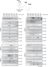
Muscle-specific poly(A)+ RNA-seq profiling. (A) Western blot analysis of MYOD1, MYOG, MEF2C, MYH6, and CKM protein expression in myoblasts grown under proliferation (GM) and differentiation (DM; 1, 3, and 5 days of differentiation [diff]) conditions. GAPDH levels were used as a control. (B and C) Validation of lncRNA expression by semiquantitative RT-PCR (sqRT-PCR) performed on total RNA extracted from GM or from myoblasts differentiated for 3 days (DM). lncRNA transcripts were grouped into upregulated (B) and downregulated (C) categories. −, RT minus control reactions. Gapdh mRNA was used as control. “a” and “b” indicate lnc-312, lnc-456, and lnc-686 splicing isoforms. PCR amplifications were performed on biological replicates, and the results of a representative experiment are shown.
FIG 2
FIG 2
Tissue specificity of lncRNAs in WT and mdx mice. sqRT-PCRs were performed on total RNA from WT and dystrophic (mdx) mouse tissue samples: brain (BRA), cerebellum (CRB), gastrocnemius (GAS), heart (HEA), lung (LUN), and tibialis (TIB). lncRNAs were grouped into several classes (a to d) according to their tissue specificity: class a, upregulated and muscle-restricted lncRNAs; class b, upregulated and heart-specific lncRNAs; class c, upregulated and ubiquitously expressed lncRNAs; and class d, downregulated and ubiquitously expressed lncRNAs. Gapdh mRNA levels were used as a control. Analysis was performed on samples from two different individuals, and results from a representative experiment are shown.
FIG 3
FIG 3
Subcellular distribution of the novel lncRNAs. Histograms show the quantifications of sqRT-PCR performed on RNA extracted from cytoplasmic (Cyt) and nuclear (N) fractions of myoblasts maintained under GM or DM (day 2) conditions. Results of a representative experiment are shown in Fig. S3 in the supplemental material. (A) Graphs show the cytoplasmic/nuclear partitioning of the lncRNAs, considering their expression conditions in GM or DM. The red shadow highlights the region of the graph occupied by lncRNAs with a bipartite subcellular distribution as defined by cytoplasmic/nuclear partitioning of control transcripts (see below). (B) Graphs show the quantification of the subcellular localization of control transcripts used to test the quality of the cytoplasmic/nuclear fractionation under GM or DM conditions: precursor (pre-Gapdh) and mature Gapdh mRNA, Rnu1-2 (U1) snRNA, and Snord55 (sno55). The red shadow defines the cytoplasmic/nuclear partition region in which no evident cytoplasmic/nuclear distribution can be assigned. Error bars represent standard deviations of data from multiple independent experiments.
FIG 4
FIG 4
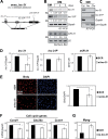
Murine lnc-31 transcript analysis. (A) Schematic representation of the genomic locus encompassing the murine lnc-31 coding region. The TSS (+1), the location of pre-miR-31, and the exon/intron structure are indicated together with the genomic coordinates. The arrow represents the position of the oligonucleotides used in the RT-PCR analyses; dashed lines denote the corresponding amplicons. (B) Analysis of murine lnc-31, lnc-31P, and miR-31 expression levels determined under GM or DM (1, 3, and 5 days of differentiation) conditions. Gapdh mRNA and Rnu2-10 (U2) snRNA were used as controls. Relative quantities (RQ) with respect to GM conditions are indicated below each lane; values are normalized to the amount of total Gapdh or U2 and represent averages of the results of three independent experiments. (C) Levels of murine lnc-31 and of its precursor lnc-31P analyzed by sqRT-PCRs performed on RNA extracted from cytoplasmic (Cyt), nucleoplasmic (Nu), and chromatinic (Chr) fractions of myoblasts under GM conditions. The oligonucleotide position for RT-PCRs is shown in panel A. Gapdh mRNA and pre-mRNA (pre-Gapdh) were used as controls (see also Fig. 3B). (D) qPCR analysis of lnc-31, lnc-31P, and miR-31 levels in C2C12 cells transfected with siRNAs against lnc-31 exonic sequences (si-lnc-31). Scramble siRNAs (SCR) were used as negative controls. lnc-31 and lnc-31P expression levels were normalized to Gapdh, whereas miR-31 expression levels were normalized to Rnu6 (U6) snRNA. (E) BrdU assay performed on C2C12 cells treated as described for panel D. Left panels show the BrdU and DAPI labeling, while the graph in the right panel shows the ratio between the BrdU-positive cells and the DAPI-positive cells under SCR and si-lnc-31 conditions. (F and G) Levels of Ccnd1, Cdc25a, Ccne1, and Myog mRNAs as measured by qPCR in C2C12 cells treated as described for panel D. Error bars represent standard errors of results from multiple independent experiments. *, P < 0.05; **, P < 0.01; ***, P < 0.001 (Student's t test).

References

    1. Edmondson DG, Olson EN. 1989. A gene with homology to the myc similarity region of MyoD1 is expressed during myogenesis and is sufficient to activate the muscle differentiation program. Genes Dev 3:628–640. doi:10.1101/gad.3.5.628. - DOI - PubMed
    1. Moncaut N, Rigby PWJ, Carvajal JJ. 2013. Dial M(RF) for myogenesis. FEBS J 280:3980–3990. doi:10.1111/febs.12379. - DOI - PubMed
    1. Perry RL, Rudnick MA. 2000. Molecular mechanisms regulating myogenic determination and differentiation. Front Biosci 5:D750–D767. doi:10.2741/Perry. - DOI - PubMed
    1. Sartorelli V, Caretti G. 2005. Mechanisms underlying the transcriptional regulation of skeletal myogenesis. Curr Opin Genet Dev 15:528–535. doi:10.1016/j.gde.2005.04.015. - DOI - PMC - PubMed
    1. Fatica A, Bozzoni I. 2014. Long non-coding RNAs: new players in cell differentiation and development. Nat Rev Genet 15:7–21. doi:10.1038/nrg3606. - DOI - PubMed

Publication types

MeSH terms

LinkOut - more resources