. 2015 Feb;35(4):747-57.
doi: 10.1128/MCB.00515-14. Epub 2014 Dec 15.

Glycolysis controls plasma membrane glucose sensors to promote glucose signaling in yeasts

Affiliations

Glycolysis controls plasma membrane glucose sensors to promote glucose signaling in yeasts

Amélie Cairey-Remonnay et al. Mol Cell Biol. 2015 Feb.

Abstract

Sensing of extracellular glucose is necessary for cells to adapt to glucose variation in their environment. In the respiratory yeast Kluyveromyces lactis, extracellular glucose controls the expression of major glucose permease gene RAG1 through a cascade similar to the Saccharomyces cerevisiae Snf3/Rgt2/Rgt1 glucose signaling pathway. This regulation depends also on intracellular glucose metabolism since we previously showed that glucose induction of the RAG1 gene is abolished in glycolytic mutants. Here we show that glycolysis regulates RAG1 expression through the K. lactis Rgt1 (KlRgt1) glucose signaling pathway by targeting the localization and probably the stability of Rag4, the single Snf3/Rgt2-type glucose sensor of K. lactis. Additionally, the control exerted by glycolysis on glucose signaling seems to be conserved in S. cerevisiae. This retrocontrol might prevent yeasts from unnecessary glucose transport and intracellular glucose accumulation.


Figures

FIG 1
FIG 1
(A) A simplified model of the Rag4-KlRgt1 glucose signaling pathway controlling RAG1 gene expression in Kluyveromyces lactis. The level of RAG1 transcription (Tx) depends on the quantity of extracellular glucose (high glucose = high transcription; no glucose = low transcription). The molecular mechanisms represented are detailed in the introduction. The dashed arrow represents the positive regulation by glycolysis on RAG1 expression that is analyzed in this study. The glucose-induced degradation of Sms1 was positioned between the nucleus and the cytoplasm because the localization of this process is uncertain. The nuclear envelope is shown as a dashed line. Phosphorylation (P) of Sms1 and Sck1 by Rag8 and KlRgt1 by an unknown kinase (kinase X) are presented. PM, plasma membrane; Cyt, cytosol; Nuc, nucleus; Tx, transcription; G-6-P, glucose-6-phosphate. (B) Table summarizing the functional conservation of the main factors of glucose signaling between K. lactis and S. cerevisiae. The molecular mechanisms of signal transduction of the two yeast species are similar. The function of each protein is indicated. A role for Tye7 and Snf2 in S. cerevisiae glucose signaling has not yet been reported. Tx, transcription.
FIG 2
FIG 2
Expression of glucose-regulated genes is downregulated in glycolytic mutants. The mRNA transcript levels of glucose permease gene RAG1 and glycolysis genes KlHXK and KlENO (A) and of glucose-signaling genes RAG4, RAG8, SMS1, and SCK1 (B) were determined by RT-qPCR in the wild-type strain (WT [MW270-7B]) and in ΔKlhxk (MWK11/F1), ΔKleno (MWK3), and ΔKlpgi (MWK12) mutants grown in YPD medium. Expression levels were normalized to the KlACT1 transcript level. For each gene, the mRNA expression level in the ΔKlhxk, ΔKleno, and ΔKlpgi mutants is presented relative to their level in the WT strain, which was set to 1 and is represented as a thick line on the graph (WT relative expression). The means of the relative-expression data from three biological replicates with standard deviation (error bars) are represented. Significance levels were determined by calculating the P value for each data set with Student's t test and are represented as asterisks: * for P < 0.05, ** for P < 0.01, and *** for P < 0.001. It should be noted that the ΔKlhxk mutant used in this figure (MWK11/F1) corresponds to a partial deletion of KlHXK coding for a nonfunctional truncated version of KlHxk. It is why KlHXK expression was still slightly observed by RT-qPCR in the ΔKlhxk strain.
FIG 3
FIG 3
KlRgt1 phosphorylation and Sms1 stability are affected in glycolytic mutants. (A) KlRgt1 phosphorylation in glycolytic mutants. WT (MWL1099), ΔKlhxk (MWL1118), and ΔKleno (MWL1121) strains expressing C-terminally 3HA-tagged KlRgt1 (KlRgt1-3HA) from the genome were grown in synthetic lactate medium to the exponential phase. One half of each culture was incubated with 2% glucose (+) for 30 min and the other was not (−) before cell collection and total protein extraction. Whole-cell lysates were analyzed for KlRgt1-HA by Western blotting (KlRgt1-3HA). (B) SMS1 is required for glycolytic control of KlRgt1 phosphorylation. KlRgt1-3HA expression in the WT (MWL1099), ΔKlhxk (MWL1118), Δsms1 (KlAS041), and ΔKlhxk Δsms1 (KlAS040) strains was analyzed by Western blotting after cells were stimulated or not stimulated with 2% glucose as described for panel A. (C) Sms1 protein level in glycolytic mutants. WT (KlAS029) and ΔKlhxk (KlAS026) strains expressing C-terminally 13myc-tagged Sms1 from the genome (Sms1-13myc) were grown and glucose stimulated as described for panels A and B. The Sms1 protein level was analyzed by Western blotting of whole-cell lysate. The percentage of Sms1 (% Sms1) was determined by densitometric analysis of the Sms1-13myc WB signal normalized to the actin signal (nd = not determined). The values are representative of the tendency observed in replica experiments. In all panels, a wild-type strain expressing untagged proteins (MWL9S1 or derivative) was used as a negative control (mock) and in-gel Coomassie staining of total protein extract and/or KlAct1 (Actin) immunodetection was used as a loading control (total extract).
FIG 4
FIG 4
Chemical glycolysis inhibition with iodoacetate affects KlRgt1 phosphorylation and Sms1 stability. (A) Iodoacetate (IA) confers a Rag phenotype. Representative examples of iodoacetate halo assays of WT cells (MWL9S1) on YPD and GAA (containing the respiratory inhibitor antimycin A) plates are shown. A 20-μl volume of water (0 μmol IA), 50 mM iodoacetate (1 μmol IA), or 100 mM iodoacetate (2 μmol IA) was dropped onto filter papers. (B) KlRgt1 is rapidly dephosphorylated upon iodoacetate treatment. Cells expressing KlRgt1-3HA (MWL1099) were grown to the exponential phase in complete synthetic medium with glycerol (Gly) or glucose (Glu) as the carbon source. Glycerol-grown cells were harvested, and glucose-grown cells were incubated with 0.25 mM iodoacetate and collected after 0, 1, 2, 5, and 10 min of treatment. Whole-cell lysates were prepared and analyzed for KlRgt1-3HA by Western blotting. Detection of total proteins by in-gel Coomassie staining was used as a loading control (total extract). (C) SMS1 deletion prevents iodoacetate-induced KlRgt1 dephosphorylation. WT (MWL1099) and Δsms1 (KlAS041) strains expressing KlRgt1-3HA were grown in glucose-containing medium (Glu) or in glycerol-containing medium (Gly) until the exponential phase. An aliquot of glucose-grown cells was treated with 0.25 mM iodoacetate for 10 and 30 min before harvest. Whole-cell lysates were prepared and analyzed for KlRgt1-3HA and actin (loading control) by Western blotting. The MWL9S1 wild-type strain was used as a mock treatment control. (D) Iodoacetate stabilizes Sms1 in glucose-grown cells. Sms1-13myc-expressing cells (KlAS029) were grown and treated as described for panel A. Total protein extract was analyzed for Sms1-13myc by Western blotting. Detection of total proteins by Ponceau staining was used as a loading control (total extract). The wild-type strain MWL9S1 was used as a mock treatment control.
FIG 5
FIG 5
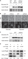
Casein kinase 1 Rag8 protein level, localization, and kinase activity in a hexokinase mutant. (A) The Rag8 protein level is stable in a glycolytic mutant. WT (MWL9S1) and ΔKlhxk (MWK11/F1) strains transformed with the pACR1 plasmid encoding an N-terminally LexA-tagged Rag8 (RAG8 promoter) were grown in lactate minimal media without uracil. Cells were then shifted (+) or not shifted (−) to 2% glucose and incubated for 30 min before cell collection. Whole-cell lysates were prepared and analyzed for LexA-Rag8 by Western blotting. MWL9S1 transformed with an empty plasmid was used as a mock treatment control. For a loading control, total proteins were analyzed by in-gel Coomassie staining (total extract). (B) GFP-Rag8 localization is not affected by the carbon source or by a glycolysis defect. WT (KlAS093) and ΔKlhxk (KlAS113) strains expressing N-terminally GFP-tagged Rag8 (PScGAL1 promoter) from the genome were grown in glucose-based (Glucose) or glycerol-based (Glycerol) minimal medium to the log phase and then observed with a fluorescence microscope. Cells were imaged using a GFP filter (GFP-Rag8) or Nomarski optics (differential inference contrast [DIC]). The acquisition times were equal for all GFP images. (C) Sck1 protein is stable in the hexokinase mutant. WT (MWL9S1) and ΔKlhxk (MWL1118) cells transformed with the pHN15 plasmid encoding LexA-Sck1 were grown in glycerol synthetic medium without uracil. Glucose was then added (+) or not added (−) to the cells for 30 min before being collected. The LexA-Sck1 protein level was then assessed by Western blotting of total protein extract. The mock treatment control corresponds to WT (MWL9S1) cells transformed with an empty plasmid. Total extracts were analyzed by Ponceau staining for a loading control (total extract). (D) Rag8 in vitro kinase activity is not affected in a glycolysis mutant. A wild-type form (WT) and a “kinase-deficient” form (K106R) of LexA-Rag8 were immunoprecipitated from WT (MWL9S1 plus pACR1 or MWL9S1 plus pACR3) and ΔKlhxk (MWK11/F1 plus pACR1) strains grown in synthetic glucose-containing medium without uracil (−ura). Kinase activity was assayed against 1 μg of recombinant His6-Sck1 expressed and purified from E. coli. Total and phosphorylated proteins were visualized by Coomassie staining (Coomassie) and autoradiography (32P), respectively. An aliquot of immunoprecipitated LexA-Rag8 was conserved for analysis of total LexA-Rag8 by Western blotting (WB). The level of Sck1 phosphorylation relative to the Rag8 quantity was quantified by densitometric analysis and normalized to the WT strain level. The specificity of the kinase activity was controlled by pretreating purified LexA-Rag8 with 1 mM ZnCl2 before performing the kinase reaction against His6-Sck1 (+ZnCl2).
FIG 6
FIG 6
The glucose sensor Rag4 is a target of glycolysis. (A and B) Rag4 protein stability is affected in a glycolysis mutant in a carbon source-dependent manner. (A) WT (MLK239) and ΔKlhxk (Δ) (ACRK101) strains expressing Rag4-TAP from its own promoter were grown in synthetic medium containing glucose (Glucose) or glycerol (Glycerol) as a carbon source. Whole-cell lysates were prepared and analyzed for Rag4-TAP by Western blotting. A wild-type strain expressing untagged proteins (MWL9S1 or derivative) was used as a negative control (mock), and in-gel Coomassie staining of total protein extract (total extract) and KlAct1 (Actin) immunodetection were used as a loading control. (B) WT (MLK191) and ΔKlhxk (ACRK106) strains expressing N-terminally 3HA-tagged Rag4 from the PScGAL1 promoter were grown and analyzed as described for panel A. (C) The Rag4 membrane distribution is impaired in a hexokinase mutant. Cell-free lysates from isogenic WT (MLK239) and ΔKlhxk (Δ) (ACRK101) strains expressing Rag4-TAP and grown in synthetic glucose-containing medium (Glucose) were subjected to differential centrifugation to separate heavy membranes (HM), light membranes (LM), and cytosolic supernatant (Sup). For all fractions, equal protein quantities were examined by Western blotting to detect Rag4-TAP, KlPma1, and KlAct1 (Actin). MWL9S1 (mock) was similarly processed to get a mock treatment control. Each fraction was analyzed by Ponceau staining for a loading control (total extract). (D) The in vivo Rag4 localization is impaired in the hexokinase mutant. WT (KlAS107) and ΔKlhxk (KlAS109) strains expressing N-terminally GFP-tagged Rag4 (PScGAL1 promoter) from the genome were grown to the exponential phase in synthetic minimal medium containing either glucose or glycerol. Cells were observed with a fluorescence microscope and imaged using a GFP filter (GFP-Rag4) or Nomarski optics (DIC). The acquisition times were equal for all GFP images. (E) Acute chemical inhibition of glycolysis by iodoacetate affects in vivo Rag4 localization. The KlAS107 strain used as described for panel C was grown to the log phase in YPD medium and then treated (+ IA) or not treated (no IA) with 0.25 mM iodoacetate. A fraction of the cells was collected periodically (0 min, 30 min, 2 h, and 6 h posttreatment) and observed under the microscope for the GFP signal (GFP-Rag4). The acquisition times were equal for all GFP images.
FIG 7
FIG 7
The S. cerevisiae glucose signaling pathway is inhibited in a hexokinase mutant. (A) The glucose-dependent phosphorylation of ScRgt1 is affected in a hexokinase mutant. WT (yAS279) and Δhxk1/2 (yAS280) S. cerevisiae strains expressing C-terminally 6HA-tagged ScRgt1 (ScRgt1-6HA) from the genome were grown in synthetic galactose medium to the exponential phase. One half of each culture was incubated with 2% glucose (+) for 30 min and the other was not (−) before cell collection and total protein extraction. Whole-cell lysates were analyzed for ScRgt1-HA (ScRgt1-6HA) and actin (loading control) by Western blotting. (B) Mth1 protein level in a hexokinase mutant. WT (yAS273) and Δhxk1/2 (yAS280) S. cerevisiae strains expressing C-terminally 9myc-tagged Mth1 from the genome (Mth1-9myc) were grown and treated as described for panel A. Whole-cell lysates were prepared and analyzed for Mth1-9myc and actin (loading control) by Western blotting. The percentage of Mth1-9myc in each lane (% Mth1) was determined by densitometric analysis of the myc signal normalized to the actin signal (nd, not determined). The values are representative of the tendency observed in replica experiments. (C) The Rgt2 protein level is impaired in a hexokinase mutant in a carbon source-dependent manner. WT (yAS275) and Δhxk1/2 (yAS276) S. cerevisiae strains expressing GFP-Rgt2 (PCUP1 promoter) from the genome were grown to the exponential phase in synthetic medium containing 0.02 mM CuSO4 (promoter induction) and glucose or galactose as a carbon source. Whole-cell lysates were prepared and analyzed for GFP-Rgt2 and actin (loading control) by Western blotting. A wild-type strain expressing untagged proteins (W303-1A) was used as a negative control (mock). A single asterisk corresponds to an aspecific band. (D) Rgt2 in vivo plasma membrane localization is impaired in the hexokinase mutant. S. cerevisiae strains and growth conditions were as described for panel C. Living cells were then observed with a fluorescence microscope and imaged using a GFP filter (GFP) or Nomarski optics (DIC). The acquisition times were equal for all GFP images.
FIG 8
FIG 8
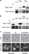
Chemical glycolysis inhibition with iodoacetate affects the S. cerevisiae glucose signaling pathway. (A) ScRgt1 is dephosphorylated upon iodoacetate treatment. Cells expressing ScRgt1-3HA (yAS223) were grown to the exponential phase in complete synthetic medium with glucose (Glu) or galactose (Gal) as a carbon source. Galactose-grown cells were harvested, and glucose-grown cells were incubated for 30 min with drug vehicle (−) or 0.25 mM iodoacetate (+). Cells were then collected, and whole-cell lysates were prepared and analyzed for ScRgt1-3HA by Western blotting. TB50a/α was used as a mock treatment control. Total proteins were analyzed by in-gel Coomassie staining for a loading control (total extract). (B) Iodoacetate stabilizes Mth1 in glucose-grown cells. Mth1-3HA-expressing cells (MLY788) were grown to the exponential phase in complete synthetic medium with glucose (Glu) or galactose (Gal) as a carbon source. Glucose-grown cells were then treated with 0.25 mM iodoacetate (+) or drug vehicle (−) for 30 min. Glucose (2% final concentration) was added to galactose-grown cells for 30 min (Gal Glu). After cell collection, total protein extracts were prepared and analyzed for Mth1-3HA by Western blotting. Mock and loading control experiments were done as described for panel B. Asterisks represent nonspecific signal. (C) Iodoacetate treatment affects Rgt2 localization at the plasma membrane. An S. cerevisiae strain expressing GFP-Rgt2 (PCUP1 promoter) from the genome (yAS233) was grown to the exponential phase in synthetic medium containing 0.1 mM CuSO4 (promoter induction) and 2% glucose (Glucose) or 2% lactate plus 0.1% glucose (Lactate). Cells were then treated with 0.25 mM iodoacetate (IA) or with water (untreated) and collected after 60 min. Cells were then observed with a fluorescence microscope and imaged using a GFP filter (GFP-Rgt2) or Nomarski optics (DIC). The acquisition times were equal for all GFP images.

References

    1. Horák J. 2013. Regulations of sugar transporters: insights from yeast. Curr Genet 59:1–31. doi:10.1007/s00294-013-0388-8. - DOI - PubMed
    1. Rodicio R, Heinisch JJ. 2013. Yeast on the milky way: genetics, physiology and biotechnology of Kluyveromyces lactis. Yeast 30:165–177. doi:10.1002/yea.2954. - DOI - PubMed
    1. Goffrini P, Wesolowski-Louvel M, Ferrero I, Fukuhara H. 1990. RAG1 gene of the yeast Kluyveromyces lactis codes for a sugar transporter. Nucleic Acids Res 18:5294. doi:10.1093/nar/18.17.5294. - DOI - PMC - PubMed
    1. Billard P, Ménart S, Blaisonneau J, Bolotin-Fukuhara M, Fukuhara H, Wésolowski-Louvel M. 1996. Glucose uptake in Kluyveromyces lactis: role of the HGT1 gene in glucose transport. J Bacteriol 178:5860–5866. - PMC - PubMed
    1. Wésolowski-Louvel M, Goffrini P, Ferrero I, Fukuhara H. 1992. Glucose transport in the yeast Kluyveromyces lactis. I. Properties of an inducible low-affinity glucose transporter gene. Mol Gen Genet 233:89–96. - PubMed

Publication types

MeSH terms

Substances