. 2014 Oct 29:11:15.
doi: 10.1186/1742-4933-11-15. eCollection 2014.

Characterization of recovery, repair, and inflammatory processes following contusion spinal cord injury in old female rats: is age a limitation?

Affiliations

Characterization of recovery, repair, and inflammatory processes following contusion spinal cord injury in old female rats: is age a limitation?

Mitra J Hooshmand et al. Immun Ageing. .

Abstract

Background: Although the incidence of spinal cord injury (SCI) is steadily rising in the elderly human population, few studies have investigated the effect of age in rodent models. Here, we investigated the effect of age in female rats on spontaneous recovery and repair after SCI. Young (3 months) and aged (18 months) female rats received a moderate contusion SCI at T9. Behavioral recovery was assessed, and immunohistocemical and stereological analyses performed.

Results: Aged rats demonstrated greater locomotor deficits compared to young, beginning at 7 days post-injury (dpi) and lasting through at least 28 dpi. Unbiased stereological analyses revealed a selective increase in percent lesion area and early (2 dpi) apoptotic cell death caudal to the injury epicenter in aged versus young rats. One potential mechanism for these differences in lesion pathogenesis is the inflammatory response; we therefore assessed humoral and cellular innate immune responses. No differences in either acute or chronic serum complement activity, or acute neutrophil infiltration, were observed between age groups. However, the number of microglia/macrophages present at the injury epicenter was increased by 50% in aged animals versus young.

Conclusions: These data suggest that age affects recovery of locomotor function, lesion pathology, and microglia/macrophage response following SCI.

Keywords: Aging; Complement; Inflammation; Locomotor function; Spinal cord injury.


Figures
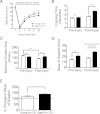
Figure 1
Figure 1
Aged rats showed greater locomotor deficits. (A) Repeated-measures ANOVA yielded a main significant effect for performance of young vs aged animals (F = 2.36, *p = 0.044) in the open-field. Post-hoc tests revealed a significant difference between age groups at 7, 21 and 28 days post-injury (Bonferroni’s multiple comparison, #p<0.05). On day 28, aged rats showed greater locomotor deficits compared to the young. (B) Using CatWalk analysis, no differences in crossing time were found between age groups at baseline, but post-SCI, aged animals demonstrated an increase in this parameter compared to young. (C) Young animals exhibited decreased contact area compared to aged animals post-injury as well as relative to their pre-injury baseline. (D-E) Since base of support was significantly different between age groups at baseline (D), post-injury data were normalized to pre-injury performance (dashed line in E) and demonstrated a significant increase in base of support of aged animals compared to young (E). Student’s t-test were used to compare between groups: ***p< 0.001; **p< 0.01, *p<0.05. Mean ± SEM is shown.
Figure 2
Figure 2
Morphometric tissue analysis revealed the critical importance of volume correction. Using direct comparisons, aged rats showed a significant increase in volume of injured tissue compared to their young counterparts (Student’s t test, *p< 0.05) (A). No age-associated differences were observed in the total lesion spread (B). Three-dimensional reconstructions of representative injured spinal cord segments (T6-T12) suggested increased length and cross-sectional area in aged animals compared to young (C).
Figure 3
Figure 3
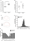
Volume correction demonstrated increased and localized pathology in aged rats compared to young at 28 dpi. Confirming observations in 3D reconstructions, stereological assessment of total spinal cord volume between T6 and T12 dorsal roots revealed a significant difference between age groups (Student’s t test, *p< 0.001) (A). Accordingly, lesion volume from Figure  3A was normalized to total cord volume (A) and expressed as a percentage. As such, aged rats showed a strong but non-significant trend for increased % lesion volume compared to young rats (Student’s t test, p= 0.056) (B). Stereologically analyzed coronal diagrams from a single section immediately caudal to the injury epicenter demonstrated potential differences in lesion area (red line) relative to cross-sectional area (black line) (C). When lesion area at each cross-section was normalized to total area at the same cross-section and quantified along the length of the spinal cord, a single point of significance was detected immediately caudal to the injury epicenter, where % lesion area of aged animals was significantly greater than the young (Student’s t test, *p< 0.05) (D). (E) Additionally, quantification of the % spared tissue along the length of the spinal cord demonstrated the same effect as % lesion area, confirming localized differences between age groups in pathology (Student’s t test, *p< 0.05). Data points represent individual animals and the horizontal line indicates group mean.
Figure 4
Figure 4
Early TUNEL-mediated cell death was selectively increased caudal to the epicenter in aged animals. Low power representative photomicrographs of coronal spinal cord sections from aged and young animals labeled for TUNEL (blue) and counter-stained with methyl green (green) are shown (A). 3D reconstructions of TUNEL-positive quantification revealed a heterogeneous distribution of TUNEL-positive cells in both age groups (B). Stereological quantification of TUNEL-positive cells showed no significant differences between the total number of TUNEL-positive cells in aged and young animals (C). Detailed distribution analysis of TUNEL-positive cells demonstrated a single point of significance where in the caudal spinal cord region, significantly more apoptotic cells were observed in aged animals compared to the young (Student’s t test, *p< 0.05) (D). Scale bar = 100 μm.
Figure 5
Figure 5
Total systemic complement activity after SCI was not influenced by age. CH50 assays showed that systemic complement activity in young animals was not significantly different from that of aged animals at 2 days post-SCI (A) or at 28 days post-SCI (B) (Student’s t test, p >0.05). Mean ± SEM is shown.
Figure 6
Figure 6
Aged rats showed an acute and localized increase in microglia/macrophage infiltration. Representative photomicrographs of coronal spinal cord sections immunolabeled for neutophils with CD43 (A-B) or microglia/macrophages with ED-1 (D-E) and counter-stained with methyl green are shown. Insets are of individual fields at higher magnification. Unbiased stereological quantification of total numbers of CD43-positive cells (C) and ED-1 positive cells (F) revealed no significant differences between age groups. Rostro-caudal distribution of CD43+ cells did not exhibit differences between groups (p > 0.05) (G). In contrast, ED-1 positive cells showed significantly increased age-associated microglia/macrophage response at the injury epicenter (H). Student's t-tests, *p< 0.05. Mean ± SEM is shown. Scale bar = 25 μm.

References

    1. Cifu DX, Seel RT, Kreutzer JS, Marwitz J, McKinley WO, Wisor D. Age, outcome, and rehabilitation costs after tetraplegia spinal cord injury. Neurorehabilitation. 1999;12(3):177–185.
    1. Dai LY. Acute central cervical cord injury: the effect of age upon prognosis. Inj Int J Care Inj. 2001;32(3):195–199. doi: 10.1016/S0020-1383(00)00176-5. - DOI - PubMed
    1. Furlan JC, Bracken MB, Fehlings MG. Is age a key determinant of mortality and neurological outcome after acute traumatic spinal cord injury? Neurobiol Aging. 2010;31(3):434–446. doi: 10.1016/j.neurobiolaging.2008.05.003. - DOI - PubMed
    1. Fehlings MG, Furlan JC. Effect of age on spinal cord injury. J Neurosurg Spine. 2007;7(3):275–276. doi: 10.3171/SPI-07/09/275. discussion 276. - DOI - PubMed
    1. Gwak YS, Hains BC, Johnson KM, Hulsebosch CE. Effect of age at time of spinal cord injury on behavioral outcomes in rat. J Neurotrauma. 2004;21(8):983–993. doi: 10.1089/0897715041650999. - DOI - PubMed