. 2014 May 31:2:11.
doi: 10.1186/2050-5736-2-11. eCollection 2014.

First experience with MR-guided focused ultrasound in the treatment of Parkinson's disease

Affiliations

First experience with MR-guided focused ultrasound in the treatment of Parkinson's disease

Anouk Magara et al. J Ther Ultrasound. .

Abstract

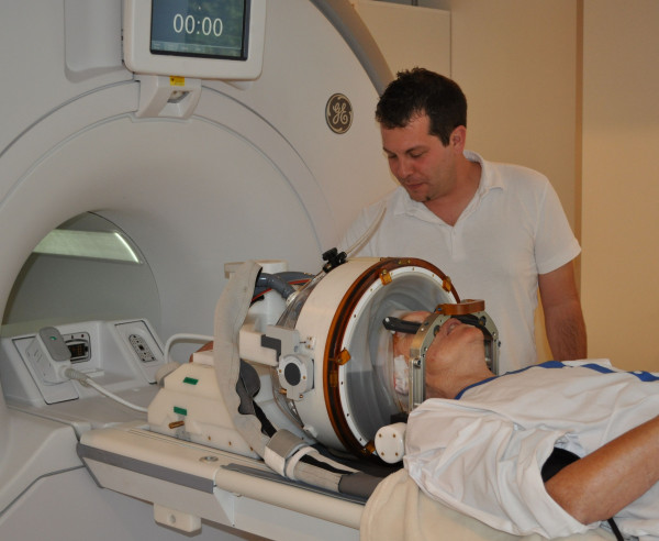
Background: Radiofrequency (RF) subthalamotomies have been proposed since the 1960s to treat patients suffering from Parkinson's disease (PD). Recently, the magnetic resonance (MR)-guided focused ultrasound technology (MRgFUS) offers the possibility to perform subthalamic thermocoagulations with reduced risks and optimized accuracy. We describe here the initial results of the MRgFUS pallidothalamic tractotomy (PTT), an anatomical and physiological update of the earlier subthalamotomies.

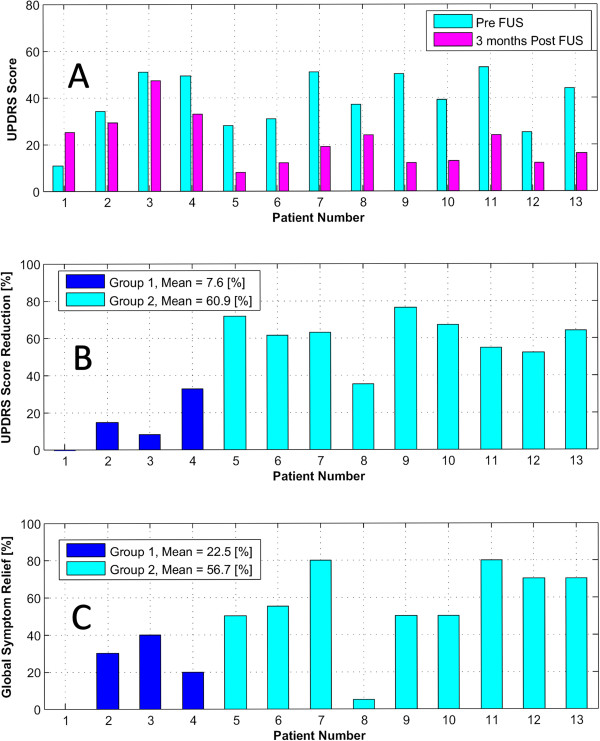
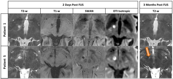
Methods: Thirteen consecutive patients suffering from chronic (mean disease duration 9.7 years) and therapy-resistant PD were treated unilaterally with an MRgFUS PTT. Primary relief assessment indicators were the score reduction of the Unified Parkinson Disease Rating Scale (UPDRS) and the patient estimation of global symptom relief (GSR) taken at 3 months follow-up. Final temperatures at target were between 52°C and 59°C. The MR examinations were performed before the treatment, 2 days and 3 months after it. The accuracy of the targeting was calculated on 2 days post-treatment MR pictures for each PTT lesion.

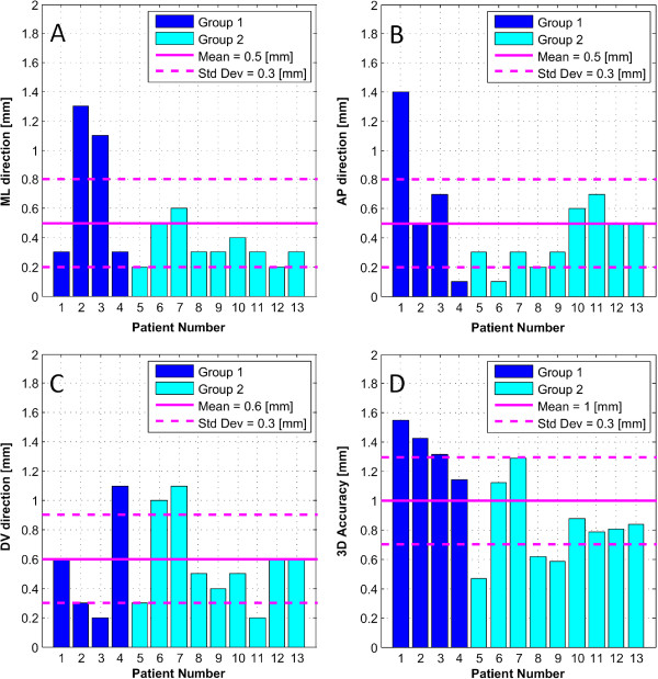
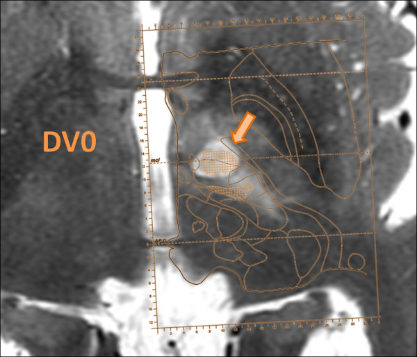
Results: The first four patients received a PTT using the lesional parameters applied for thalamotomies. They experienced clear-cut recurrences at 3 months (mean UPDRS relief 7.6%, mean GSR 22.5%), and their MR showed no sign of thermal lesion in T2-weighted (T2w) images. As a consequence, the treatment protocol was adapted for the following nine patients by applying repetition of the final temperatures 4 to 5 times. That produced thermocoagulations of larger volumes (172 mm(3) against 83 mm(3) for the first four patients), which remained visible at 3 months on T2w images. These nine patients enjoyed a mean UPDRS reduction of 60.9% and a GSR of 56.7%, very close to the results obtained with radiofrequency lesioning. The targeting accuracy for the whole patient group was 0.5, 0.5, and 0.6 mm for the anteroposterior (AP), mediolateral (ML), and dorsoventral (DV) dimensions, respectively.

Conclusions: This study demonstrated the feasibility, safety, and accuracy of the MRgFUS PTT. To obtain similar results as the ones of RF PTT, it was necessary to integrate the fact that white matter, in this case, the pallidothalamic tract, requires repeated thermal exposition to achieve full lesioning and thus full therapeutic effect.


Figures

Figure 1
Figure 1
Intra-operative patient setup. Patient lies on MR bed with the head immobilized by a metal frame and placed inside the phased-array ultrasound transducer. The space between the head and the transducer has been sealed by a silicone membrane and is being filled with cooled and degassed water.
Figure 2
Figure 2
Absolute global targeting accuracy. The bar graphs display the absolute difference between the coordinates of the center of the atlas target and of the realized target (lesion) for the mediolateral (ML) direction (A), anterioposterior (AP) direction, (B) and dorsoventral direction (C), as well as the resulting three-dimensional vector length (D). Std Dev stands for standard deviation.
Figure 3
Figure 3
Superposition of atlas map over MR imaging. Axial MR T2-weighted (T2w) series taken at 2 days follow-up with superposition of the corresponding Morel atlas plane at the dorsoventral position 0 mm (DV0, the intercommissural plane). The hypodense lesion is exactly centered on the pallidothalamic tract as displayed on the atlas.
Figure 4
Figure 4
UPDRS score and global symptom relief of the two groups. (A) Pre- and 3 months post-FUS Unified Parkinson's Disease Rating Scale (UPDRS) score of the 13 patients. (B) UPDRS score reduction (in percent) for the same 13 patients. (C) Subjective Global Symptom Relief (3 months post vs. pre-FUS), in percent, for the 13 patients.
Figure 5
Figure 5
MR imaging differences. Lesion comparison between patient number 1 (group 1) and patient number 5 (group 2) on axial T2-weighted (T2-w), three-dimensional T1-weighted (T1-w), three-dimensional SWAN and Diffusion Tensor Imaging with isotropic post-processing MR series on 2 days and 3 months (only T2-w series) post-operative follow-up. Lesion can be seen on 3 months post-FUS MR imaging only in patient of group 2 (bottom, right). All scans are located 2 mm below the intercommissural plane.

References

    1. Deuschl G, Schade-Brittinger C, Krack P, Volkmann J, Schafer H, Botzel K. A randomized trial of deep-brain stimulation for Parkinson's disease. New England J Med. 2006;355:896–908. doi: 10.1056/NEJMoa060281. - DOI - PubMed
    1. Hamani C, Richter E, Schwalb J, Lozano A. Bilateral subthalamic nucleus stimulation for Parkinson's disease: a systematic review of the clinical literature. Neurosurgery. 2005;56:1313–1321. doi: 10.1227/01.NEU.0000159714.28232.C4. - DOI - PubMed
    1. Kleiner-Fisman G, Fisman D, Sime E, Saint-Cyr J, Lozano A, Lang A. Long-term follow up of bilateral deep brain stimulation of the subthalamic nucleus in patients with advanced Parkinson's disease. J Neurosurg. 2003;99:489–495. doi: 10.3171/jns.2003.99.3.0489. - DOI - PubMed
    1. Patel N, Heywood P, O'Sullivan K, McCarter R, Love S, Gill S. Unilateral subthalamotomy in the treatment of Parkinson's disease. Brain. 2003;126:1136–1145. doi: 10.1093/brain/awg111. - DOI - PubMed
    1. Rodriguez-Oroz M, Obeso J, Lang A, Houeto J, Pollak P, Rehncrona S. Bilateral deep brain stimulation in Parkinson's disease: a multicentre study with 4 years follow-up. Brain. 2005;128:2240–2249. doi: 10.1093/brain/awh571. - DOI - PubMed

LinkOut - more resources