Review
. 2014 Nov;3(6):612-20.
doi: 10.3978/j.issn.2225-319X.2014.11.07.

SynCardia: the total artificial heart

Affiliations
Review

SynCardia: the total artificial heart

Gianluca Torregrossa et al. Ann Cardiothorac Surg. 2014 Nov.

Abstract

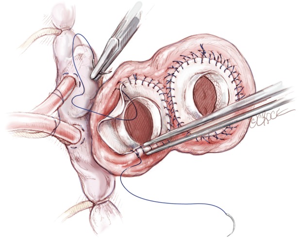
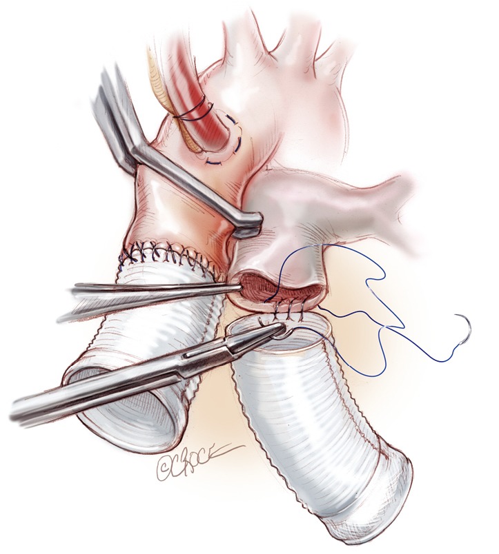
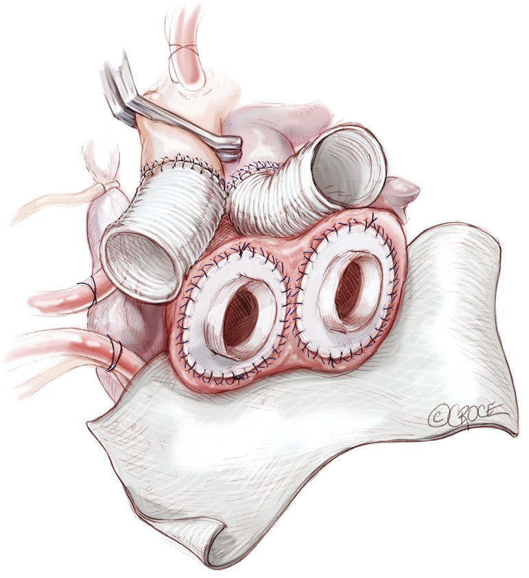
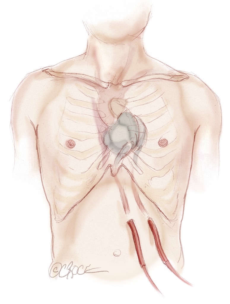
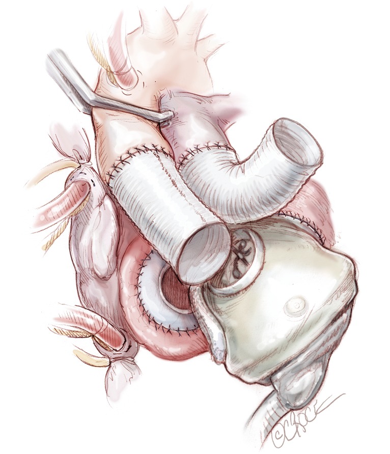
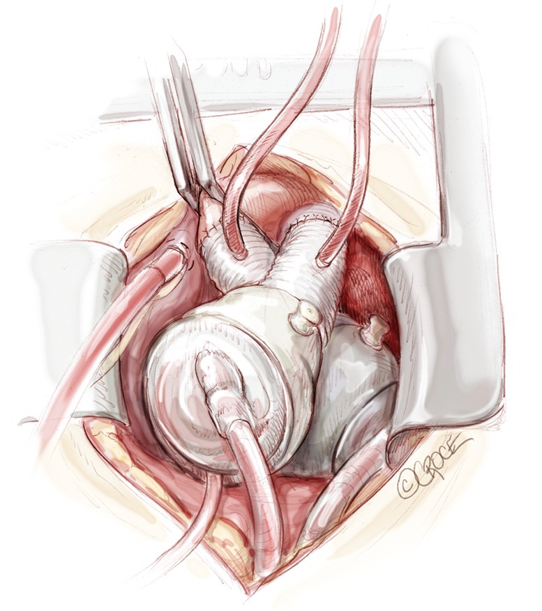
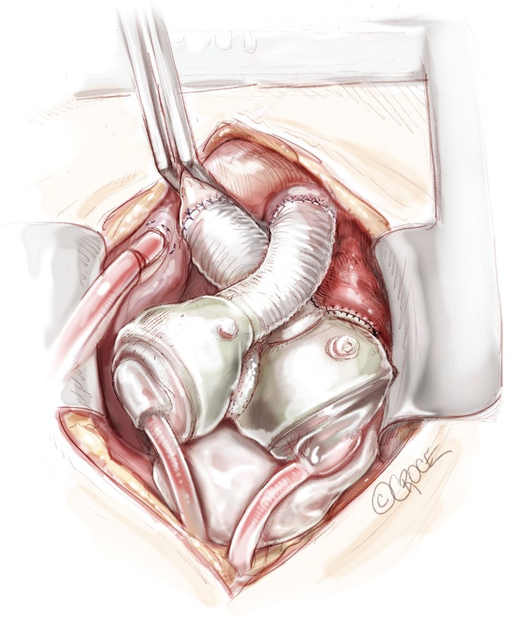
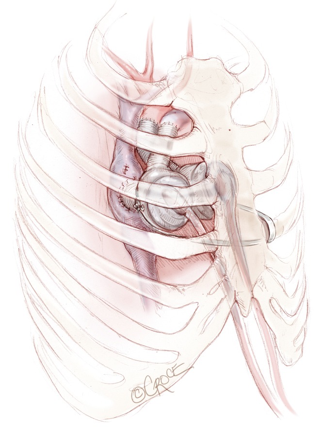
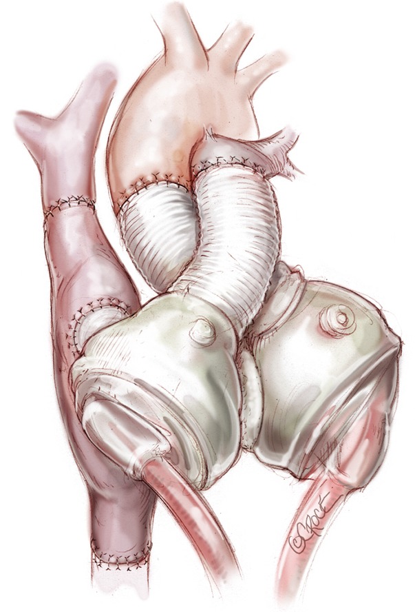
The SynCardia total artificial heart (TAH) currently provides the most definitive option for patients with biventricular failure who are not candidates for isolated left ventricular (LV) assist device placement. The techniques for implantation are adaptable to almost all patients with advanced heart failure, including those with severe biventricular cardiomyopathy, complex congenital heart disease, failed LV assist devices, failed transplantations, and acquired structural heart defects that have failed or are not amenable to conventional surgical treatment. Over the years, the implantation technique has evolved in order to minimize the surgical invasiveness of the procedure, in anticipation of additional future surgery. Meticulous hemostasis with double layer sutures, use of Gore-Tex sheets around the TAH and the pericardial cavity, and use of tissue expanders to avoid contraction of pericardial cavity around the device are discussed in detail in the following report. Additionally, we will provide our experience with implantation of TAH in various challenging scenarios, such as patients with a small chest cavity, congenital heart defects, and simultaneous use of extracorporeal membrane oxygenation (ECMO).

Keywords: SynCardia; Total artificial heart (TAH); cardiowest; implantation technque.


Figures

Figure 1
Figure 1
Device preparation: the quick connects of the atrial cuffs are trimmed and cut in a completely circular fashion leaving about three to five mm of sewing cuff. All four connectors are stretched with a clamp to facilitate later connection to the device ventricle.
Figure 2
Figure 2
Cardiopulmonary bypass institution: the ascending aorta is cannulated distally. We prefer to cannulate both cavae through the right atrium [vs. direct cannulation of the superior vena cava (SVC) and inferior vena cava (IVC)] to preserve those sites for the heart transplantation surgery.
Figure 3
Figure 3
The excision of the heart proceeds along the lines depicted in the figure. The purpose of the explantation of the heart is to leave in place both atria and 1-2 cm of ventricle muscle around each atrioventricular valve plane preserving both annuli of the mitral and tricuspid valves.
Figure 4
Figure 4
Native heart is removed. Preparation for anastomosis: the excess muscle is trimmed away around the atrioventricular junction. Coronary sinus and left atrial appendage are oversewn.
Figure 5
Figure 5
Atrial connectors are placed in the right and left cuffs with a double layer of prolene 3-0. We do not use any Felt Strip reinforcement around the atrial cuffs.
Figure 6
Figure 6
Anastomosis of the great arteries is performed. The outflow conduits are trimmed to size (3-4 cm for the aortic graft; 6-7 cm for the pulmonary one). The end-to-end anastomosis is done with a double layer 4-0 prolene.
Figure 7
Figure 7
Before placement of the ventricles, we suture two large sheets of Gore-Tex to the atrioventricular groove in order to facilitate the reentry at the time of transplantation.
Figure 8
Figure 8
The two drivelines are tunnelled under the skin and the artificial ventricle brought into the surgical field. The exit of the left driveline is seven to 10 cm below the left costal margin along the mid clavicular line. The exit of the right driveline is generally five cm medially from the left one.
Figure 9
Figure 9
The left ventricle is connected first. Careful assessment of the exact position of the ventricle and its orientation is crucial at this stage. The left atrial quick connector is grasped with two Mayo clamps and while pulling on them, the ventricle is pushed into the quick connector in its final position.
Figure 10
Figure 10
Total artificial heart in final position inside the chest. A vent is placed in the pulmonary graft and in the aortic graft before releasing the aortic cross clamp.
Figure 11
Figure 11
A breast implant is generally left in the pericardium to avoid contraction of the mediastinum around the total artificial heart (TAH) and leave space inside the chest cavity for the donor heart at the time of transplantation.
Figure 12
Figure 12
Umbilical tape or a heavy silk or polyester suture is generally wrapped two or three times around the main body of the left artificial ventricle; the two ends of the tape or suture are then tunnelled outside the chest through an intercostal space and tied around one of the rib in order to displace the left artificial ventricle.
Figure 13
Figure 13
Total artificial heart (TAH) in a Fontan physiology patient.

References

    1. DeVries WC, Anderson JL, Joyce LD, et al. Clinical use of the total artificial heart. N Engl J Med 1984;310:273-8. - PubMed
    1. El-Banayosy A, Arusoglu L, Morshuis M, et al. CardioWest total artificial heart: Bad Oeynhausen experience. Ann Thorac Surg 2005;80:548-52. - PubMed
    1. Copeland JG, Smith RG, Arabia FA, et al. Cardiac replacement with a total artificial heart as a bridge to transplantation. N Engl J Med 2004;351:859-67. - PubMed
    1. Copeland JG, Copeland H, Gustafson M, et al. Experience with more than 100 total artificial heart implants. J Thorac Cardiovasc Surg 2012;143:727-34. - PubMed
    1. Kirsch ME, Nguyen A, Mastroianni C, et al. SynCardia temporary total artificial heart as bridge to transplantation: current results at la pitié hospital. Ann Thorac Surg 2013;95:1640-6. - PubMed