. 2014 Dec 15;5(23):12331-45.
doi: 10.18632/oncotarget.2589.

Elevated endoplasmic reticulum stress reinforced immunosuppression in the tumor microenvironment via myeloid-derived suppressor cells

Affiliations

Elevated endoplasmic reticulum stress reinforced immunosuppression in the tumor microenvironment via myeloid-derived suppressor cells

Bo-Ra Lee et al. Oncotarget. .

Abstract

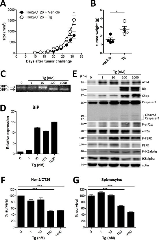
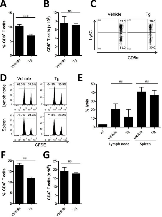
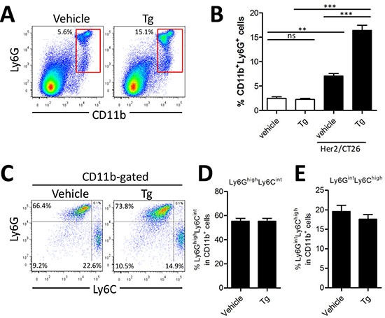
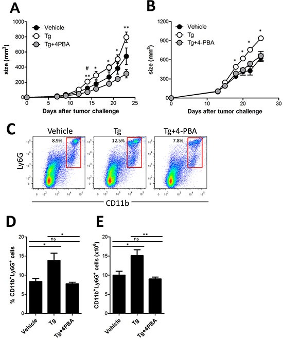
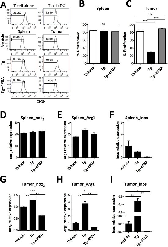
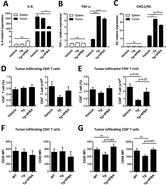
The role of endoplasmic reticulum (ER) stress in cancer has been studied in detail, and ER stress is known to increase tumor cell apoptosis, and thus, reduce tumor growth. However, in our study, persistent ER stress induced by multiple administrations of low-dose thapsigargin (Tg) accelerated tumor growth in mice. Tg-mediated ER stress increased the generation of Ly6G+CD11b+ myeloid cells, but did not alter anti-tumor effector T cells. 4-Phenylbutyric acid (4-PBA), a chemical chaperone widely used as an ER stress reducer, attenuated Tg-induced myeloid-derived suppressor cell (MDSC) expansion and tumor growth. Tg-mediated ER stress enhanced the immunosuppressive capacity of tumor-infiltrating MDSCs by increasing expression of ARG1, iNOS, and NOX2, although splenic MDSCs were not affected. Consistent with these results, 4-PBA restored the anti-tumor immune response by regulating inflammatory cytokines such as TNF-α and CXCL1/KC, and activated tumor-infiltrating CD8+ T cells that were inhibited by Tg-mediated ER stress. These results suggest that significant ER stress in a tumor-bearing host might induce tumor growth mediated by enhancement of MDSC-mediated suppression. Therefore, ER stress reducers such as 4-PBA could restore anti-tumor immunity by inhibiting suppressive MDSCs that are exacerbated by ER stress.


Conflict of interest statement

Conflict of interest

The authors declare they have no conflicts of interest.

Figures

Figure 1
Figure 1. ER stress induced by Tg accelerated tumor growth
(A) BALB/c mice were injected s.c. with 106 HER2/CT26 cells per mouse, and 100 μg/kg of Tg was administered i.p. every day before tumor challenge. Tumor growth was monitored (n = 5). (B) tumor weight at 4 weeks after HER2/CT26 injection (n = 4). Graphs show mean ± SEM. *p < 0.05, ***p < 0.001 compared with matched control group using the Student's t-test. (C) XBP1 mRNA splicing in Tg-treated HER2/CT26 cells. (D) mRNA levels of BiP from Tg-treated HER2/CT26 cells as measured by RT-qPCR. (E) immunoblot of Tg-treated HER2/CT26 cells for the PERK-eIF2α branch. (F) HER2/CT26 cells were cultured with Tg for 24 h and cell viability was analyzed. (G) splenocytes were cultured with Tg for 24 h and cell viability was analyzed. ***p < 0.001 using one-way ANOVA with Tukey's post hoc test.
Figure 2
Figure 2. Tg-mediated ER stress did not alter anti-tumor effector T cells
(A-B), at 4 weeks after tumor challenge, the percentage and absolute number of CD8+ T cells in the spleen was assessed (n = 6). (C) percentages of Ly6C+ CD8+ T cells in the splenocytes of HER2/CT26 tumor-bearing mice. (D-E) specific lysis of hP63 (TYLPTNASL) peptide-loaded CFSEhigh target cells was estimated by in vivo CTL from the spleen and lymph node of HER2/CT26 tumor-bearing mice (n = 6). (F-G) the percentage and absolute number of CD4+ T cells in the spleen of HER2/CT26-bearing mice (n = 7). Graphs show mean ±SEM. ns, not significant, **p < 0.01; ***p < 0.001 compared with matched control group using the Student's t-test.
Figure 3
Figure 3. Tg-mediated ER stress increased the generation of Ly6G+CD11b+ myeloid cells
HER2/CT26 tumor-bearing mice were treated with 100 μg/kg Tg. At 4 weeks after tumor injection, splenic MDSC populations were analyzed. (A) splenic Ly6G+CD11b+ MDSCs. (B) summary graph showing the frequency of Ly6G+CD11b+ cells (n = 6). ns, not significant, **p < 0.01, ***p < 0.001 compared with matched control group using one-way ANOVA. (C) MDSC sub-populations, monocyte-derived Ly6ChighLy6Gint and granulocyte-derived Ly6CintLy6Ghigh among the CD11b+ gated population. (D-E) summary graph of Ly6ChighLy6Gint and Ly6CintLy6Ghigh cells among the CD11b+ myeloid cells (n = 4).
Figure 4
Figure 4. (4-PBA) attenuated Tg-induced MDSC expansion and tumor growth
(A) two weeks after HER2/CT26 tumor inoculation, 100 μg/kg Tg or 10 mg/kg 4-PBA was administered every day. Growth of HER2/CT26 tumors were measured (n = 6). *p < 0.05, **p < 0.01 for Tg vs. Tg+4-PBA, #p < 0.05 for vehicle vs. Tg, using one-way ANOVA. (B) two weeks after TC-1 tumor inoculation in C57BL/6 mice, 100 μg/kg Tg or 10 mg/kg 4-PBA was administered every day. Growth of TC-1 tumors were measured (n = 6). *p < 0.05 using one-way ANOVA. (C) Ly6G+CD11b+ cells in the spleen of HER2/CT26 tumor-bearing mice treated with Tg and/or 4-PBA. (D and E) percentage and absolute number of Ly6G+CD11b+ MDSCs in the spleen of HER2/CT26 tumor-bearing mice treated with Tg and/or 4-PBA (n = 6). ns, not significant, *p < 0.05, **p <0.01 compared with matched control group using one-way ANOVA.
Figure 5
Figure 5. Tg-mediated ER stress reinforced the immunosuppressive capacity of tumor-infiltrating MDSCs
(A) MDSCs were isolated from the spleen or tumors of TC-1 tumor-bearing mice. OT-II peptide-pulsed splenic DCs and CFSE-labeled OT-II CD4+ T cells were cocultured with MDSC for 72 h. The percentages of proliferated OT-II CD4+ T cells were determined via analysis of CFSE dilution. (B) proliferation of OT-II cells cocultured with spleen-infiltrating MDSCs (n = 3). (C) proliferation of OT-II cells cocultured with tumor-infiltrating MDSCs (n = 3). (D-F) mRNA levels of Nox2, Arg1, and iNOS in spleen-infiltrating MDSCs. (G-I) mRNA levels of Nox2, Arg1, and iNOS in tumor-infiltrating MDSCs. ns, not significant, *p < 0.05, **p < 0.01, ***p < 0.001 compared with matched control group using one-way ANOVA.
Figure 6
Figure 6. 4-PBA restored anti-tumor immunity via dampening of suppressive MDSCs exacerbated by ER stress
(A-C) mRNA levels of inflammatory cytokines (IL-6, TNF-α, and KC) from spleen- or tumor-filtrating MDSCs. ns, not significant, *p < 0.05, ***p < 0.001 using one-way ANOVA. (D) the percentage and absolute number of tumor-infiltrating CD4+ T cells (n = 3). (E) the percentage and absolute number of tumor-infiltrating CD8+ T cells (n = 3, one-way ANOVA). (F) surface expression of activation markers CD25 and CD69 in tumor-infiltrating CD4+ T cells (n = 3). (G) surface expression of activation markers CD25 and CD69 in tumor-infiltrating CD8+ T cells (n = 3, one-way ANOVA).

References

    1. Peters LR, Raghavan M. Endoplasmic reticulum calcium depletion impacts chaperone secretion, innate immunity, and phagocytic uptake of cells. J Immunol. 2011;187:919–931. - PMC - PubMed
    1. Jackisch C, Hahm HA, Tombal B, McCloskey D, Butash K, Davidson NE, Denmeade SR. Delayed micromolar elevation in intracellular calcium precedes induction of apoptosis in thapsigargin-treated breast cancer cells. Clin Cancer Res. 2000;6:2844–2850. - PubMed
    1. Dubois C, Vanden Abeele F, Sehgal P, Olesen C, Junker S, Christensen SB, Prevarskaya N, Moller JV. Differential effects of thapsigargin analogues on apoptosis of prostate cancer cells: complex regulation by intracellular calcium. FEBS J. 2013;280:5430–5440. - PubMed
    1. Ko HJ, Kim YJ, Kim YS, Chang WS, Ko SY, Chang SY, Sakaguchi S, Kang CY. A combination of chemoimmunotherapies can efficiently break self-tolerance and induce antitumor immunity in a tolerogenic murine tumor model. Cancer Res. 2007;67:7477–7486. - PubMed
    1. Ko HJ, Lee JM, Kim YJ, Kim YS, Lee KA, Kang CY. Immunosuppressive myeloid-derived suppressor cells can be converted into immunogenic APCs with the help of activated NKT cells: an alternative cell-based antitumor vaccine. J Immunol. 2009;182:1818–1828. - PubMed

Publication types

MeSH terms