. 2014 Dec 16;9(12):e115140.
doi: 10.1371/journal.pone.0115140. eCollection 2014.

TAM receptors support neural stem cell survival, proliferation and neuronal differentiation

Affiliations

TAM receptors support neural stem cell survival, proliferation and neuronal differentiation

Rui Ji et al. PLoS One. .

Abstract

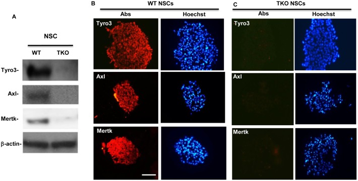
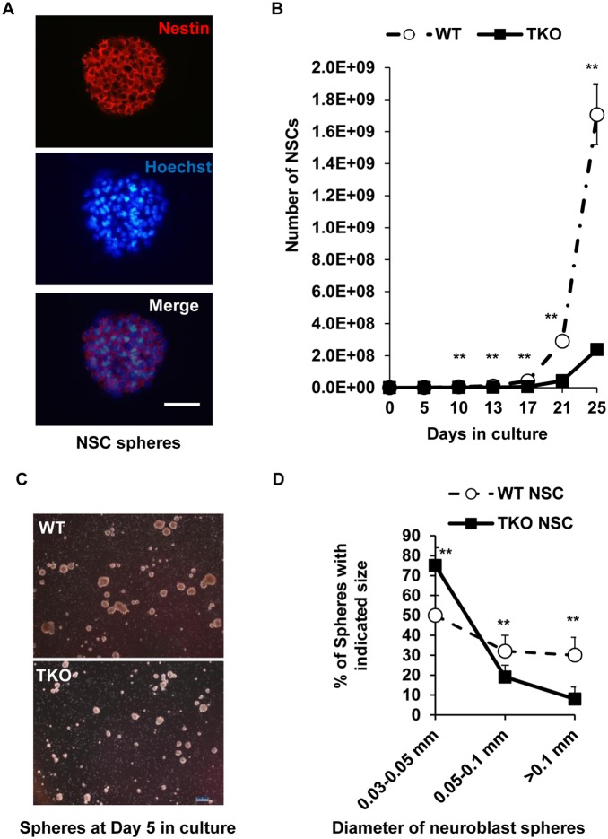
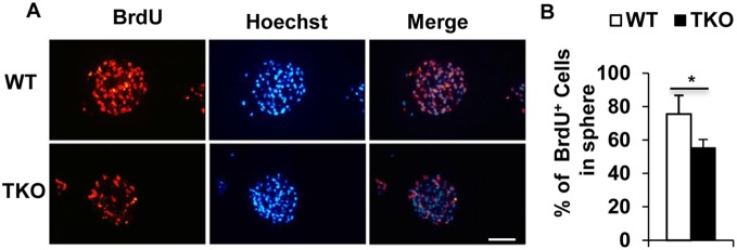
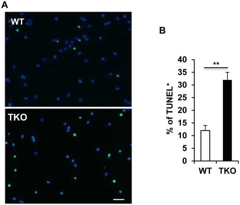
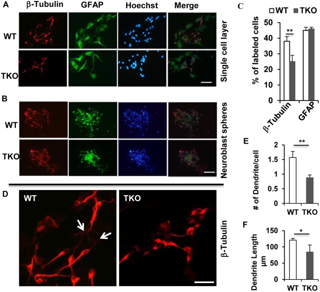
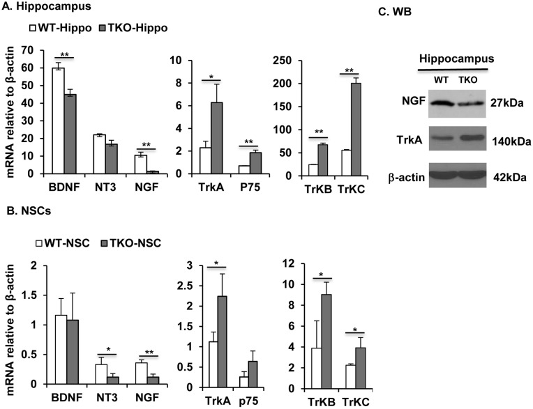
Tyro3, Axl and Mertk (TAM) receptor tyrosine kinases play multiple functional roles by either providing intrinsic trophic support for cell growth or regulating the expression of target genes that are important in the homeostatic regulation of immune responses. TAM receptors have been shown to regulate adult hippocampal neurogenesis by negatively regulation of glial cell activation in central nervous system (CNS). In the present study, we further demonstrated that all three TAM receptors were expressed by cultured primary neural stem cells (NSCs) and played a direct growth trophic role in NSCs proliferation, neuronal differentiation and survival. The cultured primary NSCs lacking TAM receptors exhibited slower growth, reduced proliferation and increased apoptosis as shown by decreased BrdU incorporation and increased TUNEL labeling, than those from the WT NSCs. In addition, the neuronal differentiation and maturation of the mutant NSCs were impeded, as characterized by less neuronal differentiation (β-tubulin III+) and neurite outgrowth than their WT counterparts. To elucidate the underlying mechanism that the TAM receptors play on the differentiating NSCs, we examined the expression profile of neurotrophins and their receptors by real-time qPCR on the total RNAs from hippocampus and primary NSCs; and found that the TKO NSC showed a significant reduction in the expression of both nerve growth factor (NGF) and brain-derived neurotrophic factor (BDNF), but accompanied by compensational increases in the expression of the TrkA, TrkB, TrkC and p75 receptors. These results suggest that TAM receptors support NSCs survival, proliferation and differentiation by regulating expression of neurotrophins, especially the NGF.


Conflict of interest statement

Competing Interests: The authors have declared that no competing interests exist.

Figures

Figure 1
Figure 1. Tyro3, Axl, and Mertk receptors are expressed in primary hippocampal NSCs.
(A) Examination of TAM receptors expression in hippocampal NSCs by western blotting. (B and C) NSC spheres were obtained and processed for immunohistochemistry following the procedures described in the Methods. The cryosections were immunostained with anti-Tyro3, -Axl or -Mertk antibodies, accordingly (B, red) and nuclei were counter-stained with Hoechst 33342 (blue). The NSC spheres from the TKO mice were used as the negative control (C). Scale bar, 100 µM representing all the scale in (B and C).
Figure 2
Figure 2. TAM receptors support primary NSCs growth and survival.
(A) The WT NSC spheres were subjected to cryostat sectioning as described in the Method. The sections were immunostained using anti-nestin antibody, and nuclei were stained blue using Hoechst 33342. Scale bar, 100 µm. (B) To maintain healthy growth of the NSCs, neuronal spheres were subcultured as described in the Method. The total cell number at each time point was calculated based on the cell number from each count and the fraction of the cells used for the next cycle of subculture. The growth curves are plotted as total cell numbers against days in culture. Data are expressed as the means ± SD at each time point. **P<0.01, n = 3. (C) NSC spheres were developed from single cell suspension of both WT and TKO NSCs in 5 days and photographed, scale bar, 200 µm. (D) The newly developed spheres in each of the WT and TKO samples were designated into three groups based on the sphere sizes of large (>0.1 mm), middle (0.05–0.1 mm) and small (0.03–0.05 mm) in diameter. The number of spheres in each group was counted and the percentage of each group among the same phenotype spheres was plotted. The data are expressed as the mean ± SD of three experiments. **P<0.01, n = 3.
Figure 3
Figure 3. TAM receptors support primary NSCs proliferation.
(A) A representative picture showing decreased BrdU incorporation into TKO NSC spheres. The proliferating stem cells were labeled with BrdU and identified with anti-BrdU antibody (red) and counter-stained with Hoechst 33342 for visualization of nuclei (blue). Scale bar, 200 m. (B) The percentage of BrdU-positive NSCs in total Hoechst stained cells are expressed as means ± SD. *P<0.05, n = 3.
Figure 4
Figure 4. TAM receptors prevent primary NSCs death from apoptosis.
The apoptotic cells in the differentiating spheres were labeled by TUNEL assay following manufacture’s instruction (Roche). Representative pictures in (A) show TUNEL staining of the differentiated WT and TKO NSCs. Scale bar, 50 µm. (B) Percentage of apoptotic cells in total differentiated NSCs. Data was presented as mean ± SD, **P<0.01, n = 3.
Figure 5
Figure 5. TAM receptors regulate primary NSCs differentiation.
Neuronal differentiation was conducted following the procedure in the Method. The differentiated cells were identified by anti-β-Tubulin and anti-GFAP antibodies and nuclei were stained by Hoechst 33342 dye. Images were observed and the photographs were taken using a Zeiss Apotome microscope. Representative pictures show the newly-differentiated neurons (β-Tubulin) and astrocytes (GFAP) in the single cell layers (A) or neuroblast spheres (B), scale bar, 200 µm. (C) The percentage of cells expressing lineage-specific markers for neuron (β-tubulin) and astrocyte(GFAP) was plotted. Data was presented as Mean ± SD **P<0.01, n = 3. (D) The β-tubulin-III positive neurons in (A) were taken at higher magnification showing neurite outgrowth in the WT but not in the TKO cells, scale bar, 50 µm. (E) and (F) show significant difference in The number of dendrite per cell (E) and the dendrite length (F) between the WT and TKO groups were significantly different. Data was Mean ± SD, **P<0.01 and *P<0.05, n = 20.
Figure 6
Figure 6. TAM receptors regulate expression of neurotrophins and their receptors.
(A, B) Real-time qPCR quantification of neurotrophins, BDNF, NT-3, NGF and their receptors, TrkA, p75, TrkB and TrkC in hippocampus (A) and NSCs (B). Data are shown as means ± SD, *P<0.05 and **p<0.01, n = 3. (C) Western blotting of NGF and TrkA level in WT and TKO hippocampus. This is one representative of three mice.

References

    1. Eriksson PS, Perfilieva E, Bjork-Eriksson T, Alborn AM, Nordborg C, et al. (1998) Neurogenesis in the adult human hippocampus. Nat Med 4:1313–1317. - PubMed
    1. Gage FH (2000) Mammalian neural stem cells. Science 287:1433–1438. - PubMed
    1. Ming GL, Song H (2011) Adult neurogenesis in the mammalian brain: significant answers and significant questions. Neuron 70:687–702. - PMC - PubMed
    1. Zhao C, Teng EM, Summers RG Jr, Ming GL, Gage FH (2006) Distinct morphological stages of dentate granule neuron maturation in the adult mouse hippocampus. J Neurosci 26:3–11. - PMC - PubMed
    1. Deng W, Aimone JB, Gage FH (2010) New neurons and new memories: how does adult hippocampal neurogenesis affect learning and memory? Nat Rev Neurosci 11:339–350. - PMC - PubMed

Publication types

MeSH terms